Case Report
Austin J Urol. 2015;2(3): 1026.
Metastatic Myoepithelioma in a Solitary Kidney Treated with Laparoscopic Microwave Ablation
Feng T¹*, Dru CJ¹, Gershman A¹ and Julien P²
¹Division of Urology, Cedars-Sinai Medical Center, USA
²Department of Radiology, Cedars-Sinai Medical Center, USA
*Corresponding author: Feng T, Division of Urology,Cedars-Sinai Medical Center, 8635 West Third Street,Suite 1070 West, Los Angeles, CA 90048, USA
Received: February 05, 2015; Accepted: April 17, 2015; Published: April 21, 2015
Abstract
Myoepithelioma is a rare entity and represents less than 1% of soft tissue tumors. We present a rare case of myoepithelioma that has metastasized to the kidney and was treated successfully with laparoscopic microwave ablation.
Keywords: Microwave ablation; Laparoscopic; Kidney; Myoepithelioma
Abbreviations
MRI: Magnetic Resonance Imaging
Case Presentation
Myoepithelioma is a rare entity and represents less than 1% of soft tissue tumors [1]. They commonly occur in salivary glands but have also been reported in the larynx, breast, and bone [2]. Genitourinary involvement by myoepithelioma is even rarer and to date, only one case report of myoepithelioma in the kidney has been documented [3].
We present a case of a 47-year-old female with metastatic myoepithelioma to the kidneys. The patient initially presented with a soft tissue mass in the right foot at the age of 20. The biopsy of the mass resulted in a tumor of myoepithelioma of soft tissue of uncertain malignant potential. She underwent wide resection of the primary lesion and did well until 2011, at which time she was diagnosed with a right kidney mass along with several lung lesions. She underwent a right radical nephrectomy and subsequent thoracoscopic wedge resections of the lung nodules. Pathology of the renal specimen and lung nodules were similar to that of the foot . Thus, it was concluded that they all represented malignant myoepithelioma. She was then followed every 3 months with chest and abdominal imaging to evaluate for recurrences.
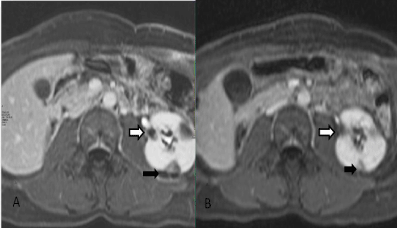
Since 2011, she has developed new metastatic lesions in her lungs and solitary left kidney. Of note, she has never had any symptoms attributed to her disease. Given that she has a solitary kidney, ablation of these renal and the lung lesion was recommended. In 2013, she underwent microwave ablation of the left lung mass as well as two left kidney lesions. One year later, she presented with a new 1-cm lesion near the left renal hilum seen on MRI (Figure 1a). Given the need for nephron sparing,another ablation procedure was recommended. However, given the location of the tumor, there was concern for potential thermal injury to the collecting system from a percutaneous method. A laparoscopic exposure of the renal system with concomitant microwave ablation was then chosen.

    Figure 1:  a) Pre-procedural axial MRI showing a 1-cm enhancing lesion in
the mid pole of the left kidney (white arrow). Black arrow shows prior site of
ablation. b) Post-procedure axial MRI showing the same lesion treated with
microwave ablation. There is no enhancement of the lesion.
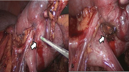
The standard exposure of the kidney was performed laparoscopically. The left ureter and renal pelvis were then dissected freely from the abutting tumor seen on the posterior medial margin of the kidney (Figure 2a). Microwave ablation was then performed with the ureter retracted away to prevent thermal injury. Complete thermal ablation of the lesion without injury to surrounding structures was confirmed at the conclusion of the procedure (Figure 2b).

    Figure 2:  Intraoperative photograph of the lesion (white arrow) and its
close proximity to the left ureteropelvic junction. b) Post thermal ablation
image of same lesion.
The patient’s postoperative course was unremarkable and she was discharged home on postoperative day one. Her post-procedure MRI at 3 months confirmed ablation of the lesion by absence of enhancement (Figure 1b) and her kidney function has remained normal.
Discussion/Conclusion
Myoepithelioma is a rare soft tissue tumor. It was recognized as a histologically distinct entity by the World Health Organization in 1991 [1]. Myoepithelial tumors represent a diverse morphological group of tumors, composed of epithelial and myoepithelial elements. Myoepithelioma of the kidney is extremely rare and only one case of report of renal myoepithelioma has been reported to date [3].We present a unique case of a primary myoepithelioma of the foot that metastasized to the lungs and kidneys, ultimately requiring multiple surgical interventions.
Review of our patient’s right nephrectomy specimen showed a kidney with malignant biphasic epithelial and myoepithelial neoplasm with chondroid, squamous, and glandular elements. The presence of nuclear atypical and together with a high mitotic rate, tumor necrosis, and immunostaining profile support the diagnosis of malignant myoepithelioma or myoepithelial carcinoma. This is consistent with the findings by Hornick et al. in their characterization of 101 myoepithelial tumors of soft tissue [4]. They classified tumors with benign morphology or mild cytologic atypia as myoepithelioma or mixed tumor and those with moderate to severe atypia as myoepithelial carcinoma. Of the 31 malignant cases, there was a 42% rate of local recurrence and a 32% rate of metastasis. This contrasts to the 18% local recurrence rate for benign myoepitheliomas.
Most cases of myoepitheliomas are treated with wide resection as our patient had with the initial primary tumor and subsequent right kidney and lung masses. However, recurrence of disease is not uncommon for malignant myoepitheliomas as described above. Our patient ultimately required additional minimally invasive urological procedures to treat recurrences in her solitary kidney. The last recurrence is noteworthy given the close proximity to the collecting system. A laparoscopic exposure for microwave ablation rather than radiographically guided ablation was employed to minimize potential thermal injury to the collecting system.
In conclusion, we present a case of a 47-year-old female with malignant myoepithelioma that has metastasized to the lungs and kidneys. We demonstrate the advantages of minimally invasive urological techniques in treatment of recurrences in a solitary kidney.
References
- Seifert G, Sobin LH. World Health Organization International Histological Classification of Tumours: Histological Typing of Salivary Gland Tumours. 1991; 20-21.
- Rekhi B, Amare P, Gulia A, Baisane C, Patil A, Puri A, et al. Primary intraosseousmyoepithelioma arising in the iliac bone and displaying trisomies of 11, 15, 17 with del (16q) and del (22q11)--A rare case report with review of literature. Pathol Res Pract. 2011; 12: 780-785.
- Gao H, Liu C, Zou H, Chun C, Cui X, Chen Y et al. Parachordoma/myoepithelioma of the kidney: first report of a myxoid mimicry in an unusual location. Int J ClinExpPathol. 2014; 7: 1258–1265.
- Hornick JL, Fletcher CD. Myoepithelial tumors of soft tissue: a clinicopathologic and immunohistochemical study of 101 cases with evaluation of prognostic parameters. Am J SurgPathol. 2003; 27: 1183-1196.