Case Report
Austin J Urol. 2015;2(3): 1028.
Paraganglioma of the Urinary Bladder: A Case Report and Review of Literature
Sriram Krishnamoorthy*, Leena Dennis Joseph, Susruthan M, Kanimozhi G, Sunil Shroff
Department of Urology, Sri Ramachandra Medical College, India
*Corresponding author: Sriram Krishnamoorthy, Additional Professor, Department of Urology, Sri Ramachandra. Medical College & RI, Chennai, India
Received: March 21, 2015; Accepted: March 23, 2015; Published: July 30, 2015
Abstract
Paraganglioma of the urinary bladder is a rare tumour with characteristic histopathological and immunehistochemical features. It is difficult to differentiate the tumour as benign or malignant based on the histological findings alone. Recognition of this tumour is important for the proper management of these patients, as these patients may or may not be having bothersome symptoms.
Extra adrenal pheochromocytomas are known as paragangliomas. Majority of these Extra adrenal tumours occur intraabdominally along the sympathetic chain. These tumours of the sympathetic nervous tissue may be non-functional or functional and when they secrete catecholamine, they cause paroxysmal hypertension, palpitation, and micturition syncope.
The current report presents a case of a 36 year old male, who was suffering from recurrent dysuria and vague supra pubic discomfort for the past six months. Clinical evaluation and imaging were normal. Cystoscopy revealed a small sessile polyp over the dome of the bladder. Histopathology showed the characteristic ‘zellballen’ pattern which was confirmed by immunohistochemical staining. The purpose of this case report is to highlight the rarity of the tumour, the various unusual modes of presentation and to stress on the need to forewarn the surgeons and the anaesthetists of the possible intra-operative hypertensive crisis during resection of such tumours.
Keywords: Paraganglioma; urinary bladder; synaptophysin
Introduction
Pheochromocytomas are tumours of the chromaffin cells, derived from the embryonic neural crest. They are most commonly located in the adrenal medulla. However 10% of these tumours are reported at Extra adrenal locations [1]. Such Extra adrenal pheochromocytomas are known as paragangliomas. Majority of these Extra adrenal tumours occur intra abdominally along the sympathetic chain. Primary paragangliomas of the urinary bladder are very rare, accounting for less than 0.05% of all bladder neoplasms and less than 1% of all pheochromocytomas[2]. Paraganglioma of the urinary bladder is a rare tumour that originates from the embryonic rests of chromaffin tissue of the sympathetic nervous system associated with the urinary bladder wall. Nearly 80% of the paragangliomas of the genito urinary tract arise from the urinary bladder, followed by a decreasing order of frequency involving the urethra, renal pelvis and the ureters [3].
These patients usually present with a symptom complex of hypertension, headache, sweating and palpitation associated with micturition. These are attributed to the catecholamine release from the tumour.
Case Report
We report a case of a 36 year old male patient, who presented with complains of recurrent mild dysuria and supra pubic discomfort for the past six months. He also complained of burning micturition. There was no history of hematuria, pyuria or lithuria. The patient did not complain of fever, chills nausea or vomiting. He is a known hypertensive for the past three years, on regular medication. There was no history suggesting a paraganglioma of the bladder like micturition syncope, headache, sweating or palpitation. He is non alcoholic, non smoker with regular bowel habits. On physical examination, his general condition was good. All the laboratory parameters were within normal limits. Urine examination was normal and urine culture was sterile. Ultrasound of the bladder and kidneys were normal and uroflow showed a satisfactory voiding pattern with insignificant residual urine.
In view of unexplained lower urinary tract symptoms, cystoscopy was done which showed a small sessile polyp measuring approximately 0.5 cms over the dome of the bladder. A clinical suspicion of urothelial lesion was suggested. The tumour was resected and sent for histopathological examination as multiple tiny fragments, which were all embedded.
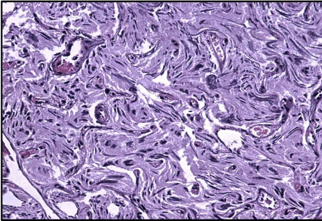
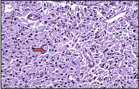
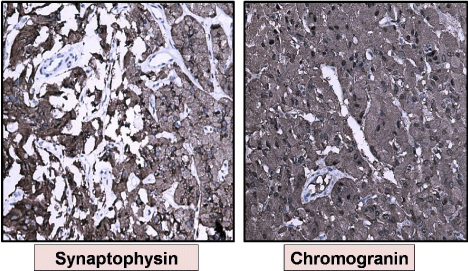
On light microscopy, the tumour cells were seen as well circumscribed nodules with a characteristic ‘zellballen’ pattern (Figure 1). The individual tumour cells were polygonal with a granular cytoplasm. There was a delicate fibro vascular stroma surrounding the tumour cell clusters (Figure 2). A provisional diagnosis of paraganglioma was given with an advice for immunohistochemical confirmation. Immunohistochemistry stains were positive for synaptophysin, vimentin and chromogranin (Figure 3). There was no invasion into the underlying muscle. There was no evidence of necrosis or abnormal mitosis. As the tumour has been completely resected, the patient was advised to come for a regular follow up.

Figure 1: Tumor Cells with a Zellballen Pattern (H&E&E×200).

Figure 2: Tumor Cells with Granular Cytoplasm (H&E&E×200).

    Figure 3:  Synaptophysin and Chromogranin positively (IHC &E× 200, seen as
brown colour).
Discussion
Paragangliomas are rare tumours arising from the chromaffin tissue of the sympathetic nervous system. Extra adrenal locations are uncommon. Paragangliomas were first reported by Zimmerman et al in 1953 [4]. These tumours of the sympathetic system may be functional or non-functional. When they secrete catecholamines they cause the characteristic symptoms in these patients. Most of these patients present with paroxysmal hypertension, palpitation and micturition syncope. Some of the patients also present with headache or blurred vision during micturition [5]. Some patients present only with hematuria. Biogenic amine levels in urine and plasma may be helpful, if classic symptoms are present. Majority of the tumours are located over the dome or the trigone of the bladder. In our patient, the tumour was located over the dome of the bladder.
Histological examination shows a classic zellballen pattern, but at times it can cause diagnostic difficulties with urothelial carcinoma [6]. The problem arises when it lacks the classic pattern, when there is invasion into the muscularis propria, when there are cautery artifacts and when classic symptoms are not present. However differentiation between the two entities is important as therapeutic modalities are different for both. In difficult situations, immunohistochemical confirmation with synaptophysin and chromogranin may be helpful to confirm the diagnosis of paraganglioma. Linnoila et.al, in their study on 120 adrenal and Extra adrenal paragangliomas, observed that Extra adrenal location, coarse nodularity of the primary tumour, confluent tumour necrosis and absence of hyaline globules were most predictive of malignancy [7].
Approximately 10% of bladder paragangliomas are malignant with local invasion, regional lymph node metastasis or distant spread. Paragangliomas have the characteristic tendency to cause local invasion which fits the condition under malignant category, but in view of a lack of cellular dissociation and absence of mitoses, they follow a benign course of disease progression [8]. Histologically there are no definitive features which can differentiate benign from malignant tumours. Nuclear pleomorphism, mitotic figures and necrosis might point towards malignant potential, but are not reliable predictors of the clinical outcome [9]. DNA ploidy has been considered to be one of the useful prognostic factors in distinguishing benign from malignant paragangliomas. Pang et.al performed DNA ploidy studies by Flow cytometry in 79 pheochromocytomas and paragangliomas and observed that non diploid tumours are considered to have an aggressive behaviour than diploid tumours [10]. However, Grignon etal, from their observation on three cases of urinary bladder paragangliomas, reported that DNA ploidy can only be used as a predictor of malignant behaviour of the tumour and cannot be considered as a diagnostic criterion for malignancies of urinary bladder paragangliomas [11].
Both CT scanning and MRI are useful in the localisation of the primary tumour and metastasis. Nuclear scan using Meta Iodine Benzyl Guanidine (MIBG) is an investigation with high sensitivity and specificity for detection of these tumours. Recognition of these tumours as an entity, in the differential diagnosis of bladder tumours is important as hypertensive crisis may be provoked by the tumour manipulation intraoperatively [12].
The classic triad of episodic hypertension, persistent hematuria and post-micturition syncope is almost confirmative of paragangliomas of urinary bladder, but is seen only with functional catecholamine secreting tumours [13]. However, as majority of them are non-functional, this triad of symptoms is relatively infrequent. Given the low incidence of malignant bladder paragangliomas, complete surgical resection of the tumour is curative. Adrenergic block preoperatively may significantly reduce perioperative mortality and morbidity. If the tumour is seen invading the muscle, then partial or radical cystectomy is the most effective treatment [14]. Lifelong clinical follow up is needed as recurrence of a malignant paraganglioma can occur very late in the clinical course. Radiotherapy and chemotherapy have limited effectiveness for treatment of bladder paragangliomas.
Conclusion
Paragangliomas of the bladder have to be considered strongly in the differential diagnosis of atypical bladder tumours. During cystoscopy and resection of the tumour, if it is a paraganglioma, the possibility of an intra operative hypertensive crisis needs to be kept in mind and handled appropriately. The whole objective of this case report is to highlight the potential rarity of such tumours, its various atypical modes of presentation and also to emphasise the need to take adequate precautions by the surgeons and the anaesthetists during trans urethral resection in order to avoid the possible intra-operative hypertensive crisis.
References
- Alderazi Y,Yeh MW,Robinson BG, Benn DE et.al. Phaeochromcytoma: current concepts. MJA. 2005; 183:201-204.
- Beilan J, Lawton A, Hajdenberg J, Rosser CJ. Locally advanced paraganglioma of the urinary bladder: a case report. BMC Res Notes. 2013; 6:156.
- Hanji AM, Rohan VS, Patel JJ, Tankshali RA. Pheochromocytoma of the Urinary bladder: a rare cause of severe hypertension. Saudi J Kidney Dis Transpl. 2012; 23: 813-816.
- Zimmerman IJ, Biron RE, Macmahon HE. Phaeochromocytoma of the urinary bladder. The New England Journal of Medicine.1953; 249: 25-26.
- Pahwa HS, Kumar A, Srivastava R, Misra S, Goel MM. Urinary bladder paraganglioma – a case series with proposed treating algorithm based on our experience and review of literature. Indian J Surg Oncol. 2013; 4: 294 - 297.
- Zhou M, Epstein JI, Young RH. Paraganglioma of the urinary bladder. A lesion that may be misdiagnosed as urothelial carcinoma in trans urethral resection specimens. Am J Surg pathol. 2004; 28: 94-100.
- Linnoila RI, Keiser HR, Steinberg SM, Lack EE. Histopathology of benign versus malignant sympathoadrenal paragangliomas: clinico pathologic study of 120 cases including histological features. Hum Pathol. 1990; 21: 1168-1180.
- Cheng L, Leibovich BC, Cheville JC, Ramnani DM, Sebo TJ, Neumann RM, Nascimento AG, Zincke H, Bostwick DG. Paraganglioma of the urinary bladder: can biologic potential be predicted? Cancer. 2000; 88: 844-852.
- Davaris P, Petraki K, Arvanitis D, Papacharalammpous N,Morakis A, Zorzos S. Urinary bladder paraganglioma. Pathol Res Pract 1986; 181: 101–106.
- Pang LC, Tsao KC. Flow cytometric DNA analysis for the determination of malignant potential in adrenal and Extra-adrenal pheochromocytomas or paragangliomas. Arch Pathol Lab Med. 1993;1142-1147.
- Grignon DJ, Ro JY, Mackay B, Ordonez NG, el-Naggar A, Molina TJ, Shum DT Paraganglioma of the urinary bladder: immunohistochemical, ultra structural, and DNA flow cytometric studies. Hum Pathol. 1991; 22: 1162-1169.
- I Mckenzie. Paraganglioma of the bladder - A case report and review. The internet Journal of Urology. 2008; 6: 1.
- Shikha Goyal, Tarun Puri, Gowthaman Gunabushanam, Anup Kumar Das, Pramod Kumar Julka. Skeletal metastases from recurrent paraganglioma of the urinary bladder. Ind J Urol. 2005; 21: 122-124.
- Ahn SG, Jang H, Han DS, Lee JU, Yuk SM. Trans Urethral Resection Of Bladder Tumour (TURBT) as an optional treatment method on pheochromocytoma of the urinary bladder. Can Urol Assoc J. 2013 ;7:130-140.