Case Report
Austin J Urol. 2016; 3(1): 1038.
Laparoscopic Renal-Sparing Radical Excision of a Perirenal Sclerosing Well-Differentiated Liposarcoma - A Case Report and Literature Review
Feng L¹, Li J¹, Zhang D¹ and Luo Y²*
¹Department of Urinary, Capital Medical University, China
²Department of Urology, University of Iowa Carver College of Medicine, USA
*Corresponding author: Yi Luo, Department of Urology, University of Iowa Carver College of Medicine, Iowa City, IA 52242, USA
Received: September 02, 2015; Accepted: January 04, 2015; Published: January 05, 2016
Abstract
A 72-year-old man presented with a 12×8×4 cm soft tissue mass located in the upper pole of the left kidney. The inferior pole of the kidney was encased by the mass. To minimize surgical damage, we performed laparoscopic renalsparing excision of the mass via transperitoneal route. In an effort to obtain negative surgical margins, the capsule of the kidney was resected along with the mass. The operation lasted 135 min and the estimated blood loss was 100 ml. No perioperative complications were encountered. Pathology confirmed the mass to be a retroperitoneal perirenal sclerosing well-differentiated liposarcoma. All surgical margins were negative. During the past 18-month follow-up, the patient reported no complaints and showed no recurrence of the tumor. Excision of the liposarcoma mass along with the renal capsule provided the patient the best opportunity to have negative surgical margins and kidney preservation. The laparoscopic renal-sparing radical excision of perirenal well-differentiated liposarcoma was safe and feasible. However, the indication should be carefully reviewed by taking into consideration the size and location of the tumors and the surgeon’s preference. The long-term outcomes of the laparoscopic surgery will need to be determined.
Keywords: Liposarcoma; Perirenal; Laparoscope; Kidney
Introduction
Liposarcoma accounts for approximately 20% of soft tissue sarcomas, which typically occurs in the lower extremities and retroperitoneum in middle to older aged males [1]. Retroperitoneal liposarcoma encasing the kidney is rare and is typically treated by complete surgical resection involving kidney removal along with excision of perirenal liposarcomas. Tumors that are large or adjacent to other organs generally require complete resection with open surgery. However, renal-sparing surgery should be considered in select patients, such as those with solitary kidneys or chronic renal insufficiency. There are limited reports on the use of laparoscopic surgery for retroperitoneal liposarcoma [2-6]. To our knowledge, there is no report on laparoscopic renal-sparing surgery for retroperitoneal perirenal liposarcomas. Here, we report a case of a 72-year-old male patient with perirenal liposarcoma who underwent laparoscopic renal-sparing radical excision of the tumor.
Case Presentation
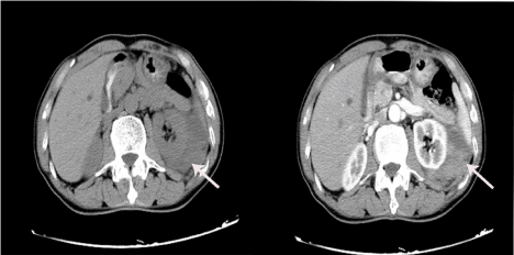
A 72-year-old Chinese male presented for a routine physical exam. Abdominal ultrasound revealed a 12×8 cm hypoechoic mass surrounding the left kidney. The patient reported no weight loss, nausea, vomiting, fevers, chills, night sweats, hematuria, and other symptoms. A CT scan of the abdomen and pelvis revealed a 12×8×4 cm soft tissue mass located in the upper pole of the left kidney. The mass was downward extended to the middle and inferior pole of the kidney. The inferior pole of the kidney was encased by the mass. The attenuation coefficient of the cyst in plain scan was -11~15 HU and 22~69 HU in contrast enhanced CT scanning (Figure 1).

    Figure 1:  CT scan showed the tumor surrounding the left kidney indicated
by arrows.
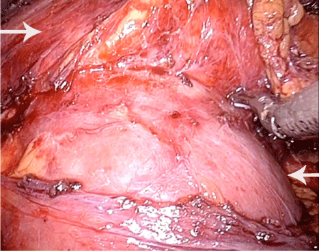
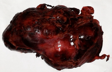
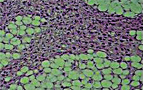
The mass was slightly enhanced and was composed largely of lipomatous mass and nonlipomatous components with a septum. The patient refused nephrectomy due to age and poor renal function, citing a 20-year history of diabetes mellitus, abnormal creatinine, and a concern about possible dialysis and recovery post-nephrectomy. Therefore, a transperitoneal laparoscopic renal-sparing excision of the perirenal liposarcoma was planned. Under general anesthesia, the patient was placed in a flank position and supported by adequate padding. 4-ports were placed, with a 10-mm camera port at the umbilicus, two 5-mm working ports at the subcostal area and midway between the umbilicus and anterior superior iliac spine, and another 10-mm tracer lateral to the rectus muscle at the level of the umbilicus. After mobilization of the descending colon from the retroperitoneum, the kidney and the well-capsulated mass were exposed. Perirenal fat and the renal capsule were opened from normal tissues to expose the renal cortex, and further dissections were performed through the plane between the renal parenchyma and the renal capsule using a blunt instrument or suction tube (Figure 2). Hemorrhagic spots on the renal parenchyma were blocked by bipolar coagulation scalpel. The specimen was removed via an extended umbilical incision using a laparoscopic homemade triangular tissueretrieval bag. A rubber drainage tube was placed and the incisions were closed subcutaneously. The operation time was 135 min and the estimated blood loss was 100ml. No perioperative complications were encountered. The patient was on a normal diet at 24 hours and was discharged 6 days postoperatively. Macro pathology showed that the mass had a capsule and was firm but pliable (Figure 3). Final pathology indicated the mass to be a retroperitoneal perirenal sclerosing well-differentiated liposarcoma (Figure 4). All surgical margins were negative. The patient was followed with CT scan every 6 months. During 18-months’ follow-up, the patient reported no symptoms with no recurrence of the tumor. Serum creatinine did not change significantly.

    Figure 2:  Excision of the tumor was performed through the plane between
the renal parenchyma and renal capsule. The top-left arrow indicates the
tumor and the bottom-right arrow indicates the renal parenchyma.

Figure 3: The tumor had a capsule and was firm but pliable in texture.

    Figure 4:  Histological H&E staining of the tumor. The tumor section showed
a moderate density of tumor cells. Some tumor cells were heteromorphic.
Sporadic fat tissues were noted in certain regions. Final pathology confirmed
the mass to be a retroperitoneal perirenal sclerosing well-differentiated
liposarcoma. Magnification: x20.
Discussion
Retroperitoneal liposarcoma can be pathologically categorized into four groups: well-differentiated liposarcoma, myxoid/round cell liposarcoma, de-differentiated liposarcoma, and pleomorphic liposarcoma [7]. Well-differentiated liposarcoma accounts for 40–45% of all liposarcomas [8,9] and can be classified as lipomalike, sclerosing, inflammatory, and spindle cell types [10]. The different histological subtypes of liposarcomas show different clinical prognosis. Well-differentiated liposarcoma is considered to be a lowgrade lesion and its 5-year survival rate can be as high as 80% [9,11]. It is considered to have minimal metastatic potential. However, the local recurrence rate of well-differentiated liposarcoma is approximately 40–60%, especially for those originating in the retroperitoneum [9]. For different subtypes of well-differentiated liposarcomas, the prognosis appears to be different. Kooby and associates have reported that patients with sclerosing liposarcomas had a better recurrencefree survival than non-sclerosing liposarcomas [11].
As surgical excision remains the dominant modality of curative therapy for liposarcomas, a wide marginal excision is strongly recommended. However, for many cases, especially when the tumors are located in deep soft-tissue sites, neurovascular components and critical organs adjacent to the tumor make a wide marginal excision impossible. Generally, for perirenal liposarcoma a standard en bloc dissection to remove all elements including the kidney and perirenal fat is needed. This kidney-loss dissection is sometimes unacceptable for patients who have solitary kidney or chronic renal insufficiency. There have been a few clinical trials on renal-sparing surgery in liposarcoma patients (Table 1). Interestingly, all patients in these reports had the same well-differentiated liposarcomas as our patient. The mean follow-up time was 22 months and no recurrence of tumor was found [12,13]. Singer and associates evaluated various factors that might contribute to the recurrence and overall survival after surgical resection of primary retroperitoneal liposarcomas [7]. They noted that surgical resection requiring nephrectomy for complete resection had no influence on disease-specific survival. Since this subtype of lipoma-like, well-differentiated liposarcoma has an optimistic prognosis [12,13], renal-sparing surgery should be considered. In order to obtain negative surgical margins, we resected the capsule of the kidney along with the tumor as reported by others [12,13]. The plane between the renal capsule and parenchyma was identified first, followed by dissections performed through the plane. There was no renal parenchyma involved, which made the renal-sparing surgery feasible with no compromise of tumor-free margins. With regard to surgical management of retroperitoneal perirenal liposarcomas, open surgery is used for most cases. There are 6 reported cases, including the current report, using laparoscopic resection for retroperitoneal liposarcomas [2-6] (Table 2).
Ref.
Age
Sex
Size (cm)
Surgical approach
Complication
Pathologic diagnosis
Follow-up months
Recurrence
12
65
Male
32×22×8
Open
Partial hemidiaphragm resection urinary retention
Sclerosing well differentiated liposarcoma
22
None
13
50
Male
12×10
Open
None
Well-differentiated
liposarcoma focal invasion
of the renal capsule24
None
13
57
Female
10×8
Open
None
Well-differentiated
liposarcoma invasion of the
renal capsule24
None
Present
72
Male
12×8×4
Laparoscope
None
Sclerosing
well-differentiated
liposarcoma18
None
Table 1: Renal-sparing surgery treatment of perirenal liposarcoma.
    Ref.
    Age
    Sex
    Size (cm)
    Location
    Operation time (min)
    Complication
    Pathologic diagnosis
    Follow-up (months)
    Recurrence


    2
    53
    Female
    10×10×7.5
    Behind the cecum
    Not stated
    Port site recurrence
    Myxoid liposarcoma
    24
    Yes


    3
    77
    Female
    9×5×4.5
    Adjacent to the left kidney
    Not stated
    None
    Well-differentiated liposarcoma
    6
    None


    4
    36
    Female
    26
    Encasing the right kidney
    Not stated
    Not stated
    Sclerosing well-differentiated    iposarcoma
    11
    None


    5
    61
    Female
    10
    Adjacent to the right kidney
    150
    None
    Well-differentiated liposarcoma
    12
    None


    6
    72
    Male
    6.3×4.8×3.8
    Behind the descending colon
    120
    None
    Well-differentiated liposarcoma
    24
    None


    Present
    72
    Male
    12×8×4
    Encasing the left kidney
    135
    None
    Sclerosing well-differentiated    liposarcoma
    18
    None
Table 2: Laparoscopic treatment of retroperitoneal liposarcoma.
Among these 6 cases, 4 were women and 2 were men, with a mean age of 61.8 years (range 36–77). In 4 cases, the tumors were located adjacent to the kidney and all were well-differentiated liposarcomas. Three patients underwent nephrectomy and our case had renalsparing surgery. Laparoscopic surgery provides an amplified intraoperative view compared to the traditional open procedure. Therefore, dissections could be safely performed through the plane between the renal capsule and parenchyma. As shown in (Table 2), the overall prognosis of laparoscopic resection of retroperitoneal liposarcomas was satisfactory and the only recurrent case was myxoid liposarcoma. It is believed that the difference in the recurrence rates by location is due to the difficulties in obtaining adequate surgical margins. Generally, patients with microscopically negative margins show higher disease-specific survival rates than those with microscopically positive margins [14]. Although our patient had a negative margin and showed no recurrence of the tumor 18 months post-surgery, long-term follow up is necessary since local recurrence of perirenal liposarcomas has been reported in some cases [9].
Conclusion
Laparoscopic renal-sparing radical excision of perirenal well- differentiated liposarcoma is safe and feasible. However, indications should be carefully reviewed, taking into consideration size and location of the tumors and the surgeon’s preference. Long-term outcomes are yet to be determined.
References
- Toro JR, Travis LB, Wu HJ, Zhu K, Fletcher CD, Devesa SS. Incidence patterns of soft tissue sarcomas, regardless of primary site, in the surveillance, epidemiology and end results program, 1978-2001: An analysis of 26,758 cases. Int J Cancer. 2006; 119: 2922-2930.
- Horiguchi A, Saito S, Baba S, Murai M, Mukai M. Port site recurrence after laparoscopic resection of retroperitoneal liposarcoma. J Urol. 1998; 159: 1296-1297.
- Ball AJ, Siddiq FM, Garcia M, Ganjei-Azar P, Leveillee RJ. Hand-assisted laparoscopic removal of retroperitoneal liposarcoma. Urology. 2005; 65: 1226.
- Lucioni A, Orvieto MA, Chien GW, Sokoloff MH, Gerber GS, Shalhav AL. Laparoscopic diagnosis and management of perinephric adipose-containing lesions. J Endourol. 2006; 20: 130-132.
- Dalpiaz O, Gidaro S, Lipsky K, Schips L. Case report: Laparoscopic removal of 10-cm retroperitoneal liposarcoma. J Endourol. 2007; 21: 83-84.
- Nomura R, Tokumura H, Matsumura N. Laparoscopic resection of a retroperitoneal liposarcoma: a case report and review of the literature. Int Surg. 2013; 98: 219-222.
- Singer S, Antonescu CR, Riedel E, Brennan MF. Histologic subtype and margin of resection predict pattern of recurrence and survival for retroperitoneal liposarcoma. Ann Surg. 2003; 238: 358-370.
- Engstrom K, Bergh P, Gustafson P, Hultborn R, Johansson H, Lofvenberg R, et al. Liposarcoma: outcome based on the Scandinavian Sarcoma Group register. Cancer. 2008; 113: 1649-1656.
- Kikuchi N, Nakashima T, Fukushima J, Nariyama K, Komune S. Well-differentiated liposarcoma arising in the parapharyngeal space: a case report and review of the literature. J Laryngol Otol. 2015; 129: 86-90.
- Bestic JM, Kransdorf MJ, White LM, Bridges MD, Murphey MD, Peterson JJ, et al. Sclerosing variant of well-differentiated liposarcoma: relative prevalence and spectrum of CT and MRI features. AJR Am J Roentgenol. 2013; 201: 154-161.
- Kooby DA, Antonescu CR, Brennan MF, Singer S. Atypical lipomatous tumor/well-differentiated liposarcoma of the extremity and trunk wall: importance of histological subtype with treatment recommendations. Ann Surg Oncol. 2004; 11: 78–84.
- Gaston KE, White RL Jr, Homsi S, Teigland C. Nephron-sparing radical excision of a giant perirenal liposarcoma involving a solitary kidney. Am Surg. 2007; 73: 377-380.
- Gupta NP, Yadav R. Renal sparing surgery for perirenal liposarcoma: 24 months recurrence free follows up. Int Braz J Urol. 2007; 33: 188-191.
- Dalal KM, Kattan MW, Antonescu CR, Brennan MF, Singer S. Subtype specific prognostic monogram for patients with primary liposarcoma of the retroperitoneum, extremity, or trunk. Ann Surg. 2006; 244: 381-391.