
Case Report
Austin J Urol. 2016; 3(2): 1041.
A Rare Case of Squamous Cell Carcinoma of Kidney in a Patient with Renal Calculus and Pyonephrosis
Sharma A*, Chaudhari R, Shaikh I, Andankar M and Pathak H
Department of Urology, TNMC & BYL Nair Hospital, India
*Corresponding author: Sharma A, Department of Urology, TNMC & BYL Nair Hospital, India
Received: January 18, 2016; Accepted: January 25, 2016; Published: January 27, 2016
Abstract
Primary squamous cell carcinoma of the renal pelvis is extremely rare with only a few cases reported in the literature. Long standing renal calculus is a known risk factor. We present a case of an elderly male who had bilateral renal calculi and presented with right-sided pyonephrosis which was managed initially by percutaneous nephrostomy and later right nephrectomy was done. The histopathological report stated squamous cell carcinoma of the pelvis with capsular and fat involvement.
Keywords: Calculus; Kidney; Pyonephrosis; Squamous cell carcinoma
Case Presentation
A 52 year-old male presented with complains of fever with chills and right flank pain of 5- 6 months duration. There were no urinary complaints and no comorbid illness. There was history of bilateral Double J (DJ) stenting done for bilateral renal calculi six months back. At admission, his vitals were stable, but he was febrile. There was a tender hard lump in right flank region. Blood investigations revealed severe anaemia and raised creatinine. X-ray Kidney Ureter Bladder (KUB) showed bilateral DJ stent in situ and a right renal radio-opaque shadow. An ultrasound revealed right gross hydronephrosis (17x13x10cm) with thinning of cortex and echogenic debris suggestive of pyonephrosis; a 2.3cm calculus in the upper pole and DJ stent in situ. There was moderate hydronephrosis (14x6cm) and lost cortico-medullary differentiation in left kidney with proximal hydroureter and DJ stent in situ. Contrast Enhanced Intravenous Urography showed Right gross hydronephrosis with a 2.6x1.7cm calculus at upper pole and DJ stent in situ; Mild to moderate hydronephrosis and upper hydroureter on left side with debris within and a 6.1mm calculi in the lower pole (Figure 1). Right percutaneous nephrostomy was done which drained 20-25 cc per day. Right nephrectomy was done (Figure 2 & 3). Histopathology report stated infiltrating squamous cell carcinoma of renal pelvis with the involvement of capsule and fat.
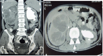
    Figure 1:  CT IVU showing Right gross hydronephrosis with a 2.6 x 1.7cm
calculus at upper pole and DJ stent in situ; Mild to moderate hydronephrosis
and upper hydroureter on left side with debris within and a 6.1mm calculi in
the lower pole.
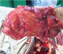
    Figure 2:  Gross specimen of right kidney suggestive of fat stranding and
bulky kidney with Percutaneous Nephrostomy Tube in situ.
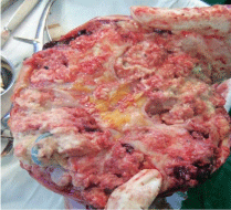
    Figure 3:  Cut-section of the gross specimen showing mass lesion in the renal
pelvis extending into the parenchyma.
Discussion
Primary Squamous Cell Carcinoma (SCC) is a rare entity and accounts for only 0.5% to 15% of all urothelial malignancies [1]. It is known to have inconclusive clinical and radiological features. The predisposing factors and causative agents include renal calculi, infection, endogenous and exogenous chemicals, hormonal imbalance and vitamin A deficiency [1,2]. It is also known to occur without any etiological factors [2]. The diagnosis is usually made at an advanced stage and the prognosis is poor.
Cancers of the kidney and renal pelvis are the ninth most common malignant cancer [3]. Transitional cell carcinoma is commoner and the SCC is rarer, seen only in 0.7% to 7% of these cases [4]. It has female preponderance and presents in the age group of 50 to 70 years [2,4].
SCC of the renal pelvis is thought to arise by the process of metaplasia of the urothelium [4]. It has recently been found associated with urinary calculi in a population-based study [5,6,7]. The pathogenesis involves tumour-supporting cytokines mediated inflammation and irritation caused by the calculi and the infection at the foci [5,8]. The other etiological factors besides calculi and infection are exogenous and endogenous chemicals, vitamin A deficiency, hormonal imbalance, schistosomiasis and smoking [4]. However, there have been cases without any apparent etiological factors [4].
The presentation of SCC of the renal pelvis is similar to that of renal calculi making the diagnosis difficult. The diagnosis heavily relies upon ultrasound, Intravenous Urography (IVU), and Contrast Enhanced Computed Tomography (CECT) Scans [5]. However, the diagnosis may not always be possible, as was with our case. The findings of SCC may be non-specific and easily missed by IVU [5].
The definitive diagnosis is made at histopathology. The diagnosis of the SCC of the renal pelvis is restricted to tumours showing extensive squamous differentiation [4]. If a significant urothelial element including urothelial carcinoma in situ is found, then the tumour should be classified as urothelial carcinoma with squamous differentiation [4]. The histologic hallmarks are pearl formation, intercellular bridges and keratotic cellular debris similar to those of SCC at any other site [4]. Except verrucous variant, most of these are moderately or poorly differentiated and are more deeply invasive than urothelial carcinoma [4].
The treatment approaches should be individualized based on the age, general condition of the patient, the grade and stage of the cancer and patient compliance [5]. Surgery, i.e., radical nephrectomy with total ureterectomy, including a bladder cuff around the ureteric orifice is the main stay of therapy in low stage patients. Systemic chemotherapy has marginal benefit [4]. Treatment with anti-EGFR therapy may be beneficial, but it needs a large number of case studies [1]. Surgery is not necessary when there are distant metastases. Radiotherapy, chemotherapy or immunotherapy could be adopted, but the effect is limited and no survival benefit has been demonstrated [5]. The prognosis of SCC of renal pelvis is very poor with a median survival of 3.5 months [4]. The poor outcome can be attributed for the advanced stage at diagnosis, but for stage for stage, the prognosis is similar for both SCC and usual urothelial carcinoma [4].
The diagnosis of renal pelvis SCC requires high index of suspicion and should be kept in mind when evaluating an elderly person with predisposing factors, especially renal calculi and infection.
References
- Sahoo TK, Das SK, Mishra C, Dhal I, Nayak R, Ali I, et al. Squamous cell carcinoma of kidney and its prognosis: a case report and review of the literature. Case Rep Urol. 2015; 2015: 469327.
- Talwar N, Dargan P, Arora MP, Sharma A, Sen AK. Primary squamous cell carcinoma of the renal pelvis masquerading as pyonephrosis: a case report. Indian J Pathol Microbiol. 2006; 49: 418-420.
- Jain A, Mittal D, Jindal A, Solanki R, Khatri S, Parikh A, et al. Incidentally detected squamous cell carcinoma of renal pelvis in patients with staghorn calculi: case series with review of the literature. ISRN Oncol. 2011; 2011: 620574.
- Bandhopadhyay R, Biswas S, Nag D, Ghosh AK. Squamous cell carcinoma of the renal pelvis presenting as hydronephrosis. Journal of Cancer Research and Therapeutics. 2010; 4: 537-539.
- Xiao J, Lei J, He L, Yin G. Renal calculus complicated with squamous cell carcinoma of renal pelvis: Report of two cases. Can Urol Assoc J. 2015; 9: 310-312.
- Verma N, Yadav G, Dhawan N, Kumar A. Squamous cell carcinoma of kidney co-existing with renal calculi: a rare tumour. BMJ Case Rep. 2011; 2011.
- Chung SD, Liu SP, Lin HC. A population-based study on the association between urinary calculi and kidney cancer. Can Urol Assoc J. 2013; 7: 716-721.
- Federico A, Morgillo F, Tuccillo C, Ciardiello F, Loguercio C. Chronic inflammation and oxidative stress in human carcinogenesis. Int J Cancer. 2007; 121: 2381-2386.