
Review Article
Austin J Urol. 2016; 3(3): 1050.
Penile Dynamic Duplex Ultrasonography
Oliveira P*, Leitao T, Oliveira T, Martinho D and Lopes T
Department of Urology, Hospital Santa Maria, Portugal
*Corresponding author: Oliveira P, Department of Urology, Hospital Santa Maria, 1649-035 Lisbon, Portugal
Received: October 04, 2016; Accepted: November 18, 2016; Published: November 23, 2016
Abstract
Ultrasonography of the penis has a well-established role in the study of penile pathology. Regarding penile anatomy, specific structures can be well identified such as the circular hyperechoic tunica albuginea or the cavernosal arteries with their parallel hyperechoic walls. A proper understanding of erection physiology, from the initial smooth muscle relaxation to the veno-occlusive mechanism at full erection, when evaluated with ultrasonography, specifically with Doppler mode, allows extrapolation of the physiological phases of erection to a spectral waveform displayed in the ultrasound screen allowing full evaluation of penile hemodynamics. A step-by-step, standardized Doppler evaluation from a flaccid state to a pharmacological induced erection represents a valuable tool in the diagnosis and management of several vascular disturbances such as erectile dysfunction, Peyronie´s disease, or Mondor´s disease. Ultrasonography is also invaluable in the correct evaluation of priapism and penile trauma.
Keywords: Penile; Doppler; Duplex; Ultrasonography
Abbreviations
US; PDDU; ED; IP; PDE5i; ICI; PSV; EDV; RI; VOD; PD; CC; CS
Introduction
Ultrasound (US) is a well-established imaging technique in the investigation of penile pathology [1]. Grayscale US allows evaluation of penile normal and pathological structures, and when combined with color and spectral Doppler provides objective and reliable evaluation of penile vasculature [2] and hemodynamics, especially with the addition of a pharmacological stimulant to produce an erection [3]. This technique, called Penile Dynamic Duplex Ultrasonography (PDDU), although not mandatory in the era of highly effective orally active agents for the treatment of Erectile Dysfunction (ED), might be necessary in primary ED (not caused by organic disease or psychogenic disorder), young patients with a history of pelvic or perineal trauma, who could benefit from potentially curative vascular surgery, patients with penile deformities which might require surgical correction (e.g., Peyronie´s disease) and for medico-legal reasons [4,5].
PDDU is a time consuming technique that should be performed in a standardized way in order to achieve an accurate clinical diagnosis [5].
Penile Anatomy
The penis consists of three cylinders [2,6], two dorsal hypoechoic Corpora Cavernosa (CC) surrounded by the thick fibrous sheath of the tunica albuginea [7,8] and the ventral Corpus Spongiosum (CS) containing the urethra (Figure 1), often compressed and difficult to visualize optimally from the ventral aspect [9]. The corpora cavernosa consist of multiple smooth muscle and endothelial-lined sinusoids, which are capable of considerable volume expansion. The albuginea is a 2-layer tunica with outer longitudinal fibers and inner circular fibers visualized as a linear hyperechoic structure [9], generally less than 2 mm thick [6]. The inner layer constitutes the intracavernosal septum, which is generally complete proximally and becomes fenestrated along the dorsal aspect in the mid-distal part. This anatomy is advantageous, making an injection to just one of the corporal bodies, enough for vasoactive medication to circulate to the contralateral side [10]. The three corpora are surrounded by the more superficial Buck´s fascia.
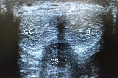
    Figure 1: Transverse mid-shaft dorsal scan showing both Corpora Cavernosa
(CC) surrounding by hyperechoic tunica albuginea (white arrow) and the
more ventral Corpus Spongiosum (CS).
The arterial supply of the penis arises from the branches of the common penile artery, which is the direct continuation of the internal pudendal artery. This artery branches into 3 named arteries. The bulbourethral artery enters de spongiosum superiorly and supplies the urethra, the spongiosum and the glans penis [10]. The dorsal artery that courses between the dorsal vein and penile nerves. The cavernosal arteries enter and supply the corpora cavernosa via numerous helicine arteries, which supply the sinusoids via arterioles (Figure 2).These are recognized by their parallel hyperechoic walls [7] (Figure 3). Anatomical variation in the penile arterial distribution may be as high as 50% [6]. Emissary veins pierce the tunica albuginea to drain into the deep dorsal vein, via the spongiosal, circumflex, and cavernosal veins.
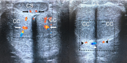
    Figure 2: Transverse mid-shaft ventral scan showing both cavernosal
arteries (white arrows), both bulbourethral arteries (black arrows), both dorsal
arteries (black arrow´s heads) and deep dorsal vein (white arrow head).
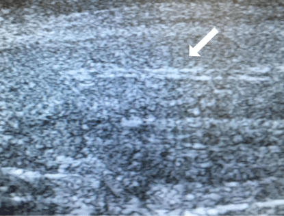
Figure 3: Cavernosal artery showing parallel hyperechoic walls (white arrow).
Physiology of Erection
In a flaccid state, the resting smooth muscle tonicity of the cavernosal arterioles and sinusoids is high with consequential low volume inflow and outflow. After appropriate neural stimulation, relaxation of the smooth muscle occurs through increased parasympathetic drive from the sacral nervous plexus, resulting in nitric oxide release from endothelial cells within the cavernosa, promoting a high inflow to the corporeal bodies. The resulting engorgement of the cavernosal sinusoids produces penile lengthening and tumescence. As the tunica albuginea elongates and expands, it begins to occlude the emissary veins between the inner circular and the outer longitudinal layers, leading to passive limitation or cessation of venous outflow. This is known as the “veno-occlusive mechanism” [6]. The high inflow and restricted outflow rapidly increases Intracorporeal Pressure (IP), and when this pressure nears systolic pressure reduction in inflow will also normally occur.
Ultrasound Technique
The examination should be performed in a confortable, quiet environment in order to reduce anxiety and allow cavernous smooth muscle relaxation [3,7]. A full explanation of the procedure should be provided, including information about the low risk of priapism, and informed consent should be obtained [7].
Patient is placed in a supine position and the sonography starts in the flaccid state, using a high-frequency (7.5-12 MHz) linear probe. The gray scale US can be conducted by either the dorsal or ventral approach and should include both transversal and longitudinal views. In the ventral approach, the penis is held lying on the abdominal wall (anatomical position). A survey scan is performed, including views at the base of the penis, mid-shaft and distal shaft [6,11], observing cavernosal homogeneity, the presence of plaques, fibrosis, echogenicity or calcification [5] and the anatomy of the cavernosal arteries.
After prepping the lateral aspect of the penile shaft with an alcohol or povidone-iodine prep pad, an injection of a vasoactive agent is administered into the corpora cavernosa. Several pharmacological agents are available including prostaglandin E1 (alprostadil), papaverin, bimix (combination of papaverin and phentolamin), or trimix (combination of prostaglandin E1, papaverin, and phentolamin). Although there is no consensus on the drug or doses that should be used, alprostadil 10 μg has been considered as a reasonable initial injection protocol [5,13]. Dose adjustments are patient specific, according to patient´s age, concurrent medical diseases (diabetes, hypertension, atherosclerosis), and previous Doppler US examinations [6]. For example a patient presenting with no erections after a radical prostatectomy that used to have normal erections prior to his procedure will be given a low dose (i.e., alprostadil 10 μg). A patient, however, with significant cardiovascular disease with no erections would be given a higher dose (i.e., alprostadil 20 μg) [11]. Alprostadil is a metabolite of arachidonic acid and is a potent smooth muscle relaxant and vasodilator. It also has a a2- adrenergic blocking effect and hence has the potential of reducing the sympathetic overtone in patients with psychogenic erectile dysfunction [12]. The use of Phosphodiesterase type 5 inhibitors (PDE5i) given 1 hour prior to the examination as been advocated. Nevertheless better clinical responses are seen with Intracavernosal Injection (ICI) including patients unable to reach full erections with PDE5i.Therefore, oral administration of PDE5i should not be used to replace ICI [14].
The injection is administered into one of the corpora cavernosa, in the distal two thirds of the penile shaft. The side is unimportant as the drug will diffuse across the fenestrations in the septum between the two corpora cavernosa [7,11]. Care should be taken to avoid the urethra ventrally and the neurovascular bundle dorsally. Therefore, the injection should be given laterally.
After switching to color and spectral Doppler mode, the examination is performed with the penis in the anatomical position, placing the probe longitudinally in the ventral aspect of the penile shaft, and measurements are made at each cavernosal artery, separately. Due to the velocity gradient that exists between the base and the tip of the penis, velocity measurements are most accurate and reproducible if measured consistently at the base [7,8,15].
Parameters that have been utilized in PDDU to render an overall vascular diagnosis include Peak Systolic Velocity (PSV), End-Diastolic Velocity (EDV) and Resistive Index (RI). Cavernosal artery diameter was used for evaluating arterial function, but due to inconsistency in predicting an adequate rigidity, it is no longer routinely measured [13,16].
Some author´s advocate that penile ultrasonography in the flaccid state may predict the presence of vascular abnormalities during erection [17-19], avoiding an invasive ICI, and reducing time and cost. Nevertheless, flaccid state evaluation can´t detect venous abnormalities, the cavernosal arteries are difficult to visualize prior to injection [7], and PDDU allows for more precise characterization of anatomical and pathological variations [13].
Once pharmacological erection is induced, the examination must follow systematic measurements at 5-minutes intervals until the maximal PSV and minimal EDV have been reached [7,8]. Measurements are made at 5, 10, 15, and 20 minutes at each cavernosal artery, reserving a 30-minutes measurement in cases when a steady state has not been previously achieved [5]. Regarding spectral waveform, early in tumescence, we can see increased systolic and diastolic velocities. As IP increases, diastolic pressure diminishes and the systolic waveform narrows. When IP exceeds diastolic pressure, diastolic waveform will reverse as veno-occlusion occurs. At full rigidity, IP is equal or greater than systolic pressure, producing further narrowing of the systolic peak or eventually interruption to systolic flow, prior to the detumescense phase as diastolic flow returns [1,8]. Patients should be asked about the degree of tumescence and rigidity during the examination and how it compares with the maximal erection he actually is able to attain and maintain at home without pharmaco stimulation [5,6]. When a good quality erection is not achieved (i.e., patient´s anxiety) or when there is a venous leakage pattern on Doppler US, redosing might be considered [6,13]. The incidence of priapism in the context of maximal pharmacological stimulation for diagnostic US can vary from 1% (alprostadil alone) to 10% (trimix). Absence of cavernosal artery flow or a RI greater than 1.0 (absent or reversed diastolic flow) has been shown to be highly specific in predicting priapism and, if noted during the examination, should prompt either immediate treatment or prolonged observation [6]. If the erection exceed 3- to 4-hours, injection of 1 mL phenylephrine solution 200 μg/mL, is given every 3 to 5 minutes until detumescence. The patient should be monitored for symptoms such as hypertension, headache, reflex bradycardia, tachycardia, palpitations or cardiac arrhythmia [5,10].
Anxiety or psychological inhibition frequently hinders the optimal response to ICI. In these cases, sexual stimulation may have a role as an adjunct to ICI, either self-stimulation [6] or audio-visual stimulation [20].
Once full erection is attained, another gray scale scan should be performed in order to best visualize penile morphology, anatomy, and abnormalities (i.e., Peyronie´s plaque and curvature).
One significant technical consideration is the Doppler angle. The angle of insonation is inversely related to Doppler shift. As the angle of insonation increases, approaching 90°, the Doppler shift decreases and the calculated blood flow velocity decreases to 0. For example, arterial velocity in the same vessel will be calculated as 20, 25 or 203 cm/sec as the probe-vessel angle changes from 30° to 45° and then to 85Z° [3]. For this reason, to obtain accurate and reproducible measurements the operator must put the angle correction to no more than 60° (Figure 4).

    Figure 4: Probe placed longitudinally in the ventral aspect of the penile shaft (A). Before angle correction, the angle is almost perpendicular to the cavernosal artery
(80°) (white arrow) and the Doppler box is straight (black arrow) (B). After correcting the angle to no more than 60° (white arrow), by obliquate the Doppler shift,
an oblique Doppler box is seen (black arrow) (C).
Erectile Dysfunction
Erectile dysfunction is defined as the persistent inability to attain and maintain an erection sufficient to permit satisfactory sexual performance [4]. The etiology can be grouped into organic causes, psychogenic causes or both. Organic causes may be vasculogenic, neurogenic, anatomical, hormonal, drug-induced or traumatic [4,6]. Vasculogenic causes can be arterial, venogenic, or some combination of the two [10]. The principal role of imaging in patients with ED is to differentiate between vascular and nonvascular causes [6].
PSV is the most accurate measure of arterial disease as the cause of ED. The measurement of PSV allowed researchers to accept “normal” lower limits for the PSV between 25 and 30 cm/s [5] (Figure 5). A PSV of 30 cm/s or greater, after adequate pharmacological stimulation, indicates arterial sufficiency [4,5], whereas a PSV below 25 cm/s is diagnostic of arterial insufficiency as the cause of ED (Figure 6). A PSV less than 25 cm/s have a sensitivity of 100% and a specificity of 95% [10]. Intermediate values are not specific. Angiographic correlation has shown that a velocity threshold above 25 cm/s is 92% accurate in the diagnosis of arterial integrity [5]. Asymmetry of PSV greater than 10 cm/s from the contralateral side, indicate unilateral cavernous arterial insufficiency [10].
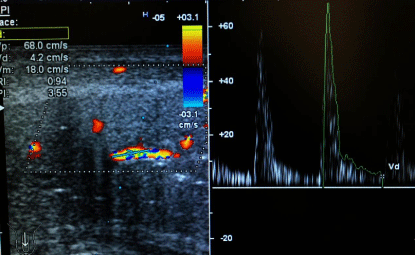
    Figure 5: Longitudinal images with color and spectral Doppler of the left
cavernosal artery15 min after administration of 10 μg of alprostadil. PSV is
68.0 cm/s and EDV is 4.2 cm/s, representing a “normal” pattern.
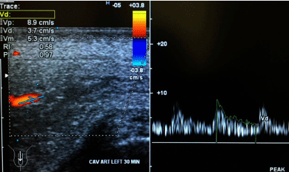
    Figure 6: Left cavernosal artery 30 min after administration of 20 μg of
alprostadil. PSV is 8.9 cm/s and EDV is 3.7 cm/s, consistent with arterial
insufficiency.
Veno-Occlusive Dysfunction (VOD) or venous leak can only be diagnosed in cases of ED where the patient was confirmed to have appropriate arterial function as measured by PSV. The EDV and the corresponding semi-quantitative measurement of the RI may be informative about penile veno-occlusion [5,13]. At full erection, the EDV is expected to be close to zero. EDV < 3 cm/s and a RI > 0.8 are generally considered normal [4]. An EDV >5 cm/second is accepted as the measurement at which a venous leak is present [13] (Figure 7), with a sensitivity of 90% and a specificity of 56% [21]. The main limitation of this parameter is its lack of specificity for venous leak in the presence of arterial insufficiency. In this subset of patients, calculation of RI may be used with improved diagnostic accuracy to determine VOD [22]. On PDDU an RI of less than 0.75, measured at 20 minutes following maximal pharmacostimulation, has been found to be associated with a VOD in 95% of patients [7,10]. Adequate arterial inflow with a semi rigid erection of a short duration with persistent EDV > 5 cm/second is suggestive of VOD. The gold standard method for the diagnosis of VOD is dynamic infusion cavernosometry and cavernosography [5].
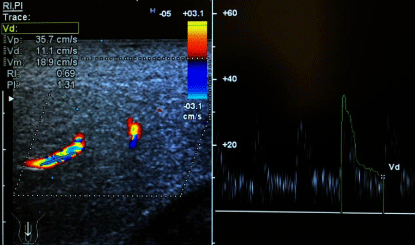
    Figure 7: Left cavernosal artery 30 min after administration of 20 μg of
alprostadil. PSV is 35.7 cm/s, EDV is 11.1 cm/s, and RI is 0.69, consistent
with venous leak.
Incomplete smooth muscle relaxation due to sympathetic discharge (i.e., anxiety) may lead to a false diagnosis of arterial insufficiency and/or VOD. Although redosing may overcome anxietyassociated erectile failure, there is currently no scientifically validated method to definitively ensure that complete smooth muscle relaxation was attained during PDDU. Therefore, Teloken et al. concluded that, in selected men, especially young men with the diagnosis of VOD, PDDU should be repeated [23].
Peyronie´s Disease
Peyronie´S Disease (PD) is a benign disorder characterized by the formation of plaques of fibrous tissue in the tunica albuginea. These plaques may be associated with ED and pain on erection.
Both arterial insufficiency and VOD have been identified as the underlying cause of ED on PD [7].
Although PDDU is not a first-line examination in PD, it can give information about the plaque size and location, which appears as a hyperechoic or isoechoic lesion with through shadowing. The pharmacologically induced erection also gives an idea of the severity and direction of penile curvature.
PDDU is indicated in PD to assess the presence of ED because it can determine if the patient is ultimately a candidate for a penile implant or one of the various incision/plication/grafting techniques available [10].
Priapism
Priapism is defined as a full or partial erection that continues for more than 4 hours. It can be differentiated as low-flow (ischemic) or high-flow (arterial) using PDDU. In the setting of priapism, no injectable medication should be used. High-flow priapism is commonly a result of pelvic or perineal trauma which results in arterial fistulization between the cavernosal artery and the sinusoids of the corpora cavernosa. PDDU reveals a high PSV and often a high diastolic flow [10]. Low-flow priapism is a medical emergency with risk of tissue necrosis resulting from sinusoidal thrombosis and venoocclusion. PDDU reveals the absence of cavernosal blood flow with high resistance, low velocity trace from the cavernosal artery [7].
Mondor´s disease
Mondor´s disease is a rare form of superficial thrombophlebitis, which involves the superficial dorsal vein of the penis [24]. It is a benign and usually self-limited process, presenting with a subcutaneous, tender, cord-like induration [24,25]. The etiology is unknown [25]. PDDU reveals an enlarged superficial dorsal vein with no flow. The presence of low-flow, high-resistance arterial waveforms in cavernosal arteries (priapism-like) has also been reported [25], suggesting a form of venous out-flow disturbance due to compression of the deep dorsal vein by an enlarged superficial dorsal vein and adjacent soft tissue edema caused by thrombophlebitis [25].
Penile trauma
PDDU is useful both in the acute setting and in long-term followup after penile trauma. In the acute setting it may reveal a penile hematoma and the rupture point in the tunica albuginea with blood flow. In long-term follow-up it may be useful to identify sequelae resulting in ED such as venous leak, fibrous plaque formation and post-traumatic fistula formation [7,26].
Conclusion
Penile dynamic Doppler ultrasound is a time-consuming and technically demanding technique in the study of penile pathology. It requires a thorough understanding of relevant penile anatomy and functional physiology. When performed by experienced hands and in a standardized fashion it provides a real-time reliable imaging modality assessing static anatomic features and vascular dynamics. It is a vital tool in the office assessment of erectile dysfunction, Peyronie´s disease and other vascular abnormalities and it can be used to diagnose and guide the patient and the practitioner to the best treatment options.
References
- Wilkins CJ, Sriprasad S, Sidhu PS. Colour Doppler ultrasound of the penis. Clin Radiol. 2003; 58: 514-523.
- Bertolotto M, Neumaier CE. Penile sonography. Eur Radiol. 1999; 407-412.
- Aversa A, Bruzziches R, Spera G. Diagnosing erectile dysfunction: the penile dynamic colour duplex ultrasound revisited. International journal of Andrology. 2005; 61-63.
- Hatzimouratidis K, Giuliano F, MoncadaI, Muneer A, Salonia A, Verze P. Guidelines on male sexual dysfunction: erectile dysfunction, premature ejaculation, penile curvature and priapism. European Association of Urology. 2016.
- Sikka SC, Hellstrom WJ, Brock G, Morales AM. Standardization of vascular assessment of erectile dysfunction: standard operating procedures for duplex ultrasound. J Sex Med. 2013; 10: 120-129.
- Mihmanli I, Kantarci F. Erectile dysfunction. Semin Ultrasound CT MRI. 2007; 28: 274-286.
- Halls J, Bydawell G, Patel U. Erectile dysfunction: the role of penile doppler ultrasound in diagnosis. Abdom Imaging. 2009; 34: 712-725.?
- Patel DV, Halls J, Patel U. Investigation of erectile dysfunction. The British Journal of Radiology. 2012; 85: 569-578.
- Bhatt S, Kocakoc E, Rubens DJ, Seftel AD, Dogra VS. Sonographic evaluation of penile trauma. J Ultrasound Med. 2005; 24: 993-1000.
- LeRoy TJ, Broderick GA. Doppler blood flow analysis of erectile function: who, when and how. Urol Clin N Am. 2011; 38: 147-154.
- Gupta N, Herati A, Gilbert BR. Penile doppler ultrasound predicting cardiovascular disease in men with erectile dysfunction. Curr Urol Rep. 2015; 16: 16.
- Bari V, Ahmed MN, Rafique MZ, Ashraf K, Memon WA, Usman MU. Evaluation of erectile dysfunction with color doppler sonography. J Pak Med Assoc. 2006; 56: 258-260.
- Aversa A, Sarteschi LM. The role of penile color-duplex ultra- sound for the evaluation of erectile dysfunction. J Sex Med. 2007; 4: 1437-1447. ?
- Yang Y, Hu J-I, Ma Y, Wang H-x, Chen Z, Xia J-g, et al. Pharmaco-induced erections for penile color-duplex ultrasound: oral PDE5 inhibitors or intracavernosal injection? International Journal of Impotence Research. 2012; 24: 191-195.
- Pagano M, Stahl PJ. Variation in penile hemodynamics by anatomic location of cavernosal artery imaging in penile duplex doppler ultrasound. J Sex Med. 2015; 12: 1911-1919.
- Roy C, Saussine C, Tuchmann C, Castel E, Lang H, Jacqmin D. Duplex doppler sonography of the flaccid penis: potencial role in the evaluation of impotence. J Clin Ultrasound. 2000; 28: 290-294.
- Corona G. Penile doppler ultrasound in patients with Erectile Dysfunction (ED): role of peak systolic velocity measured in the flaccid state in predicting arteriogenic ED and silent coronary artery disease. J Sex Med. 2008; 5: 2623-2634. ?
- Kahveciog N, Kurt A, Ipek A, Yaziciog KR, Akbulut Z. Predictive value of peak systolic velocity in the flaccid penis. Advances in Medical Sciences. 2009; 54: 233-238.
- Sen J, Godara R, Singh R, Airon RK. Colour doppler sonography of flaccid penis in evaluation of erectile dysfunction. Asian J Surg. 2007; 30: 122-125.
- Kuo YC, Liu SP, Chen JH, Chang HC, Tsai VFS, Hsieh JT. Feasability of a novel audio-visual sexual stimulation system: an adjunct to the use of penile duplex doppler ultrasonography for the investigation of erectile function. J Sex Med. 2010; 7: 3979-3983.
- Kadioglu A, Tefekli A, Erol H, Cayan S, Kandirali E. Color Doppler ultrasound assessment of penile vascular system in men with Peyronie´s disease. Int J Impot Res. 2000; 12: 263-267.
- Yafi FA, Libby RP, McCaslin IR, Sangkum P, Sikka SC, Hellstrom WJG. Failure to attain stretched penile length after intracavenosal injection of a vasodilator agent is predictive of veno-occlusive dysfunction on penile duplex doppler ultrasonography. Andrology. 2015; 3: 919-923.
- Teloken PE, Park K, Parker M, Guhring P, Narus J, Mulhall JP. The false diagnosis of venous leak: prevalence and predictors. J Sex Med. 2011; 8: 2344-2349.
- Dell Atti L. Role of ultrasonography with color-doppler in diagnosis of penile Mondor´s disease. J Ultrasound. 2014; 17: 239-241.
- Han HY, Chung DJ, Kim KW, Hwang CM. Pulsed and color doppler sonographic findings of penile Mondor´s disease. Korean J Radiol. 2008; 9: 179-181.
- Kim SH, Kim SH. Post-traumatic erectile dysfunction: doppler US findings. Abdom imaging. 2005; 31: 598-609.