Case Report
Austin J Urol. 2017; 4(1): 1051.
Genito-Urinary Tuberculosis – A Great Mimicker and Its Atypical Manifestations: An Interesting Case Series
Krishnamoorthy S*, Gopalakrishnan G and Kekre NS
1Department of Urology, Sri Ramachandra Medical College & RI, India
2Department of Urology, Vedanayagam hospital, India
3Department of Urology, Christian Medical College, India
*Corresponding author: Krishnamoorthy S, Department of Urology, Sri Ramachandra Medical College & RI, India
Received: October 04, 2016; Accepted: December 28, 2016; Published: January 04, 2017
Abstract
Tuberculosis continues to be a major health problem in the developing world. Renal tuberculosis is the most common site of extra – pulmonary tuberculosis. It comprises 20% of all extra pulmonary tuberculosis. The accurate incidence and prevalence of Genito Urinary Tuberculosis (GUTB) is difficult to be figured out because a very large varieties of atypical presentations. It is often called as a “Great Mimicker” because of the variety of other conditions that it might mimic. A vast majority of patients remain asymptomatic and present in late stages as the disease is not thought of in these asymptomatic patients. Moreover the urine can become sterilized relatively rapidly after the initiation of chemotherapy, further adding on to the diagnostic difficulties.
In this manuscript, we report a series of GUTB cases that presented in an atypical manner, causing diagnostic and management dilemma. The purpose of this manuscript is to highlight the variety of atypical manifestations of GUTB and also to stress on the need for a high index of clinical suspicion in identifying such cases at an earlier stage in order to prevent the disease from progressing to non- salvageable state.
Keywords: Tuberculosis; Genitourinary tuberculosis; Kidney; Anti tuberculosis therapy; Nephrectomy
Background
Tuberculosis is a major public health problem in India. Every year 1.8 million new cases of TB are diagnosed of which 0.8 million are smear positive [1]. The emergence of drug resistance strains adds to the complexity of the situation in our country. The urinary tract is one of the commonest sites of extra-pulmonary tuberculosis [2]. While in the majority the symptoms are cast- iron, in a not insignificant number, the presentation is atypical. This naturally results in delayed diagnosis. In this article we wish to highlight some of the atypical presentations of genito-urinary tuberculosis and focus on the need for constant vigil in establishing the diagnosis.
Case 1
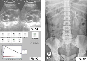
A 35 year old female with primary infertility was evaluated for low grade fever and vague abdominal discomfort. She also had a decrease in appetite and a loss of weight. On evaluation, she was diagnosed to have abdominal tuberculosis with thickened omentum. Omental biopsy confirmed her disease. She did not have any loin pain or obstructive or irritative urinary complaints. As the ultrasound also showed right hydronephrosis (Figure 1A), she was further evaluated. Her serum Creatinine was 1.0 mg%. Her urine AFB was negative. Intra-venous Urography suggested non-visualized right kidney (Figure 1B). Isotope Renogram confirmed a very poorly functioning right kidney (Figure 1C). She was started on Anti-Tuberculous Therapy (ATT) and subsequently underwent right Nephrectomy. Prior to Nephrectomy, she had a cystoscopy and retrograde ureterogram. The bladder was of good capacity. The ureteric orifices and bladder surface were normal. Retro-grade study demonstrated a complete cutoff at the level of right lower ureter, about 2 centimeters (cm) proximal to the ureteric orifice. The kidney, on cut-section was found to be grossly hydronephrotic with multiple cavitations. The histopathology report was suggestive of tuberculosis pyelonephritis. Following ATT and Nephrectomy, her constitutional symptoms improved significantly.

    Figure 1: Ultrasound shows right hydronephrosis, non visualized right kidney
and functioning right kidney.
Case 2
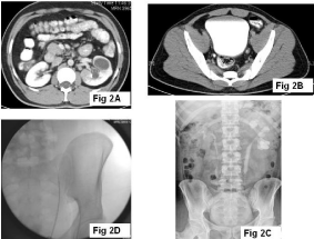
A 33 year old male presented with recurrent bouts of total painless hematuria with passage of amorphous clots for the past 12 years. He also had associated fever and left loin pain. The fever was predominantly low grade, with an evening rise of temperature. He had occasional mild dysuria, but had no other Lower Urinary Tract Symptoms (LUTS). He also had a swelling of the right testis, associated with pain, which subsided with analgesics and antibiotics, but with a residual thickening and induration. General examination suggested evidence of chronic right epididymitis. His renal function was normal. CT abdomen revealed a left upper calyceal infundibular stenosis with a grossly dilated upper calyx and a normal capacity urinary bladder (Figure 2A,2B). Cystoscopy revealed a diffusely congested bladder with adequate capacity. The ureteric orifices were normal in location and shape. Intravenous Urogram showed multiple infundibular stenois, small capacity renal pelvis and multiple ureteric strictures (Figure 2C). On retrograde evaluation, the whole ureter was found to be very narrow, that could admit only a 5 Fr ureteric catheter to pass through (Figure 2D). The individual calyces were dilated, with evidence of multiple infundibular stenoses. DJ stenting was done, but on stent removal, his symptoms recurred. He then had a balloon dilatation of the ureteric strictures but ultimately had a left upper polar Nephrectomy and an ileal replacement of the left ureter as the symptoms and obstruction persisted.

    Figure 2: CT abdomen revealed a left upper calyceal infundibular stenosis
with a grossly dilated upper calyx and a normal capacity urinary bladder.
Case 3
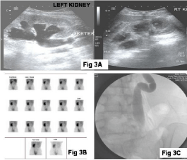
A 31 year old lady presented with recurrent attacks of febrile urinary tract infections for the past 2 years. On evaluation, she was diagnosed to have urinary tuberculosis with a serum creatinine of 4.0 mg%. On ultrasound evaluation, the right kidney showed multiple infundibular stenoses with left lower ureteric obstruction and left hydro-uretero nephrosis (Figure 3A). Isotope Renogram showed a very poorly functioning right kidney (Figure 3B). Following a left percutaneous nephrostomy, her Creatinine reached a nadir of 1.3 mg%. Her bladder was of a very small capacity. Left nephrostogram revealed a stricture at the level of left lower ureter (Figure 3C). She underwent right nephro-ureterectomy with augmentation cystoplasty using ileo-caecum and left ureteric reimplantation. The histo pathology report was suggestive of right tuberculosis pyonephrosis.

    Figure 3: On ultrasound evaluation, the right kidney showed multiple
infundibular stenoses with left lower ureteric obstruction and left hydrouretero
nephrosis.
Case 4
A 55 year old male presented with a painless swelling of the right testis of four months duration. He had recurrent low grade fever for the past one month, not associated with chills or rigor. He did not have any loin pain or bothersome voiding difficulties. He was not treated for tuberculosis in the past. No one in the family had received treatment for TB. There was no trauma to the scrotum. General examination was unremarkable. On examination, there was an 8 x 4 cms swelling in the right testis. It was hard, non tender with an irregular surface. The left testis, both epididymes and the cord structures were clinically normal. The ESR was 45 mm in one hour, total WBC count of 3400 per cub mm with differential count 72 polymorphs and 25 lymphocytes. Urine microscopy revealed microscopic haematuria of 10-12 RBCs/high power field. The urine culture was sterile. Serum LDH was 947 U/L but beta HCG (<1 mIU/ millilitre) and AFP (1.71 IU/millilitre) were normal. The chest X- ray was normal. Urine examination for acid fast bacilli (done after the diagnosis was made) was negative. Ultrasonogram of the scrotum revealed a 7x4.5 cms hypoechoic lesion in the right testis. The left testis and both epididymes were normal. Ultrasound abdomen was normal. A clinical diagnosis of testicular tumour was made and he underwent right inguinal high orchidectomy. Histopathological examination revealed a caseating granulomatous lesion, consistent with tuberculosis orchitis. Testicular parenchyma was replaced by caseating granulomas composed of epithelioid histiocytes, lymphocytes and Langhans’ multinucleate giant cells and surrounded by lymphocytes and plasma cells. There was interstitial fibrosis. The granulomatous infiltrate extended onto the albuginea. Occasional atrophic seminiferous tubules were present.
Discussion
About 8-15% of patients with pulmonary tuberculosis develop urinary tuberculosis [3]. Classically the bladder has been called the “sounding board” for the diagnosis of urinary tuberculosis. Severe LUTS in a patient with sterile pyuria would herald a diagnosis of urinary tuberculosis. In the series of patients described in this article, none of them had LUTS. The diagnosis of tuberculosis was made in hindsight after obtaining histology during the definitive procedure. Patients in this series presented with incidental loin mass, hematuria, renal failure, and a testicular mass.
In a vast majority, the diagnosis of urinary tuberculosis is made based on clinical history, backed by radiologic imaging and microbiological confirmation. The yield of AFB in urine is around 30% and hence there is a need to send multiple samples for culture [4]. In view of the generally poor yield on culture most clinicians make the decision to treat for tuberculosis based on history and radiologic imaging. At times cystoscopic biopsy can be helpful in coming to a conclusion.
There have also been reports of idiopathic ureteric strictures in patients with no other symptoms of LUTS except loin discomfort. These lesions are very common and many a time manifest only after the diagnosis is made and during the anti-tubercular therapy [5]. In the era of minimally invasive treatments, some of these patients would undergo endourological procedures with no tissue for histology. It is conceivable that some of these strictures could have a tuberculosis etiology, which would be missed. Hematuria occurs in about 37% of patients [6] and can occur independent of LUTS as seen in one of our cases. Failure to keep this fact in mind will result in the diagnosis being missed. Some patients present primarily with renal failure. In tuberculosis, renal failure in most instances is secondary to obstructive uropathy. It may also be due to parenchymal destruction from the tuberculosis process [7]. In the latter situation the patient will require renal replacement therapy.
It has been mentioned that when the bladder is involved in the tuberculosis process, the kidneys will almost always be involved [8]. The converse need not be true. Hence in these patients classical LUTS is absent while the kidney has suffered irreparable damage.
Genital tuberculosis usually involves the epididymis. Traditionally it has been said that in patients with epididymal tuberculosis, the urinary tract is also involved and hence the recommendation is to do an IVU or other imaging to identify renal/ureteric involvement. Isolated involvement of either the testis or the epididymes is extremely rare. In an earlier publication from this institution patient with testicular tuberculosis was found to have normal a urinary tract [9]. Rarely as seen in the case presented in this article, the testis can be involved in isolation and this differential needs to be kept in mind in dealing with patients who have testicular swellings.
Conclusion
No disease process is as atypical as a typical tuberculosis process [10]. The manifestations of tuberculosis are greatly variable. It can cause a variety of clinical patterns that can mimic other diseases. Simultaneously, atypical presentations need to be kept in mind. The purpose of this article is to highlight these atypical presentations thereby instituting early and appropriate treatment.
References
- Chauhan LS, Tonsing J. Revised National TB control programme in India. Tuberculosis. 2005; 85: 271-276.
- Mochalava TP, Starikov IY. Reconstructive Surgery for treatment of Urogenital Tuberculosis: 30 years of observation. World J. Surg 1997; 21: 511-515.
- Hemal AK, Gupta NP, Rajeev TP, Rajeev Kumar, Dar L, Seth P. Polymerase chain reaction in clinically suspected genitourinary tuberculosis: comparison with intravenous urography, bladder biopsy and urine acid fast bacilli culture. Urology. 2000; 56: 570-574.
- Kapoor R, Ansari MS, Mandhani A, Gulia A. Clinical presentation and diagnostic approach in cases of genitourinary tuberculosis. Indian J Urol. 2008; 401-405.
- Claridge M. Ureteral obstruction in tuberculosis. Br J Urol. 1970; 42: 688-692.
- Gokce G. Genitourinary tuberculosis: A review of 174 Cases. Scand J Infect Dis. 2002; 34: 338-340.
- Gupta NP. Reconstructive Surgery for the Management of Genitourinary Tuberculosis: A single Center Experience. J of Urol. 2006; 175: 2150-2154.
- Cek M. EAU Guidelines for the Management of Genitourinary Tuberculosis. European Urology. 2005; 48: 353-362.
- Paul J, Krishnamoorthy S, Teresa M, Kumar S. Isolated tuberculous orchitis: A mimicker of testicular malignancy. Indian J Urol. 2010; 26: 284-286.
- Cooper HG, Robinson EG. Treatment of Genitourinary Tuberculosis: report after 24 years. J of Urol. 1972; 108: 136-142.