Research Article
Austin J Urol. 2022; 8(2): 1078.
Split Appendix Malone and Mitrofanoff for the Management of Neurogenic Bowel and Bladder
Khan MI¹ and Khan MK²*
¹Assistant Professor of General surgery, Khyber Teaching Hospital, Pakistan
²Assistant Professor Pediatric Urology, Institute of Kidney Diseases, Pakistan
*Corresponding author: Muhammad Kamran Khan, Assistant Professor Pediatric Urology, Institute of Kidney Diseases, Pakistan
Received: September 25, 2022; Accepted: October 25, 2022; Published: November 01, 2022
Abstract
Purpose: To share our experience of neurogenic bowel and bladder management by creating Malone Antegrade Continent Enema (MACE) procedure and Continent Urinary Diversion (Mitrofanoff VQZ technique) at the same time by using split appendix.
Material and Methods: Between July 2017 and December 2021, 30 patients; 5 to 15 years old (mean 9.54±2.65 years) underwent VQZ Mitrofanoff and MACE using split appendix with or without bladder augmentation forneurogenic bladder and bowel management secondary to myelomeningocele. Proximal part of appendix was used for MACE by taking 3 antireflux stitches by wrapping seromuscular layer of cecum around base of appendix whereas distal part of appendix is used for Mitrofanoff creating VQZ stoma. The average length of appendix taken was 9.5 cm (8-12 cm).
Results: All the patients were kept clean and dry throughout and were followed up for an average of 15 months (12 to 30 months). Only one patient was reported with MACE stoma stenosis because she lost to follow up and didn’t use the stoma for a year, stoma revision was performed on her. Procedure time is also reduced in split appendix MACE and Mitrofanoff as compared to if monti tube or cecal flap is reconstructed for MACE.
Conclusion: MACE and Mitrofanoff with or without bladder augmentation are very invaluable procedures for the management of neurogenic bowel and bladder. Split appendix is an ideal channel for both Mitrofanoff and MACE.
Introduction
Fecal and urinary incontinence in spina bifida patients is one of the most devastating conditions involving children. It has both social and psychological implications and decreased quality of life along with other co morbidities. Most of these patients are treated with various types of enemas to clean out the colon for bowel management and clean intermittent catheterization with or without anticholinergic medications for bladder management.
Bowel management program usingenemas can be administered with Foley's balloon catheter per rectal or alternatively by The Peristeen trans-anal irrigation system which is a relatively much effective, safe, non-operative alternative in children with fecal incontinence [1]. With growing age, most children cannot tolerate use of rectal enemas on a daily basis. In 1990, Malone et al. [2] proposed that the Antegrade Continence Enema could be administered via appendix used as a conduit. A one-way valve mechanism was created in order to allow catheterization of appendix via abdominal wall for colonic irrigation and to prevent stool leakage simultaneously. It also allows the self-administration of enema easy and make the patient’s independent [3].
In cases where conservative measures and clean intermittent catheterization fail tomanage neurogenic bladder then surgical treatment is opted which includes botulinum toxin injected to the detrusor muscle and continent catheterizable conduit with or without bladder augmentation [4-6].
Simultaneous Malone and Mitrofanoff procedures using split appendix is performed in patients with long appendix (greater than 9cm) [7]. When appendix is short or absent then other procedures like split appendix with cecal extension, appendiceal Mitrofanoff with cecal flap for antegrade continent enema procedure or Monte procedure are adopted for these continent conduit channels. Cecal extension of the appendix seems to be a good option when the appendix is too short for a simple split procedure [8]. For Mitrofanoff procedure we use right iliac fossa for external stoma whereas umbilicus is our usual site for Malone procedure.
The aim to conduct the retrospective study was to convey our experience of neurogenic bowel and bladder management by creating Malone Antegrade Continent Enema (MACE) and continent urinary diversion (Mitrofanoff VQZ technique) at the same time by using split appendix. The safety and efficacy of the technique and the complications associated with the procedure were to be drawn from the results of this study.
Material and Methods
We conducted a retrospective study that included 30 patients who underwent split appendix Malone and Mitrofanoff; with or without bladder augmentation for the management of neurogenic bowel and bladder due to spina bifida, from July 2017 till December 2019. The hospital ethical committee agreed to the approval before the study was started. Proximal part of appendix was used for MACE by taking 3 antireflux stitches by wrapping seromuscular layer of cecum around base of appendix whereas distal part of appendix was used for Mitrofanoff creating VQZ stoma. The average length of appendix taken was 9.5 cm (8-12 cm).
Pediatric patients with urinary and fecal incontinence due to neurogenic bladder and bowel secondary to spina bifida were included in the study. These patients either didn’t want per urethral intermittent catheterization and per rectal enemas or had failed conservative treatment for the condition. Complete blood picture, serum electrolytes and renal function tests were done as a part of preoperative evaluation along with radiological evaluation including ultrasound Kidney, Ureter and Bladder (KUB), Voiding Cystourethrography (VCUG), urodynamic analysis and a nuclear renal scan with 99m Technetium Dimercapto-Succinic Acid (DMSA).
Surgical Technique
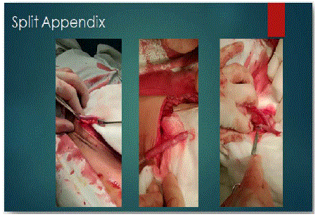
Lower midline access was preferred in the study since it allowed an access to the bladder as well as the ileo-cecal junction thereby to the appendix and ileum if required. An intact and good size (almost 8 cm) appendix with its mesentery was isolated carefully and was divided from the cecum with a 3 cm stump left behind while on the other hand, the distal part of appendix was mobilized on its mesentry and 12Fr nelaton tube was introduced to ensure its patency. Three (3) antireflux stitch of silk 3/0 are taken around the base of proximal appendix and by wrapping seromuscular layer of cecum (Figure 1).

Figure 1: Shows a) appendix on its mesentry b) divided appendix c) antireflux stitches around the proximal part of appendix.
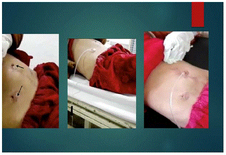
The distal end of the appendiceal Mitrofanoff channel was attached by either extra vesical tunneling into the native bladder or by intravesical technique in cases of bladder augmentation. An adequate length of tunnel was created whereby the internal opening of the tunnel was secured to the muscle and mucosa of the bladder with absorbable sutures. Smooth catheterization of the tunnel made was ensured effectively. The abdominal end of the conduit made was brought out through the abdominal wall, and a stoma was fashioned by the VQZ technique (Figure 2). Post operative suprapubic catheter along with a Mitrofanoff catheter was placed for 3 weeks for the purpose of bladder drainage. Malone stoma was brought out at the umbilicus with 10Fr nelaton tube secured in it.

    Figure 2: a) VQZ Mitrofanoff and MACE umbilical stoma b) VQZ Mitrofanoff

stoma with catheter c) MACE umbilical stoma with catheter.
Follow up
Post operatively patients were followed at3 weeks for initiating clean intermittent catheterization through Mitrofanoff and daily enema through MACE. In first year, the follow-up was planned at 6 months interval however after the first year it was done yearly. The mentioned follow-up was planned with ultrasound KUB, serum electrolytes, renal function tests, urodynamic studies and urine routine examination. DMSA was performed when episodes of UTI occurred in children who had a mild degree of reflux or dilation of the ureter.
Statistical Analysis
The parameters analyzed were age, gender, augmentation cystoplasty needs, the duration of surgery in minutes, hospital stay in days, per operative and postoperative complications. Data was analyzed using SPSS version 22. The categorical variables such as gender and complications were presented in the form of frequency and percentage, while continuous variables such as age, operative time and hospital stay were presented in the form of mean ± Standard Deviation (SD).
Results
The study was retrospective involving 30 patients who underwent split appendix Malone and Mitrofanoff for the management of neurogenic bowel and bladder secondary to spina bifida, from July 2017 till December 2019; having mean age of 9.54±2.6 years(5-15 years) (Table 1). The study included 19 males (63%) and 11 females (37%).
    Number

    30


    Male

    19(66%)


    Female

    11(34%)


    Age(years)

    9.54±2.65(5-15)


    Need for BA

    24(80%)


    Operation time,    minlaverage)

    212 min(105-260    min)


    Operated time    for BA

    237±11 min


    Operated time    without BA

    111±3.8 min


    Hospital stay

    4±212-6)


    Final continence

    29/29(96.5%)
Table 1: Demographic.
Augmentation cystolplasty was performed in 24 children (80%) while 6 patients (20%) did not require bladder augmentation. Mean operative time in minutes was 212+-52 (105-260). Operative time was significantly less for those patients without bladder augmentation as compared those who required bladder augmentation (111+- 3.8 minutes vs 237+-11 minutes). Mean hospital stay was 4.1+- 1.3 (2-6 days) and similarly it was significantly reduced in patients who did not require bladder augmentation (2 days vs 4.8+-0.8 days).Among post-operative complications; wound infections were noted in 2 (6.6%) patients, which was managed successfully with antibiotics and dressings. Post-operative fever was reported in only one patient (3.3%). Stomal stenosis at one year was noted in 3 patients (10%) whereas 2 were Mitrofanoff stoma and one was MACE stoma, which required stoma revision. UTIs and wound infections occurred only in patients with bladder augmentations. Appendiceal stoma revision was conducted in 2(6.6%) patients only (Table 2).
    Name of    complication

    No;s %


    wound infection

    2(6.6%)


    fever

    1(3.3%)


    stomal stenosis    at 1 year

    3(10%)


    UTI

    7(23.33%)
Table 2: Complications.
Discussion
Continent catheterizable conduit is created in order to facilitate effective emptying of bladder by means of Clean Intermittent Catheterization (CIC). It is recommended for protection of the upper urinary tract and even improves continence whereas MACE is created for the smooth administration of enema for the effective bowel management and therefore, makes the patient independent. For a successful Mitrofanoff procedure, there is a requirement for a good capacity with low pressure reservoir bladders. Concomitant bladder augmentation is preferred in situations where bladder is having a small capacity and low compliance. It has been found that children using Malone and Mitrofanoff stomas for the bowel and bladder management are more compliant with the treatment and have a better quality of life [9].
The main complications encountered so far have been prolapsed and incontinent stoma. The others includedifficulty incatheterization, typically due to channel stricture/stenosis and false passage. The incidence of the discussed complications varies widely depending on the series, the type of channel used, and the length of follow-up. In general, the incidence of stomal prolapse ranges from 2–5% while stomal incontinence 1–47%, difficulty in catheterization 5–32% and overall rates of surgical revision range from 18–58% [10-13]. We did not notice any mucosal prolapse and incontinence in our series but two patients (6.6%) had Mitrofanoff stomal stenosis and one patient (3.3%)had Malone stenosis. Whereas according to A.J Renseng et al [14]. the incidence of Malone stenosis was 49 % , which is very high as compare to our study and other studies too, one reason may be there criteria for stomal stenosis was inability to catheterize the stoma whereas in our case a stomal stenosis is labelled only when it requires stomal dilatation or revision. They have also long average followup of almost 5 years as compare to 15 months follow up in our study.
The only patient with Malone stoma stenosis in our study did not use the stoma after the nelaton tube removal for enema administration. Stomal stenosis is difficult to treat and has high recurrence rate. it can be treated with stomal dilatation, stomal revision and minimally invasive methods like stomal triamcilone injection [15]. According to PP Reddy stomal triamcilone injection results in ease of catheterization in about 72 % of patient with difficultcatherization which ia comparable to stomal revision. We kept the Malone stomal catheter for a month initially and then administered enema through intermittent catheterization. Prolong initial Malone catheter placement may be a reason for decreased incidence of stomal stenosis in our study group. Probably the reason of 100 percent continence in these neurogenic patients might be that we use fascial bladder neck slings in such patients to enhance chances of urinary continence.
The channels created from split-appendix technique hold outcomes and revision rates comparable with those of other described techniques. However, it has the benefit of avoiding a bowel resection and its accompanying risks in those patients who do not require bladder augmentation as was the case in 20% of our series. The split appendix technique is associated with significantly reduced operative time in cases with simultaneous MACE and Mitrofanoff procedures and save the time of bowel resection, anastomosis, Monti tube creation or cecal flap and tube creation.
Limitations of this study are its retrospective nature, Small study cohort, short follow-up data and a single center study. These patients need long term follow-up to know about the long term outcomes of the procedure.
References
- Pacilli M, Pallot D, Andrews A, Louiza D, Willets D. Use of Peristeen® transanal Colonic irrigation for bowel management in children: A singlecenter experience. Journal of pediatric surgery. 2014; 49: 269-72.
- Malone PS, Ransley PG, Kiely EM. Preliminary report: The antegrade continence enema. Lancet. 1990; 336: 1217–18.
- Bischoff A, Levitt MA, Peña A. Bowel management for the treatment of pediatric fecal incontinence. Pediatric Surgery International. 2009; 25: 1027- 1042.
- Clark T, Pope JC, Adams MC, Wells N, Brock JW. Factors that influence outcomes of the Mitrofanoff and Malone antegrade continence enema reconstructive procedures in children. The Journal of urology. 2002; 168: 1537-1540.
- Thomas JC, Dietrich MS, Trusler L, DeMarco RT, Pope JC, Brock JW, et al. Continent catheterizable channels and the timing of their complications. The Journal of urology. 2006; 176: 1816-1820.
- Farrugia M, Malone PS. Educational article: The Mitrofanoff procedure. Journal of pediatric urology. 2010; 6: 330-337.
- Kajbafzadeh AM, Chubak N. Simultaneous Malone antegrade continent enema and Mitrofanoff principle using the divided appendix: report of a new technique for prevention of stoma complications. J Urol. 2001; 165: 2404-9.
- Kudela G, Smyczek D, Springer A, Korecka K, Koszutski T. No Appendix is Too Short—Simultaneous Mitrofanoff Catheterizable Vesicostomy and Malone Antegrade Continence Enema (MACE) for Children with Spina Bifida. Urology. 2018; 116: 205-207.
- Szymanski KM, Whittam B, Misseri R, et al. Long-term outcomes of catheterizable continent urinary channels: What do you use, where you put it, and does it matter?. J Pediatr Urol. 2015; 11: 210.e1-7.
- Süzer O, Vates TS, Freedman AL, Smith CA, Gonzalez R. Results of the Mitrofanoff procedure in urinary tract reconstruction in children. British journal of urology. 1997; 79: 279-282.
- Leslie B, Lorenzo AJ, Moore K, et al. Long-term follow-up and time to event outcome analysis of continent catheterizable channels. J Urol. 2011; 185: 2298-302.
- Welk BK, Afshar K, Rapoport D, MacNeily AE. Complications of the catheterizable channel following continent urinary diversion: their nature and timing. The Journal of urology. 2008; 180: 1856-1860.
- Polm PD, Kort LMOD, Jong TPVMD, Dik P. Techniques Used to Create Continent Catheterizable Channels: A Comparison of Long-term Results in Children. Urology. 2017; 110: 192-195.
- Rensing AJ, Koenig JF, Austin PF. Pre-operative risk factors for stomal stenosis with Malone antegrade continence enema procedures. Journal of pediatric urology. 2017; 13: 631.e1-631.e5.
- Reddy PP, Strine AC, Reddy T, Noh PH, Defoor Jr WR, Minevich E, et al. Triamcinolone injection for treatment of Mitrofanoffstomal stenosis: Optimizing results and reducing cost of care. J purol. 2017; 131: 371-75.