Case Report
Austin J Vasc Med. 2014;1(1): 4.
Endovascular Management of an Isolated External Iliac Artery Dissection after Pelvic Fracture
Steven Deso1, Ducksoo Kim2 and Nii-Kabu Kabutey2,3*
1Department of Radiology, Stanford University Medical Center, USA
2Department of Radiology, Boston University Medical Center, USA
3Department of Surgery, UC Irvine Medical Center, USA
*Corresponding author: Nii-Kabu Kabutey, Department of Radiology & Surgery, UC Irvine Medical Center 101 The City Drive South Orange, CA 92868, USA
Received: September 10, 2014; Accepted: October 08, 2014; Published: October 10, 2014
Abstract
Purpose: To describe the successful endovascular repair of an isolated external iliac artery dissection in the setting of an unstable pelvic fracture.
Case Report: A 48-year-old man presents with left pelvis pain after intentionally jumping from a 30 foot balcony. On Computed Tomography (CT) imaging, he is found to have a vertical shear type pelvic fracture and an isolated left external iliac artery dissection. He undergoes successful endovascular repair with placement of an Abbott 9mm x 6cm self-expanding stent. Follow-up angiography demonstrates brisk flow across the stent with no further filling of the false lumen.
Conclusion: Endovascular treatment is a safe and viable alternative to open vascular repair in patients with an isolated external iliac artery dissection in the setting of blunt pelvic trauma.
Keywords: External iliac artery dissection; External iliac artery injury; Stent; Pelvic fracture; Endovascular repair
Introduction
Isolated External Iliac Artery (EIA) injuries are rare but serious findings in some pelvic fractures [1]. The implications of such injuries are considerable and include increased lower extremity amputations and death among those affected [2]. The presence of multiple injuries in these patients can make open vascular repair technically difficult and dangerous given the risk of hemorrhage and fecal contamination [3-5]. Here we present a case of an isolated external iliac artery dissection in the setting of a pelvic fracture that was successfully managed with the endovascular placement of a self-expanding stent.
Case Report
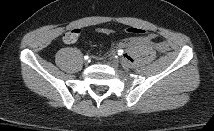
Institutional review board approval is not required for this type of report at our institution. A 48-year-old man presented with right leg and left pelvis pain after an unwitnessed intentional jump from a 30- foot balcony. The patient was hemodynamically stable with a Glasgow Coma Scale (GCS) of 15 and a negative Focused Assessment with Sonography for Trauma (Fast) on presentation. Contrast-enhanced Computed Tomography (CT) was performed which showed a comminuted fracture involving the left ala of the sacrum, fractures of the left superior and inferior pubic rami, and diastasis of the symphisis pubis consistent with a vertical shear type pelvic fracture. A hematoma was noted to extend from the left aspect of the pelvis superiorly to the left retroperitoneum without active extravasation. Focal dissection of the proximal left External Iliac Artery (EIA) was also noted (Figure 1). Given the potential risks of open vascular repair, a decision was made to pursue endovascular treatment.
Figure 1: Contrast-enhanced axial CT image showing a comminuted fracture involving the left ala of the sacrum with associated hematoma and focal dissection of the proximal left EIA (arrow).
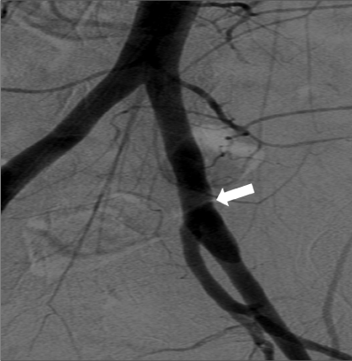
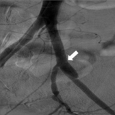
A right common femoral approach was used, and an aortogram was obtained using a 5-French Omniflush catheter (Angiodynamics, Latham, NY). The Antero-Posterior (AP) images showed no evidence of active extravasation, both common iliac arteries appeared normal in contour and caliber, and a non-flow limiting dissection was noted in the proximal left EIA (Figure 2). Right Anterior Oblique (RAO) images were obtained which clearly showed dissection of the left EIA (Figure 3). Digital Subtraction Angiography (DSA) of the left lower extremity showed patent left superficial femoral and popliteal arteries with three vessel run-off to the foot.
Figure 2: AP angiogram showing no evidence of contrast extravasation, normal contour of the CIAs bilaterally and a non-flow limiting dissection of the proximal left EIA (arrow).
Figure 3: RAO angiogram showing dissection of the proximal left EIA (arrow).
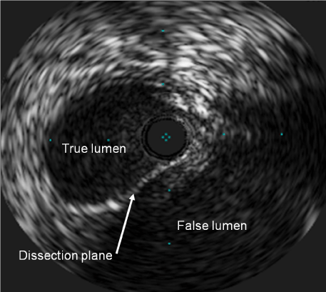
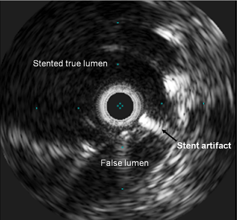
An Intravascular Ultrasound (IVUS) probe (Volcano, San Diego, CA) was advanced over a V-18 control wire (Boston Scientific, Miami, FL) to the left EIA where it was used to delineate the extent of the dissection. A clear dissection plane was apparent on IVUS (Figure 4) starting at the level of the left CIA bifurcation and extending 3-4cm distally down the left EIA. A decision was made to forego conservative management and treat primarily given the patient’s unstable pelvic fracture, the likelihood of further non-physiological stresses to the vascular wall prior to definitive pelvic stabilization, and the potential for rapid evolution into flow limiting dissection.
Figure 4: IVUS image of the left EIA showing a clear dissection plane with true and false lumens.
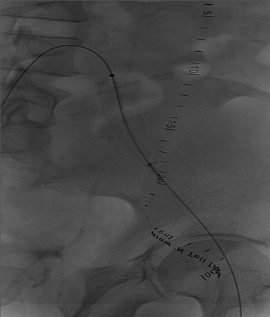
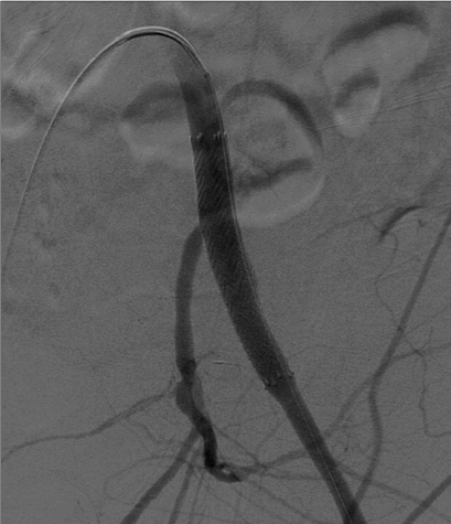
The IVUS probe was exchanged for a 9mm x 6cm self-expanding stent (Abbott, Abbott Park, IL). The stent was deployed with the proximal end positioned at the level of the left iliac arterial bifurcation to ensure complete obliteration of the lesion (Figure 5). Post-deployment angioplasty was performed with a 9mm x 6cm balloon (Bard Medical, Covington, GA). Repeat IVUS imaging showed the stented left EIA to be patient with obliteration of the false lumen (Figure 6). Follow-up angiography demonstrated brisk flow across the stent with no further filling of the false lumen (Figure 7). Selective angiography was performed on the right common femoral artery and a 6-French Perclose device (Abbott, Abbott Park, IL) was used to obtain hemostasis. The procedure was well tolerated with no immediate complications. A left femoral traction pin was placed by the orthopedics team, with subsequent closed reduction and percutaneous pinning of the left pelvic fracture in the OR. The patient required five units of blood during the first 48hr, but ultimately did very well. He was discharged to sub-acute rehab 5½ weeks later.
Figure 5: Abbott 9mm x 6cm self-expanding stent deployed with the proximal end positioned at the level of the iliac artery bifurcation.
Figure 6: IVUS image of the left EIA taken after stenting showing obliteration of the false lumen.
Figure 7: Angiogram showing brisk flow through the stented left EIA, preserved patency of the left IIA, and no further filling of the false lumen.
Discussion
EIA injuries after pelvic fractures are rare and can include laceration, dissection, and crush injuries leading to arterial occlusion [3,6-8]. Proposed biomechanical mechanisms behind these injuries include direct compression of the iliac artery against the bony pelvis and transmission of tensile or shearing forces throughout the vessel wall [1,9]. A large retrospective study of 6377 patients with pelvic fractures by Cestero et al [2] placed the incidence of all blunt iliac artery injuries at 3.5%. However, isolated EIA injuries only account for a fraction of these pooled incidents. A retrospective study of 224 patients with pelvic fractures by Carrillo et al [1] noted CIA or EIA injuries in 4% with isolated EIA injuries in just 1.3%. Similarly a study of 53 patients with Malgaigne vertical-shear fractures like the one presented here showed concomitant arterial injury in8% with only one instance of isolated EIA injury (2%) [10].
Among all isolated EIA injuries associated with pelvic fracture, isolated EIA dissection is exceedingly rare and has only been described twice in the literature [6,7]. Lankford et al [6] reported a case of a 43-year-old man with a pelvic fracture and bilateral EIA dissection following a crush injury. The patient was treated surgically with excision of the EIAs and placement of interposition grafts between the common iliac and common femoral arteries bilaterally. Teebken et al [7] subsequently reported a case of an 18-year-old man with bilateral complex pelvic fractures and bilateral EIA dissections following a motorcycle accident. The patient’s left side was treated conservatively, and the right was treated surgically with an autologous reversed saphenous vein bypass from the iliac bifurcation to the proximal common femoral artery.
To our knowledge, there have been no reports of successful endovascular repair of an isolated EIA dissection in the setting of a pelvic fracture. There have been several reports detailing the successful endovascular repair of spontaneous EIA dissections that were not associated with pelvic trauma [11,12]. Additionally, the endovascular management of blunt arterial trauma in various anatomical locations has been well described [4]. After pelvic fracture in particular, there have been two reports of successful endovascular repair of CIA dissections [5,9]. Similarly there have been several reports illustrating the successful endovascular management of isolated EIA lacerations and occlusions after pelvic fracture [3,8]. In these cases, endovascular repair seems to be well suited to this particular patient population given the presence of multiple injuries which can make open vascular repair technically difficult and even dangerous given the risk of hemorrhage and fecal contamination [3-5].
Alternative treatment options include conservative management or an open surgical repair. Due to the rarity of this condition there is limited knowledge regarding non-operative management strategies for traumatic isolated external iliac artery dissections. Careful short term and long term surveillance would be required for potential complications of occlusion and aneurysmal dilatation. Open surgical revascularization can be performed via and intraabdominal or retroperitoneal approach. Potential complications include bleeding, infection and thrombosis. Open surgical repair can be more challenging in the presence of hematoma and proximal aortic or common iliac artery cross clamping may be required for hemorrhagic control.
EIA stent placement is associated with high patency and is an acceptable minimally invasive method of arterial dissection treatment especially for patients with concomitant injuries. The presence of an intraabdominal hematoma, may not allow for post-operative therapeutic anticoagulation that may be required during or after open surgical revascularization. Despite excellent long-term patency rates, stent placement in relatively young patients should still be given careful consideration. Proper placement of a stent or stent graft does not necessarily preclude open surgical revascularization if the stent becomes occluded in the future.
Although rare, iliac artery injuries after a pelvic fracture can have serious implications on both the treatment and outcome [2]. When compared to patients with pelvic fractures alone, patients with pelvic fractures and concomitant iliac artery injuries have a significantly greater number of lower extremity amputations (7.7% vs 1.5%), complications (24% vs 12.4%), and overall mortality (40.3% vs 15.4%) [2]. Prolonged periods of time between presentation and diagnosis can also have deleterious consequences. Carrillo et al [1], noted that of the 8 patients with pelvic fractures and iliac artery injuries, two were diagnosed with arterial injuries greater than 6hr after the initial accident, and both required amputation. As a result, hemorrhage control and immediate restoration of vascular perfusion must be the primary goals of treatment in these patients [1].
Conclusion
Injury to external iliac artery can occur in the setting of blunt pelvic trauma. Although rare, these injuries may require timely diagnosis and effective intervention to reduce their overall morbidity and mortality. Although open surgical repair has been the established treatment for these injuries, endovascular repair is a viable alternative that can provide a durable solution without the inherent risks of conventional surgery.
References
- Carrillo EH, Wohltmann CD, Spain DA, Schmieg RE Jr, Miller FB, Richardson JD. Common and external iliac artery injuries associated with pelvic fractures. J Orthop Trauma. 1999; 13: 351-355.
- Cestero RF, Plurad D, Green D, Inaba K, Putty B, Benfield R, et al. Iliac artery injuries and pelvic fractures: a national trauma database analysis of associated injuries and outcomes. J Trauma. 2009; 67: 715-718.
- Balogh Z, Vörös E, Süveges G, Simonka JA. Stent graft treatment of an external iliac artery injury associated with pelvic fracture. A case report. J Bone Joint Surg Am. 2003; 85-85A: 919-22.
- Brandt MM, Kazanjian S, Wahl WL. The utility of endovascular stents in the treatment of blunt arterial injuries. J Trauma. 2001; 51: 901-905.
- Shah SH, Ledgerwood AM, Lucas CE. Successful endovascular stenting for common iliac artery injury associated with pelvic fracture. J Trauma. 2003; 55: 383-385.
- Lankford A, Senkowski CK. Bilateral external iliac artery dissections after pelvic fracture: case report. J Trauma. 1999; 47: 784-786.
- Teebken OE, Lotz J, Gänsslen A, Pichlmaier AM. Bilateral iliac artery dissection following severe complex unstable pelvic fracture. Interact Cardiovasc Thorac Surg. 2008; 7: 515-516.
- Sternbergh WC, Conners MS, Ojeda MA, Money SR. Acute bilateral iliac artery occlusion secondary to blunt trauma: successful endovascular treatment. J Vasc Surg. 2003; 38: 589-592.
- Lyden SP, Srivastava SD, Waldman DL, Green RM. Common iliac artery dissection after blunt trauma: case report of endovascular repair and literature review. J Trauma. 2001; 50: 339-342.
- Semba RT, Yasukawa K, Gustilo RB. Critical analysis of results of 53 Malgaigne fractures of the pelvis. J Trauma. 1983; 23: 535-537.
- Hirai S, Hamanaka Y, Mitsui N, Isaka M, Kobayashi T. Spontaneous and isolated dissection of the external iliac artery: a case report. Ann Thorac Cardiovasc Surg. 2002; 8: 180-182.
- Kwon SH, Oh JH. Successful interventional treatment of a spontaneous right common iliac artery dissection extending retrogradely into the left external iliac artery. J Vasc Interv Radiol. 2006; 17: 717-721.
