
Special Article - Vascular Imaging
Austin J Vasc Med. 2016; 3(1): 1017.
Pathogenesis, Diagnosis and Treatment of Splenic Artery Aneurysms
Sulkowski L¹*, Szura M², Pasternak A², Matyja M² and Matyja A²
¹Department of General and Vascular Surgery, Czestochowa, Poland
²Department of General Surgery, Jagiellonian University, Poland
*Corresponding author: Leszek Sulkowski, Department of General and Vascular Surgery, Regional Specialist Hospital, Poland
Received: May 29, 2016; Accepted: July 12, 2016; Published: July 13, 2016
Abstract
Splenic Artery Aneurysm (SAA) remains rare pathology. Although rare, the SAA is the most common visceral artery aneurysm with 4:1 female-to-male predominance, commonly located in the distal third of the splenic artery (75% of cases). It remains asymptomatic in over 95% of patients.
Patients with this type of aneurysm often present without symptoms and the SAAs are often found during investigation of other abdominal diseases. Multislice SC angiography can clearly and accurately display the location of the SAA. Because of the associated potentially fatal consequences, prompt management of SAA is essential. Open and endovascular management strategies can be applied.
Keywords: Splenic artery; Visceral artery; Aneurysm; Splenic artery aneurysm; Visceral artery aneurysm
Introduction
Pathogenesis
Visceral artery aneurysms are rare and represent 0,1-0,2% of all aneurysms [1]. Splenic Artery Aneurysm (SAA), first described in 1770 by Beussier [2], is the most common visceral artery aneurysm (60%), followed by hepatic (20%), superior mesenteric (5,9%) and celiac (4%) artery aneurysms [3,4]. SAA is third most common intraabdominal aneurysm, following aortic and iliac arteries aneurysms [3]. The prevalence of SAA is unknown due to its asymptomatic nature [5]. It remains asymptomatic in over 95% of patients. SAA may be missed during the examination due to the location of the splenic artery. Most SAA cases are undiagnosed until surgery following rupture. SAA ruptures are rare, but associated with a high mortality rate [6-8]. The risk of rupture is increased with pregnancy, portal hypertension, liver cirrhosis and liver transplantation [8].
Since uncommon occurrence, SAA is rare considered in the differential diagnosis of abdominal diseases. However, rupture can be a catastrophic event [9] with high associated mortality [7,8].
SAA is predominantly located in the distal third of the splenic artery (75%) (Figure 2) [8]. 20% of SAAs are located in the middle third of the splenic artery.
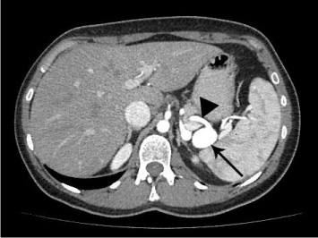
    Figure 1:  Contrast-enhanced CT scanning demonstrating the saccular
aneurysm of the splenic artery (arrow).
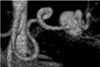
    Figure 2:  Contrast-enhanced CT scanning with tridimensional reconstruction
demonstrating the tortuous splenic artery and the saccular SAA.
Most cases of SAA are associated with atherosclerosis, although there were reported a few cases of fibromuscular dysplasia [10].
Patients with SAA often present without symptoms and the SAAs are found during investigation of other abdominal diseases [3].
True SAAs are usually not greater than 3cm in diameter. Mural thrombus and calcifications frequently occurs. Splenic artery pseudo-aneurysms are even rarer than true aneurysms, with a slight male predominance, probably due to pancreatitis and presence of pancreatic pseudo-cysts [8,5].
SAAs association with pregnancy
SAAs occur predominantly in younger women (58%) of childbearing age and has 4:1 female:male predominance. Almost a century after the SAA was first described, Corson reported the association between pregnancy and rupture of SAA [3,11]. The rupture is frequently associated with pregnancy [7,12]. Up to 95% of SAAs present during pregnancy. Among them over 95% were found following rupture. SAA rupture during pregnancy is associated with disproportionately high maternal (64-75%) and fetal (72,5-95%) mortality rates [4,6,12]. Pregnancy and increasing parities are factors associated with SAA [10].
Diagnosis
Multi-slice CT angiography can accurately and clearly display the location of visceral artery aneurysms. Aneurysm’s shape, extend, aneurysmal wall, course of the artery and relationship with other vessels can be revealed (Figures 1-3) [13]. However, since the SAAs are often asymptomatic they are found during investigation of other diseases or when rupture occurs [3].
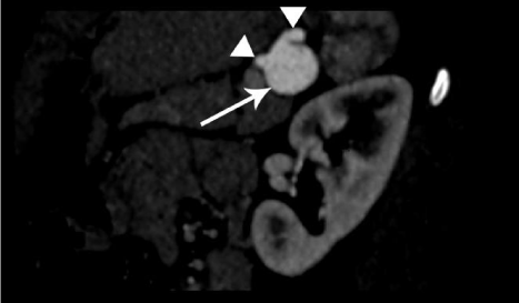
    Figure 3:  Contrast-enhanced CT scanning demonstrating the aneurysm
of the splenic artery (arrow) and splenic antery proximally and distally
(arrowheads).
Treatment
The first described case of successful surgical repair of the SAA was in 1940 by McLeod [3,14]. Treatment should be offered to patients suffering from the SAA greater than 2cm in diameter, growing or associated with inflammation; women of childbearing age or pregnant (prior to the 3rd trimester of gestation); symptomatic patients and in case of portal hypertension [3,15].
SAA is associated with operative mortality rate of 25% when ruptured, comparing to 0,5% for non-ruptured cases [3,4]. When rupture occurs during pregnancy the mortality rate became disproportionately high, with maternal mortality rate of 64-75% and fetal mortality rate of 72,5-95% [5,7,16]. Because of the associated potentially fatal consequences, prompt management of SAA is essential [4,7]. Different management modalities can be applied: open surgery and endovascular approach.
Open surgical repair (Figure 4) includes excision and repair (patch closure or primary anastomosis) of SAA located at distal third of splenic artery. Postoperatively CT scanning should reveal patent splenic artery (Figures 5 and 6). When SAA is located in proximal and medial third of splenic artery proximal and distal ligation of aneurysm sac should be considered [8].
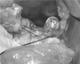
    Figure 4:  Open exposure of the SAA (ligations on splenic antery proximally
and distally from the saccular aneurysm).
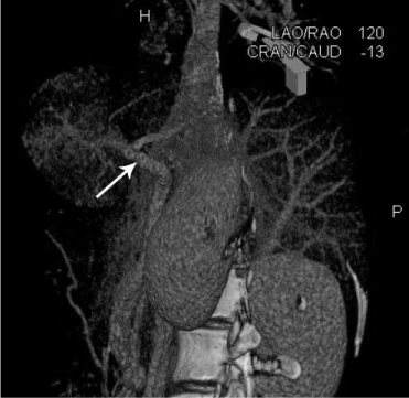
    Figure 5:  Contrast-enhanced CT scanning with tridimensional reconstruction
revealing patent splenic artery postoperatively (arrow).
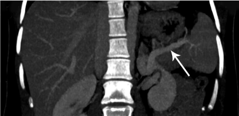
    Figure 6:  Contrast-enhanced CT scanning revealing patent splenic artery
postoperatively (arrow).
The other options for open management are ligation of the SAA located in proximal third of splenic artery without resection of the aneurysm sac and with preservation of splenic function [9] as well as aneurysm resection with splenectomy for the SAA in distal third of splenic artery [17].
Another option is endovascular repair under local anesthesia. It has a lower success (95,2% vs 98,6%) and periprocedure mortality rates (0,6% vs 5,0%) as well as higher late complications (9,2% vs 3,3%) and reinterventions rates (8,0% vs 2,9%) compared with open surgery [8,15,18,19].
Covered stents, bare stents, coils, microcoils and N-Butyl Cyanoacrylate Glue (NBCA) can be useful in management [8,19-22]. Peripheral or cardiac stent-graft deployment leads to exclusion of the aneurysm sac. Stent graft implantation is safe and effective. The effectiveness of multilayer stents or multiple overlapping bare stents in patient at high surgical risk was also proved. Stents can be deployed alone or followed by a trans-stent coiling. The result is physiologic exclusion of an aneurysm, thrombus formation and aneurysm shrinkage. No late rupture and no ischemic damage of spleen were observed [8,20,21]. Covered stents due to being less flexible and requiring larger access vessels may not be possible to deliver.
The embolization of choice is use of coils and microcoils. Embolization of ruptured SAA ensure immediate control of bleeding. This technique can be applied regardless of the SAA location, etiology and clinical presentation as well as patient’s general health [18]. Success rate of endovascular embolization is 95,2% [15]. In case of procedure failure the subsequent endovascular repair at a later date leads to successful ablation of the aneurysm sac. Minor complication (abdominal pain, fever) may follow endovascular repair as a manifestation of postembolization syndrome [15,18,19].
Nevertheless, due to possible ineffective or incomplete coil embolization an increasing role of N-Butyl Cyanoacrylate Glue (NBCA) is observed. Benefits of NBCA are reduction of procedure time (compared with coil embolization) and enabling embolization of distal, impossible to cathaterizate vessels. However training and experience are of prime importance to avoid complications [19,22].
Endovascular treatment (among which transcatheter embolization is preferred) remain a treatment of choice for both elective and acute, ruptured SAAs. In case of unfavorable anatomy for the endovascular repair (elongated and tortuous splenic artery) there is a room for open surgery [8,15,18,19,22,23].
Conclusion
The SAA is an uncommon asymptomatic pathology, becoming a life-threatening, when a rupture occurs.
Due to high mortality rate in case of aneurysm rupture, even asymptomatic SAAs should be treated.
Rapid diagnosis and treatment is of prime importance.
Open, endovascular and laparoscopic approach can be applied in the SAA management.
Endovascular treatment (among which transcatheter embolization is preferred) remain a treatment of choice for most of elective and acute cases.
References
- Olsen AB, Ralhan T, Harris JH Jr, Evani V. Superior mesenteric artery pseudoaneurysm after blunt abdominal trauma. Ann Vasc Surg. 2013; 27: 674-678.
- Beaussier M. Sur unaneurisme de l'artèrespléniquedont les parois se sont ossifies. J Med Clin Pharm. 1770; 32: 157.
- GuillaumonI AT, Chaim EA. Splenic artery aneurysm associated with anatomic variations in origin. J Vasc Bras. 2009; 8: 177-181.
- Sadat U, Dar O, Walsh S, Varty K. Splenic artery aneurysms in pregnancy-a systematic review. Int J Surg. 2008; 6: 261-265.
- Agrawal GA. Splenic artery aneurysms and pseudoaneurysms Clinical distinctions and CT appearances. Am J Roentgenol. 2007; 188: 992-999.
- Ha JF, Phillips M, Faulkner K. Splenic artery aneurysm rupture in pregnancy. Eur J Obstet Gynecol Reprod Biol. 2009; 146: 133-137.
- Oakley E, Ho JD, Johnson V, Vancamp J, Melson T, Hick JL. Splenic artery aneurysm: an important cause of hemoperitoneum and shock. J Emerg Med. 2014; 46: 65-67.
- Mohan IV, Stephen MS. Peripheral arterial aneurysms: open or endovascular surgery? Prog Cardiovasc Dis. 2013; 56: 36-56.
- Wei YH, Xu JW, Shen HP, Zhang GL, Ajoodhea H, Zhang RC, et al. Laparoscopic ligation of proximal splenic artery aneurysm with splenic function preservation. World J Gastroenterol. 2014; 20: 4835-4838.
- Unlu C, van den Heuvel DA, Leeuwis JW, de Vries JP. Ruptured aneurysm of the splenic artery associated with fibromuscular dysplasia, Ann VascSurg. 2014; 28: 15-18.
- Lynch MJ, Woodford NW. Rupture of a splenic artery aneurysm during pregnancy with maternal and foetal death: a case report. Med Sci Law. 2008; 48: 342-345.
- Boumans D, Weerink LB, Rheineck Leyssius AT, Swartbol P, Veneman TF. Splenic artery rupture during pregnancy concealed by a pancreatic lymphangioma: a rare co-occurrence. Ann Vasc Surg. 2013; 27: 1-4.
- Ming WD, Li XG, Xue HD, Jin ZY. Role of multi-slice computed tomography angiography in the diagnosis of visceral artery aneurysms. Zhongguo Yi XueKeXue Yuan XueBao. 2014; 36: 296-299.
- Macleod D, Maurice T. Rupture of a branch of the splenic artery: associated with pregnancy. The Lancet. 1940; 924-925.
- Hogendoorn W, Lavida A, Hunink MG, Moll FL, Geroulakos G, Muhs BE, et al. Open repair, endovascular repair, and conservative management of true splenic artery aneurysms. J Vasc Surg. 2014; 60: 1667-1676.
- Dawson J, Fitridge R. Update on aneurysm disease: current insights and controversies: peripheral aneurysms: when to intervene - is rupture really a danger? Prog Cardiovasc Dis. 2013; 56: 26-35.
- Chen JS, Chuang SC, Wang SN, Chang WT, Kuo KK, Lee KT, et al. Natural course of splenic artery aneurysm with associated spontaneous splenorenal shunt in non-cirrhotic liver: an 18-year observational follow-up and review of literature. Kaohsiung J Med Sci. 2013; 29: 55-58.
- Loffroy R, Guiu B, Cercueil JP, Lepage C, Cheynel N, Steinmetz E, et al. Transcatheter arterial embolization of splenic artery aneurysms and pseudoaneurysms: short- and long-term results. Ann Vasc Surg. 2008; 22: 618-626.
- Lakin RO, Bena JF, Sarac TP, Shah S, Krajewski LP, Srivastava SD, et al. The contemporary management of splenic artery aneurysms. J Vasc Surg. 2011; 53: 958-964.
- Künzle S, Glenck M, Puippe G, Schadde E, Mayer D, Pfammatter T. Stent-graft repairs of visceral and renal artery aneurysms are effective and result in long-term patency. J Vasc Interv Radiol. 2013; 24: 989-996.
- Zhang L, Yin CP, Li HY, Bao JM, Zhao ZQ, Qu LF, et al. Multiple overlapping bare stents for endovascular visceral aneurysm repair: a potential alternative endovascular strategy to multilayer stents. Ann Vasc Surg. 2013; 27: 606-612.
- Madhusudhan KS, Gamanagatti S, Garg P, Shalimar, Dash NR, Pal S, et al. Endovascular Embolization of Visceral Artery Pseudoaneurysms Using Modified Injection Technique with N-Butyl Cyanoacrylate Glue. J Vasc Interv Radiol. 2015; 26: 1718-1725.
- Pulli R, Dorigo W, Troisi N, Pratesi G, Innocenti AA, Pratesi C. Surgical treatment of visceral artery aneurysms: A 25-year experience. J Vasc Surg. 2008; 48: 334-342.