
Review Article
Austin J Vet Sci & Anim Husb. 2015; 2(2): 1013.
Present Status on the Taxonomy and Morphology of Echinococcus Granulosus: A Review
Rahman WA¹*, Elmajdoub LE², Noor SAM² and Wajidi MF³
¹School of Food Science and Technology, University Malaysia Terengganu, Malaysia
²School of Biological Sciences, University Sains Malaysia, Malaysia
³School of Distance Education, University Sains Malaysia, Malaysia
*Corresponding author: Wahab A Rahman, School of Food Science and Technology, University Malaysia Terengganu, 21300 Kuala Terengganu, Malaysia
Received: May 09, 2015; Accepted: September 04, 2015; Published: September 09, 2015
Abstract
Echinococcus granulosus has been researched extensively and previously discussed in detail as it is the species that is most widely distributed throughout the world. It is a cyclozoonotic infection which has important global implication as it is found in many animals and humans and involves a large group of countries. The high incidence and wide distribution of this parasitic infection that man shares with animals make the disease one of the most serious zoo noses in many parts of the world. Epidemiological studies indicate that the sheep strain of E. granulosus is the leading agent of cystic echinococcosis in humans. The common intermediate host of E. granulosus is many domesticated mammals such as sheep, cattle, pigs, goats and camels. Sheep, which harbor the most fertile hydatid cysts, are the most important intermediate host and represent the most important source of infection to dogs through the feeding of infected offal, giving rise to high prevalence rates as shown in many countries of the world.
Keywords: Taxonomy; Morphology; Echinococcus granulosus
Introduction
Echinococcosis or hydatid disease, caused by the larvae of the closely related species Echinococcus granulosus and E. multilocularis is the most serious tapeworm infection occurring in man and domesticated animals. This is not because it is particularly common but because of the lack of an effective chemotherapeutic agent and the difficulty of surgery often results in a poor prognosis. The two species have been clearly differentiated and the presence of strains (regarded by some as subspecies, and of each, adapted to various hosts) had been established as early as the 1950s’, but until today there is still much confusion in various aspects of the disease, especially epidemiology. The present review is an attempt to re-visit some important nonmolecular aspects of the disease.
History of Hydatid Disease
Hydatidosis is a disease with an extremely long history as it has been known since the time of Hippocrates, and it is caused by the tapeworm Echinococcus granulosus. According to [1] Echinococcus granulosus has been known since ancient times, for example, Aretaeus and Galen were familiar with hydatid cysts. The generic name Echinococcus is from the Greek words “echinos”, which means hedgehog and “kokkos” which means berry, while the species term granulosus is from the Latin word “granulum” which means little grain. The term hydatid is from the Greek word “hydatis” which means a drop of water [1,2].
At present Echinococcus granulosus, the causative agent of hydatidosis is almost ubiquitous. It was Redi in 1684, Hartmann in 1685, and Tyson in 1691 who first suspected their animal and the bladder were really worms. In 1766, Pallas first mentioned the similarity of hydatids in man and other mammals and Goeze in 1782 first studied the protoscoleces of the metacestoda and recognized their relationship to those of taenial origin and differentiated the hydatid cyst from the cysticercus. In 1695, Hartman first observed the adult worms in the dog’s intestine, and later Rudolphi in 1808 studied details of the adult worms. Then Von Siebold in 1852 followed by Haubner, Leuckart, Kuchenmeister and Nettleship fed protoscoleces of cysts on domestic animals to dogs and observed the development of the adult worms in the intestines of the hosts. Later Naunyn, 1863 in Germany, Krabbe, 1863 in Iceland, and Thomas, 1885 in Australia obtained adults worms in dogs from protoscoleces of human origin [2].
Taxonomy of Echinococcus Granulosus
According to Yamaguti, Braithwaite [3,4] and Thompson (1986), the tapeworms whose larval stage causes hydatid cysts belong to the following:
Phylum: Platyhelminthes.
Superclass: Eucestoda.
Class: Cestoidea.
Subclass: Cestoda.
Order: Cyclophyllidea Ben; Braun, 1900.
Family: Taeniidae Ludwig, 1886.
Genus: Echinococcus Rud, 1801.
The genus Echinococcus was established for the larval cestoda which was earlier designated Taenia visceralis socialis granulosus by Goeze in 1782 obtained from the liver of sheep. In 1786, Batsch adapted the binary system of nomenclature of Linnaeus and gave the name Hydatigene granulosus to the taxon described by Goeze [5]. In 1963, Rausch and Nelson reviewed the species of Echinococcus and concluded that the status of E. felidis Ortlepp, 1937 and E. patagonicus Szidat, 1960 was uncertain, but that both were possibly co-specific with E. granulosus. In addition, E. cameroni Ortlepp, 1934; E. intermedius Lopez-Neyra and Soler, 1943; E. longimanubrius Cameron, 1926; E. lycaontis Ortlepp, 1934; E. minimus Cameron, 1926, and E. Ortlepp Lopez-Neyra and Soler 1943 were considered synonyms of E. granulosus Rausch and Nelson 1963.
Strains of Echinococcus Granulosus
Echinococcus granulosus has been divided into several strains according to the host [6]. Echinococcus granulosus has been researched extensively and discussed in detail as it is the species that is most widely distributed throughout the world. For example, it has six strains which have been identified as G1 genotype (common sheep strain), G2 genotype (Tasmanian sheep strain), G4 genotype (horse strain), G3, G5 genotypes (cattle strain), G6 genotype (camel strain), G7 genotype (pig strain) [7,8]. In addition, Haag et al. [9] recorded five potential hybrid genotypes between cattle and sheep strains found only in Southern Brazil.
Distribution of Echinococcus Granulosus
According to Belding, Beck and Davies, Beaver et al., Schantz et al., Shambesh [1, 10-12] E. granulosus has a worldwide distribution. Indeed as Abdou, Matossian et al., Polydorou [13-15] point out, hydatidosis is a cyclozoonotic infection which has important global implication as it is found in many animals and humans and involves a large group of countries. For example, hydatid disease is considered endemic in humans and animals in North Africa, while in the rural areas of Libya, Tunisia, Algeria and Morocco, it is classified as hyper endemic. In addition, it is prevalent in countries in Latin America, the Middle and Near East, the Mediterranean littoral, a number of African countries and several European ones. The high incidence and wide distribution of this parasitic infection that man shares with animals make hydatidosis one of the most serious zoonoses not only in Egypt, but also in the Middle East, Cyprus, Iraq, Jordan, Kuwait, Lebanon, Iran and Syria.
Epidemiological studies indicate that the sheep strain of E. granulosus is the leading agent of cystic echinococcosis in humans. It has been extensively studied in a number of different areas and is now present in Asia, Africa, South and Central America and the Mediterranean region and also common in parts of Europe, especially the United Kingdom and Australia [11,12]. Those regions with the most extensive and intensive E. granulosus infections are sheep and cattle-raising countries, including Tanzania and the southern half of South America and Paraguay [16]. In addition, human infection occurs frequently throughout northern, southern and eastern Europe, Siberia, Turkestan and Mongolia as illustrated in (Figure 1).
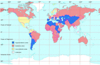
Figure 1: Worldwide Distribution of E. granulosus.
Morphology of Echinococcus Granulosus
The adult E. granulosus lives attached to the mucosa of the small intestines of dogs and related animals. According to Sweatman and Williams, Belding, Beaver et al. it is a minute worm measuring 2 to 9 mm in length [1,17,18]. It consists of three or four segments and has a globular scolex of 0.3 mm in diameter containing a rostellum and four cuplike oval suckers. The rostellum is armed with a double crown of large and small hooklets. The total number of hooklets ranges from 25-44 [17], 28-40 [1], 28-50 [2], and 32-40 [18]. The majority of protoscoleces have two rows of hooks with an equal number of large and small hooks. The scolex is followed by a short neck and usually only one or two immature segments. The mature segment has fully developed reproductive organs consisting of testes and ovary. The gravid segment is the broadest and longest. In the gravid unit, the uterus contains as many as 500 eggs, which are discharged into the faeces from the ruptured segment (Figure 2). The subspheric egg is 34 to 41 μm in diameter, has a brown hexacanth embryo, and is similar in appearance to those of other taenial worms [1] (Figure 3).
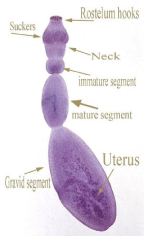
Figure 2: Morphology of Adult Worm of E. granulosus.
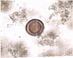
Figure 3: Morphology of egg of E. granulosus.
Life cycle of Echinococcus Granulosus
Final host
According to Lewall [16], the final hosts of E. granulosus are dogs, wolves, jackals, dingoes, coyotes and foxes. Feline species are seldom infected naturally, but the parasite has been reported in cats, wild cats and leopards, which can also serve as hosts, but with low efficiency.
Intermediate host
The common intermediate host of E. granulosus is many domesticated mammals such as sheep, cattle, pigs, goats and camels. Sheep, which harbor the most fertile hydatid cysts, are the most important intermediate host and represent the most important source of infection to dogs through the feeding of infected offal, [19]. In Canada, deer, moose, bison, wild boar and horse are often infected and can serve as hosts. Animal hosts differ from area to area [10]. Humans are accidental intermediate hosts.
Development
The intermediate host ingests eggs of E. granulosus, which are passed in the faeces of the final host and are immediately infective. They are resistant to external conditions and are capable of development even after months outside the body [1]. Following ingestion by an intermediate host, they hatch in the small intestine and the resulting oncospheres invade the blood vessels of the wall. The hatching process uses the disaggregation of the keratin-like blocks of the embryophore by pepsin, pancreatic enzymes, etc., followed by the activation of the contained oncospheres [20].
The liberated muscular oncosphere uses its six hooks to attach itself to the microvilli of the jejunal region of the small intestine [21] and enters the lymphatic or mesenteric venules and becomes lodged in various organs. This probably in turn affects the differential distribution of the metacestodes between the liver and lungs. Within twelve hours after ingestion, it arrives at the liver, where if not destroyed by phagocytic cells, it develops into a hydatid cyst [1]. They then settle in the liver, forming a respective cyst, which is the origin of an Echinococcus. However, the liver is not always the primary filtration site [22]. Inhaled eggs can also cause pulmonary hydatid disease as it has been shown that eggs administered to sheep by a tracheostomy develop into lung cysts [16]. The larvae of E. granulosus may also develop in the heart, spleen, kidneys, brain, eyes and long bones (Dunn, 1978). The speed of development of the embryo to the fully formed metacestoda is slow, and during the first 10-14 days, it involves cellular proliferation, degeneration of the oncosphere hooks, and formation of a central cavity and development of the laminated and germinal layers. For example, the time taken to produce the first brood capsules from eggs is reported to be not less than 7 months in mice [23], 10-12 months in pigs [24], and 10-48 months in sheep [21].
When a final host ingests or inhales these hydatid cysts, the protoscoleces project their heads and secure a firm hold among the villi of the small intestine and mature into adults in 40-50 days. Since each worm stays in a host for 5-29 months, the eggs of E. granulosus are liberated after detachment of a gravid segment, which occurs every two weeks. Thus, the number of eggs produced by an adult is less than those for other cestodes [18]. In addition, the small intestine may finally become full of adults as a result of repeated infections or intake of a large number of cysts. Each scolex that is ingested is capable of developing into an adult worm in the intestinal tract in about seven weeks [10]. In heavily endemic areas, 50% of the dogs are infected with adult worms, and up to 90% of sheep and cattle and 100% of camels may be infected with hydatid cysts [25] as illustrated in Figure 4. Borrie et al. recorded the mode of transmission to be a direct transfer of eggs by hand or unwashed vegetation to the mouth [26]. However, experimental evidence has been reported that echinococcosis can become established in the lungs of sheep from eggs without their prior ingestion through intratracheal route.
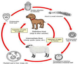
Figure 4: Life Cycle of E. granulosus.
The pathology caused by the presence of a single unilocular cyst depends very much on its site in the body. In the liver, the cyst may cause compression of the liver cells, leading to biliary stasis and cholangitis. Lung cysts are always intracapsular and their rupture may result in protoscoleces being coughed up in the sputum. Hydatid cysts in the brain or spinal cord are usually smalland may cause symptoms earlier than in other sites. Renal cysts sometimes occur and, if they burst into the kidney pelvis, may become secondarily infected.
Unilocular Hydatid Cyst
The hydatid cyst in E. granulosus is usually unilocular; a spherical cyst the size of a child’s head or larger. The larval growth occurs through evagination with hollow protuberances of the cyst wall, and by exogenous budding of solid and bare extensions of the germinal membrane with progressive invasion of the organ [27]. The cyst increases in size, attaining in the course of its growth a diameter of about one mm in one month, 10 mm in five months, and 200 mm after several years. The final cyst usually grows to a maximum of 10- 12 cm in diameter [1]. By the fourth day, larvae reach a diameter of 40μm, when they begin to develop a cystic cavity. By the end of three weeks the young cyst has attained a diameter of 250 μm [2]. The cyst is filled with pale-yellow fluid and comprises three layers: a thick connective tissue capsule formed by the reaction of the host, a median laminated layer and an internal thin germinal membrane about 22- 25 μm thick which produces the brood capsules containing protoscoleces.
The laminated layer can be up to five mm thick; with nonnucleated hyaline supporting curricula, also a brood capsule is a spherical body of about 0.15 mm in diameter [28]. From the germinal layer, masses of cells are budded out into the cystic cavity. These cells become vacuolated and later stalked [2]. The thin-stalked brood capsules often break away from the germinal layer and may rupture, releasing invaginated protoscoleces, which settle down to add to the brood capsules and sediment also containing separated hooks and other components. It is called hydatid sand [29].
Daughter cysts which are free in the cavity of the main hydatid cyst appear to be quite common in certain hosts, such as humans. The endogenous daughter cyst with a thin, transparent wall develops in the cystic fluid and at times may produce granddaughter cysts [28]. The number of protoscoleces thus formed within a mother Echinococcus varies, and may be as numerous as two million. Each protoscolex can develop in the canine final host into a new egg-producing adult tapeworm in 5-8 weeks [5]. These cysts are usually described as fertile cysts. However, hydatid cysts often fail to produce brood capsules or protoscoleces, and such sterile (infertile) cysts are quite common. For example, Thompson (1977) found 27% of horse cysts and 51% of sheep cysts to be sterile. Some become sterile by bacterial invasion or calcification [2].
Hydatid cysts can survive for the entire lifespan of the host, and maximum longevity of 16 years has been recorded for horse cysts [30] and 53 years in humans [31]. Although much larger cysts containing several liters of hydatid fluids have been recorded [32], Echinococcus lacking protoscoleces may sometimes arise, and the brood capsules never produce protoscoleces. Such incomplete cysts are called acephalocyst and are seen mainly in cattle [18]. According to Elamin et al., a sterile cyst was seen in a 4-year-old female camel, which showed a large vesicle on its back, and when excised, the cyst weighed 1750 g and contained 2.5 L of fluid [33].
In addition, a variety of unilocular cyst is osseous hydatid, that, it is found in the bones. Osseous hydatid in confined by dense osseous tissue and the laminated layer is poorly developed. The osseous hydatid migrates along, for example, the bony canal as a naked protoplast, frequently eroding the surrounding osseous tissue [2]. According to Mahmoud and El-Garhy the cyst wall contains a carbohydrateprotein substrate complex, collagen and possibly calcium. Calcium is also reported in protoscoleces of hydatid sand [34].
Diagnosis of hydatid cysts
As most E. granulosus infections remain asymptomatic for many years before the increasing number of cysts are able to cause symptoms in the infected organs, different methods have been widely used in the diagnosis of hydatid disease including imaging techniques in humans, immunodiagnostics in livestock, final hosts and humans [35].
Hydatid fluid (hydatid sand) aspirated from a cyst through the opening of a cyst may show the presence of protoscoleces. Normally the protoscoleces are invaginated. The presence of protoscoleces is diagnostic and based on morphology of cyst [36]. The presence of protoscoleces indicates activity of the cyst. However, if they are absent, they are an indication for growth of cyst or degeneration [36].
Prevalence of hydatid cysts in livestock
The eggs adhere to the coat of the final host, which are usually dogs, and can be found concentrated particularly on the hair near the mouth and anus. In addition, they are also found in dust and vegetation with a particularly high concentration around kennels, but in water, they sink rapidly. Apart from those activities associated with dogs by which eggs spread other methods by which eggs are spread include wind, flies and beetles [37]. Also, Dowling et al. reported that studies have shown that taeniids eggs can be dispersed over a wide area by blowflies and birds [38].
Sweatman and William and Gemmell reported that eggs of E. granulosus might remain infective for several weeks, although sunlight and heat have been reported to be lethal during the first 3 weeks [17,37]. The eggs are still infective after they had been kept in tap water at 6 °C for 7 months.In addition, Gemmell observed that eggs of E. granulosus were made harmless by boiling water, but because heat penetrates dog faeces slowly, infective material should be boiled at least 5 minutes to render the eggs harmless [37].
North Africa
In Tunisia, Deve reported hydatid cysts in 20.6% of sheep, 16% of goats, 30% of camels and in all the cattle examined at different abattoirs in that country [39]. In addition, Benosman [40] found a 6.9% infection rate in sheep, 19.4% in cattle, 28.8% in pigs, 66.2% in camels and 5% in horses while Brahmi reported a prevalent rate of infection of 2.8% in sheep, 8% in cattle, 19% in pigs and 17.5% in camels [41].
In Morocco, little was published on the incidence of hydatid disease until recently when Pandey et al. reported an 80% infection rate in 35 camels [42]. In 1988, Pandey et al reported that 5.3% of sheep and 44.6% of cattle examined from Quarzazate in southern Morocco had hydatid cysts.
In Algeria, Jore [43] found 21.9% of sheep and 30% of cattle infected in different Algerian abattoirs, while Larbaoui et al. found that 42.1% of a large sample of camels had hydatid cysts [44].
In Egypt, hydatid cysts have been reported in different livestock hosts. For instance, Elkordy recorded 31% infection in camels, 10% in cattle, and 1.5% in sheep at a Cairo abattoir [45]. Moreover, he observed calcified cysts in 36.4% of animals examined. At the same abattoir, Abdou recorded 30% infection in camels, 10% in cattle and 5% in sheep, with slightly lower levels at an Alexandria abattoir [13]. Furthermore, Elrafaie et al. found 3.4% of camels at Cairo abattoir infected with hydatid cysts [46]. In another survey on the incidence of hydatid disease in slaughtered camels, it was found that 26.5% were infected at Assiut abattoirs during 1985-1989 [47]. All the surveys also revealed that the liver was the most commonly infected organ at 64% of the infected cattle, and 75% of the infected sheep. Lung infections represented 36% in cattle and 4.5% in sheep. There were a few cysts in the spleen and other organs of some of the animals. The highest levels of fertile cysts occurred camels at 68.4% and sheep at 63.6%, and the lowest from those in cattle at 11.3%.
In Libya, data on the disease and its epidemiology is still extremely limited, although the problem is acknowledged as serious in both humans and livestock [48]. Hydatid cysts infection in livestock was first reported by Medulla [49], who found that hydatid infections were common in camels and other livestock animals. In 1961, Cicogna investigated the prevalence of unilocular hydatid cysts in livestock and found that 40% of sheep and 70% of cattle were infected. Gusbi et al. [50] recorded studies in Libya that show that livestock animals drink from open water sources and have free access to vegetables gardens, which were often unfenced, thus this is one of the reasons that help in the prevalence of infection hydatid disease.
However, apart from incidental information collected from previous records, at abattoirs during routine work at meat inspection nothing is known about the prevalence of hydatid disease in farm animals. For example, Dar and Taguri simply collected incidental abattoir data for the period, (1975-1977) reporting prevalence of 3.1- 63.4% in sheep, 5.5-18.1% in goats, 1.4-34.7% in camels and .7-4.9% in cattle [51]. In addition, Gusbi et al. [50] reported high prevalence in sheep and Gusbi et al. [52] found that 5.4% of cattle at 14 different areas were infected, and 35.9% of camels were infected. Tashani et al. [48] recorded the infection rate in Benghazi city at 20% in sheep, 3.4% in goats, 11% in cattle and 13.6% in camels. Aboudaya [53], reported a larger collection of data from 46 abattoirs, covering the months of 1976 from June to December. The prevalence of hydatid disease was 6.6% in cattle, 4.3% in sheep and 27.25% in camels. Finally, Gusbi et al. [52] recorded the intensity of infection in camels from Benghazi, Alsaa, Sebha, and Tripoli. Of 193 cysts obtained, 68 (35.2%) were in the liver and 125 (64.8%) in the lung. Finally, Elmajdoub et al. [54] reported the fertility rates in the Misurata region in Libya were 62.7% in sheep and 60.3% in camels, but all examined cysts from cattle were sterile.
Other Locations
Hydatidosis is common in the sheep population with the prevalence in some areas as Kazakhstan reaching over 50% in old sheep [55]. Similarly, Mobedi et al. [56] and Motakef et al. (1976) recorded that sheep were commonly infected, and act as important intermediate host of E. granulosus and the prevalence of infection and cyst fertility rates were high. However, although a high proportion of cattle were infected with E. granulosus, the fertility rate of hydatid cysts was low. In the case of Iran, hydatid cysts are most common in camels, and the cysts have a high fertility rate. For example, Hosseini [57] recorded the prevalence of hydatid cysts in some regions from Iran 1.5-16.8% in sheep, 6-14.7% in cattle and 11.3%-64.0% in camels. In addition, Hosseini [57] recorded fertility rates of 88% in sheep, 83.7% in cattle, and 13% in camels and the viability rates of protoscoleces were 60% in sheep, 75% in cattle, and 60% in camels in Iran. In the case of Jordan, Abo-Shehada [58] reported that the infection rates for sheep, cattle and camels were 1.3-71.1%, 1.3-12.9%, and 8.8-11.1% respectively, while Abdel-Hafez and Al-Yaman [59] reported the infection rate in spleen from sheep was 7.6% in Jordan. Molan [60] recorded infection rates of 4.5- 44% in sheep, 4.3-13.9% in cattle and 20.4-72% in camels from different regions in Iraq. Hydatid cyst is endemic in Iraq, but in spite of the high incidence of this disease in livestock [61]. Lyagoubi et al., [62] suggested that hepatic cysts developed at a slower rate than pulmonary cysts, and this has been attributed to a lower allowance of hydatid cyst development in pulmonary tissue. The intensity of E. granulosus infection in sheep increased with age in a linear pattern, and this implies that E. granulosus is in an endemic fixed state with no evidence of protective immunity in the intermediate host (Torgerson et al., 1998). The distribution of parasites followed a negative binomial distribution [63].
In New Zealand, hydatid disease is still a major problem although the incidence rate has been reduced, with about 70 new clinical cases per year. The most heavily infected countries in Europe appear to be upper Greece, Italy, and Spain. Also, throughout southwestern Asia and northern Africa where sheep, goats and camels and to a lesser extent cattle and pigs are slaughtered hydatid infection in the people remains a health problem [64].
References
- Belding MD. The super family Taeniodea: The genus Echinococcus in: Textbook of Parasitology. 3rd edn. London, Appetion- Century-Crofts. 1965: 626-644.
- Beaver PC, RC Jong, and EW Cupp. Cyclophylliidean Tapeworms in Clinical Parasitology, 9th ed. Philadelphia, Leo and Febiger, 1984; 527-543.
- Yamaguti S. Cestodes of mammals in Systema Helminthum. 1959; 361, 435. Chancery Lane, London, England.
- Braithwaite PA. Hydatid disease: epidemiology and pathology. Aust N Z J Surg. 1983; 53: 203-209.
- Thompson RCA. Biology and systematic of Echinococcus 1986. In “Biology of Echinococcus and Hydatid disease” (R.C.A. Thompson, ed): 5-43 George Allen and unwin, London.
- Thompson R C A. Biology and systematic of Echinococcus and Hydatid disease. Thompson R.C.A and Laymbery, A.J. (eds). CAB International Wallingford, Oxen; 1995: 1-37.
- Rosenzvit MC, Zhang LH, Kamenetzky L, Canova SG, Guarnera EA, McManus DP . Genetic variation and epidemiology of Echinococcus granulosus in Argentina. Parasitology. 1999; 118 : 523-530.
- Lymbery A J. Genetic diversity, genetic differentiation and speciation in the genus Echinococcus. Rudolphi, 1801, in: Thompson R.C.A and Laymbery, A.J. (ed) Echinococcus and Hydatid disease. CAB International Wallingford, Oxen; 1995: 51-88.
- Haag KL, Araújo AM, Gottstein B, Siles-Lucas M, Thompson RC, Zaha A. Breeding systems in Echinococcus granulosus (Cestoda; Taeniidae): selfing or outcrossing? Parasitology. 1999; 118 : 63-71.
- Schantz PM, JChai PS, Craig J, Eckert DJ, Jenkins CNL, Macpherson , et al. Epidemiology and control of hydatid disease, 1995. In Thompson R.C.A and Laymbery, A.J. (ed) Echinococcus and Hydatid disease. CAB International Wallingford, Oxen; 233-331.
- Shambesh MK. Human cystic echinococcosis in North Africa in: Compendium on cystic echinococcosis by F. I. Andersen, H. Ouhrlli and Kachani. Brigham Young University. 1997; pp: 234.
- Abdou AH. Incidence and public health importance of hydatidosis in the Middle East with special reference to U.A.R. Journal of Veterinary Science of United Arab Republic. 1965; 2: 125-134.
- Matossian RM, Rickard MD, Smyth JD. Hydatidosis: a global problem of increasing importance. Bull World Health Organ. 1977; 55: 499-507.
- Polydorou K. Echinococcosis/ Hydatidosis control in Libya. Report UNOP/WHO, Mediterranean Zoonoses Control Center. 1981; Pp: 1-11.
- Lewall DB. Hydatid disease: biology, pathology, imaging and classification. Clin Radiol. 1998; 53: 863-874.
- sweatman Gk, Williams Rj. Comparative Studies On The Biology And Morphology Of Echinococcus Granulosus From Domestic Livestock, Moose And Reindeer. Parasitology. 1963; 53: 339-390.
- Miyazaki I. Echinococcosis in helminthic zoonoses, pp: 247-267. Toyo-Kaiji Bldg., No.6; 7-2, Shinbashi 4-chome, Minato-ku, Tokyo, Japan. 1991.
- Torgerson PR, Karaeva RR, Corkeri N, Abdyjaparov TA, Kuttubaev OT, Shaikenov BS. Human cystic echinococcosis in Kyrgystan: an epidemiological study. Acta Trop. 2003; 85: 51-61.
- Beck JW and JE Davies. The somatic tapeworms in: Medical Parasitology, 3th, ed. London, Mosby Company. 1981; pp: 215-223.
- Lethbridge RC. The Biology of the onchosphere of cyclophyllidean cestodes. Helminthological Abstracts, A. 1980; 49: 59-72.
- Heath DD. The migration of oncospheres of Taenia pisiformis, T. serialis and Echinococcus granulosus within the intermediate host. Int J Parasitol. 1971; 1: 145-152.
- Matsaniotis N, Karpathios T, Koutoyzis J, Nicolaidou P, Fretzayas A, Papadellis F, et al. Hydatid disease in Greek children. Am J Trop Med Hyg. 1983; 32: 1075-1078.
- Colli CW, Schantz PM. Growth and development of Echinococcus granulosus from embryophores in an abnormal host (Mus musculus). J Parasitol. 1974; 60: 53-58.
- Slais J, Vanĕk M. Tissue reaction to spherical and lobular hydatid cysts of Echinococcus granulosus (Batsch, 1786). Folia Parasitol (Praha). 1980; 27: 135-143.
- Noble RN and AN Noble. Chapter 9, 10 in : Parasitology, the biology of animal parasites. 3rd ed, Lea and Febiger. 1974; pp: 205-206, 244-250. London.
- Borrie J, Gemmell MA, Manktelow BW. An experimental approach to evaluate the potential risk of hydatid disease from inhalation of echinococcus ova. Br J Surg. 1965; 52: 876-878.
- Vogel H. [How does the alveolar echinococcus grow? (author's transl)]. Tropenmed Parasitol. 1978; 29: 1-11.
- Brown HW and FA Neva. Extraintestinal larval tapeworms of human beings in Basic Clinical Parasitology. 5th Edition, Judith Warm Steining, USM, 1983; PP: 191-198.
- Faust EC and RR Russel. Cyclophyllidean tapeworms of man. In: Faust`s Clinical Parasitology 7th, ed., London. 1964; pp 678- 697, Kimpton.
- Ronéus O, Christensson D, Nilsson NG. The longevity of hydatid cysts in horses. Vet Parasitol. 1982; 11: 149-154.
- Spruance SL . Latent period of 53 years in a case of hydatid cyst disease. Arch Intern Med. 1974; 134: 741-742.
- Soulsby EJL. Helminths, Arthropods and Protozoa of domesticated animals. Pp 119-122, 7th edition. London, Bailliere. Tindall, London, 1982.
- Elamin EA, Ramadan RO, Fatani AE, Al Atiya SA, Abdin-Bey MR. Prenatal infection with a hydatid cyst in a camel (Camelus dromedarius). Vet Rec. 2001; 149: 59-60.
- Mahmoud LH, el-Garhy MF. A histochemical study on the hydatid cyst and electron microscopy of the hydatid sand of Echinococcus granulosus. J Egypt Soc Parasitol. 2002; 32: 647-656, 2 p following 656.
- Craig PS. Echinococcus granulosus: immunodiagnosis and vaccination, a perspective. Parassitologia. 1997; 39: 345-347.
- Craig PS, MT Rogan and JC Allan. Hydatidosis and cysticercosis larval cestodes.1995 In: Medical Parasitology: A practical approach, Eds Gillespie, S.H. & Hawkey, P.M. IRL Press (London); pp: 209-237.
- Gemmell MA. The Styx field-trial. A study on the application of control measures against hydatid disease caused by Echinococcus granulosus. Bulletin of the World Health Organization. 1968; 39: 73-100.
- Dowling PM, Abo-Shehada MN, Torgerson PR. Risk factors associated with human cystic echinococcosis in Jordan: results of a case-control study. Ann Trop Med Parasitol. 2000; 94: 69-75.
- Deve F. Enquete etiologique sur l` echinococcose en Tunisie. Archives de I` Institut Pasture de Tunis. 1923; 12: 353-386. (Cited from Anderson, F.L., Ouhell, H. and Kachani, M. (1997): Compendium on cystic Echinococcosis in Africa and in Middle Eastern countries with special Ref reference to Morocco ed. Brigham Young University, USA).
- Benosman F. Considerations epidemiologique sur l` hydatidose animals en Tunisie. Archives de l` Institue Pasteur. Tunis. 1965; 3-4: 409- 418.
- Brahmi C. L`hydatidose humaine et animale en Tunisie. These Ecole Natiomale Veterinaire de lyon. 1970; P.104. (Cited from Andersen, F.L., Ouhell, H. and Kachani, M. (1997): Compendium on cystic echinococcosis in Africa and in middle eastern countries with special Ref reference to Morocco ed., Brigham Young University, U.S.A).
- Pandey VS, Ouhelli H, Ouchtou M. Hydatidosis in sheep, goats and dromedaries in Morocco. Ann Trop Med Parasitol. 1986; 80: 525-529.
- Jore D`Arces M. La situation sanitaire de Alger`a a regard des maladies parasitoses. Bulletin of International Epizootiology. 1955; 43: 117-187.
- Labraoui D, Alliulia R, OsiĬskaia LV, OsiĬskiĬ IIu, Benel'muffok M. [Echinococcosis in Algeria]. Med Parazitol (Mosk). 1980; 49: 21-23.
- El-kordy MI. On the incidence of hydatid disease in domestic animals in Egypt. Journal of the Royal Egypt Medical Association. 1946; 29: 265-279.
- el-Refaie SA, Bassiouny GA, Marie NA, Moris E. Concomitant hepatic Fasciola and hydatid infections in animals. J Egypt Soc Parasitol. 1984; 14: 421-427.
- Ahmed LS. Incidence of hydatid disease in camels slaughtered at Assiut abattoir. Assiut Veterinary Medical Journal. 1991; 24: 274-278.
- Tashani OA, Zhang LH, Boufana B, Jegi A, McManus DP. Epidemiology and strain characteristics of Echinococcus granulosus in the Benghazi area of eastern Libya. Ann Trop Med Parasitol. 2002; 96: 369-381.
- Medulla C. La ciranaica del punto di vista sanitario. Archivio italino di Scienze Mediche Coloniale. 1931; 12: 391-418.
- Gusbi AM, Awan MA, Beesley WN. Echinococcosis in Libya. II. Prevalence of hydatidosis (Echinococcus granulosus) in sheep. Ann Trop Med Parasitol. 1987; 81: 35-41.
- Dar FK and SC Taguri. Epidemiology and epizootiology hydatidosis in Libya, and recommendations for programmer of surveillance and control of the disease. Garyonis Medical Journal. 1979; 2: 11-15.
- Gusbi AM, Awan MA, Beesley WN. Echinococcosis in Libya. IV. Prevalence of hydatidosis (Echinococcus granulosus) in goats, cattle and camels. Ann Trop Med Parasitol. 1990; 84: 477-482.
- Aboudaya MA. Prevalence of Echinococcus granulosus among domestic animals in Libya. Trop Anim Health Prod. 1985; 17: 169-170.
- Elmajdoub L O, K Elhoti and N Haded. Prevalence of Hydatid Disease in Slaughtered Livestock Animals from Misurata Abattoirs, (Libya). Journal of Union of Arab Biologists Cairo. 2007; 28A: 163-174.
- Torgerson PR, Shaikenov BS, Baitursinov KK, Abdybekova AM. The emerging epidemic of echinococcosis in Kazakhstan. Trans R Soc Trop Med Hyg. 2002; 96: 124-128.
- Mobedi I, Madadi H, Arfaa F. Camel, Camelus dromedarius, as intermediate host of Echinococcus granulosus in Iran. J Parasitol. 1970; 56: 1255.
- Hosseini SH. Slughtered house survey of hydatid cyst and determination of fertility and viability rates of protoscoleces in sheep, cattle and camel, 2nd Iranian congress of veterinary Medicine students. 1995.
- Abo-Shehada MN. Prevalence of hydatidosis in donkeys from central Jordan. Vet Parasitol. 1988; 30: 125-130.
- Abdel-Hafez SK, al-Yaman FM. Spleen hydatidosis in sheep from north Jordan. Vet Parasitol. 1989; 30: 191-196.
- Molan AL. Epidemiology of hydatidosis and echinococcosis in Theqar Province, southern Iraq. Jpn J Med Sci Biol. 1993; 46: 29-35.
- Molan AL, Baban MR. The prevalence of Echinococcus granulosus in stray dogs in Iraq. J Trop Med Hyg. 1992; 95: 146-148.
- Lyagoubi M, S Mouline, A El-Mesnaoui, A El-Aziz, and M Cadi Soussi. Surgical procedures used in Morocco for removal of hydatid cysts. In Anderson, F. L. Ouhell, H. and Kachani, M (1997): Compendium on cystic Echinococcosis in Africa in Middle Eastern countries with Special Ref reference to Morocco ed., Brigham Young University, U.S.A. 1997; pp. 186-193.
- Lahmar S, Kilani M, Torgerson PR, Gemmell MA. Echinococcus granulosus larvae in the livers of sheep in Tunisia: the effects of host age. Ann Trop Med Parasitol. 1999; 93: 75-81.
- Wood ED. The horse and the hydatid tapeworm. Mear Hygenist. 1977; 23: 10,12, 14-15.