
Case Report
Austin J Vet Sci & Anim Husb. 2016; 3(2): 1025.
Chronic Left Abomasal Displacement Following Bronchopneumonia in a Calf
Katsoulos PD*, Karatzia MA, Boscos C and Karatzias H
Clinic of Farm Animals, Faculty of Veterinary Medicine, Aristotle University of Thessaloniki, Thessaloniki, Greece
*Corresponding author: Katsoulos PD, Clinic of Farm Animals, School of Veterinary Medicine, Aristotle University of Thessaloniki, 11 Stavrou Voutyra str. 54627 Thessaloniki, Greece
Received: October 17, 2016; Accepted: November 29, 2016; Published: November 30, 2016
Abstract
Left abomasal displacement was detected in an 8 month old calf during clinical examination for bronchopneumonia. Abomasal relocation was attempted twice with the rolling procedure but the displacement relapsed. Two and a half months later the calf was re-evaluated and the occurrence of chronic left abomasal displacement was confirmed. The calf had similar growth rates with its herd mates despite the left sided ventral abdominal distention for more than two months, representing the position of the displaced abomasum. Diagnosis was based on the detection of audible “ping” during the simultaneous auscultation and percussion over the site of distention and was confirmed by determination of pH at the fluid collected by abomasocentesis. The last attempt for conventional abomasal relocation was unsuccessful and the animal was slaughtered about two months later due to reduced appetite for some days and an increase in the size of distention, according to the farmer.
Keywords: Calf; Abomasal displacement; Chronic; Left; Diagnosis
Case Presentation
Abomasal Displacement (AD) is one of the most important gastrointestinal diseases in cattle and represents the most common reason for abdominal surgery [1]. Although the disease in practice is mainly diagnosed in adult cattle, AD does occur in calves as well. The prevalence of Left Abomasal Displacement (LDA) in calves is unknown but there is agreement among practitioners about its common occurrence [2]. Clinical signs are not as typical as in adults and the calves are usually presented because of in appetence, poor weight gain and chronic or intermittent bloat [3-5]. The atypical symptoms, the lack of suspicion for AD in calves and the management factors that contribute to less careful observation compared to milking cows in modern dairy herds allow many cases to pass undetected and to be presented in chronic form [3]. At the current report, a case of LDA in a calf following bronchopneumonia that despite the timely diagnosis was progressed to chronic is presented. Additionally, the clinical manifestation of the disease in calves, the diagnostic approach and the treatment options are discussed.
An 8 month old dairy Holstein male calf raised in a feedlot was presented to our ambulatory clinic because of two days’ in appetence. The calf was reared in a straw bedding pen with other 16 calves of the same age. The daily ration was consisting of wheat straw ad libidum, 0.8kg corn, 0.2kg wheat bran, 1kg soyabean meal, 1kg barley and 0.1kg of a mixture of vitamins and trace minerals. The calf was well grown, had dry and raised hair coat, low quantity of dry feces, bilateral mucous-mucopurulent nasal and slight serous eye discharge, increased respiratory rate (35breaths/min) and abdominal effort. Clinical examination revealed fever (40.1oC), moderately increased heart rate (82beats per min), and increased respiratory but not abnormal sounds at the auscultation of the lungs and hypomotility of the rumen (1 contraction per 2min). “Ping” sounds were detected during the simultaneous auscultation and percussion on the left at lower levels of the last three ribs. No such sounds were detected elsewhere on the left or the right side and there was no pain response at the tests for detection of anterior abdominal pain. Blood glucose was 4.6mmol/l (reference range: 1.84-4.17mmol/l) and blood b-hydroxyboutyrate 0.8mmol/l, as determined on farm with a handheld devices [6,7].
The clinical diagnosis set was bronchopneumonia and LDA. Two doses of 10mg/kg tilmicosin (Micotil 300®; Elanco Animal Health, USA) subcutaneously 72h apart and 3 daily doses of 2.2mg/kg flunixin meglumin (Finixin®; Schering-Plaugh Animal Health Corp., USA) intravenously were suggested for the treatment of bronchopneumonia. Surgical treatment after the stabilization of the calf was suggested for the correction of the AD. After the owner’s refusal for this approach, the rolling procedure was attempted following the administration of medication. The calf was casted onto his right side and he was rolled over towards his left side with concurrent percussion and balloting in order to track the movement of the gas filled abomasum following the “pings”. After the relocation of the abomasum, the calf was kept recumbent on his left side until 10 litres of warm water with a mixture of vitamins and minerals (SuperBiovit®; Dor.Ky D&D Ltd., Israel) and rumenotorics (Cevalactyl®; Ceva Hellas, Greece) were administered. Then, the calf was allowed to stand and the correct relocation of the abomasum was confirmed once again.
Two days afterwards, the calf was reevaluated. Bronchopneumonia was markedly improved and the animal had regained appetite according to the farmer. However, “ping” sounds were audible again on the left side and the aforementioned rolling procedure was repeated successfully. Due to the high possibility of another relapse, it was suggested that the animal should be slaughtered, taking into consideration the withdrawal periods of the medications used, if reduced appetite and poor growth rates occurred.
There was no follow-up of the case until two and a half months later. At that visit, the calf was well grown compared to its herd mates of the same age (body condition score 3 vs. 2.75-3.5 respectively) but, as shown in Figure 1, a distinct asymmetry of the abdominal silhouette was observed at the left ventral quadrant when evaluated from behind [8]. The distension started to be visible about 5 days after the second conventional attempt for abomasal relocation, according to the farmer. A “ping” sound was detectable during the simultaneous auscultation and percussion at the site of distension but was less high pinched than the previous times. The auscultation of the rumen at the hollow of the left flank revealed normal rumen sounds and motility (3 contractions per 2min). Based on these findings chronic LDA was strongly suspected and was confirmed by the determination of the pH value at the fluid obtained by abomasocentesis at the lowest level of the audible pings, using a 16G 2 in hypodermic needle after aseptically preparation. The pH value was 3.7 as determined by a hand-held pHmeter device. The attempt to relocate the abomasum by rolling was unsuccessful.
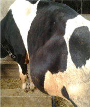
    Figure 1:  Left sided ventral abdominal distention lasting for two and a half
month in a 10 months old calf with chronic left abomasal displacement.
The animal was slaughtered about two months later due to reduced appetite for some days and increase in the size of distention, according to the farmer.
Discussion
The predominated clinical signs at the initial presentation of this calf were those of bronchopneumonia without any indication of AD. The diagnosis of AD was based on the detection of the characteristic “ping” at the simultaneous auscultation and percussion during the routine clinical examination of the calf. However, in practice this part of hands-on clinical examination is often neglected in calves, especially if there are obvious clinical signs of another disease, and many cases of abomasal displacement pass undetected.
Although the etiopathogenesis of the disease in calves has not been determined in detail, it is generally accepted that, as in adult cattle, any factor causing atony or hypotony of the abomasum, gas accumulation and distention of the organ predisposes to AD [1]. The introduction to high concentrate diets [2] and, most commonly, the concurrent presence of other diseases, in particular bronchopneumonia [4,9,10], are considered as predisposing factors. At the present case, it is believed that endotoxemia induced by bronchopneumonia [11,12] was the cause of AD. Endotoxemia, among others, inhibits ruminal smooth muscle contraction [13] and decreases abomasal emptying rate in a dose-dependent manner [14] Indicative clinical signs of endotoxemia at this case were, apart from fever, ruminal hypomotility and slight hyperglucemia [15]. In order to ameliorate these effects and, among them, the abomasal hypomotility, a non-steroidal antiinflammatory drug, flunixin meglumin, was prescribed as an adjunct at the treatment of bronchopneumonia.
The surgical approach is the treatment of choice for AD [1]. The farmer rejected this option, so the rolling procedure was selected. The immediate relocation of the abomasum at its normal position suggests that there were no adhesions and the displacement of the abomasum was recent. The reoccurrence of LDA after rolling was not unexpected due to the low permanent cure rates, less than 60%, of this procedure [1,3,16].
After the second failed relocation of the abomasum, the chronic form of LDA occurred. The abomasum was located caudally to the rib cage and extended distended dorsally to the paralumbar fossa. This is the typical position of the LDA in calves [3], causing left sided abdominal distension [9,17]. This is in contrast to the findings in adult cattle where the abomasum is positioned in the rib cage causing sunken left flank due to the displacement of the rumen to the right [3]. This is probably related with anatomic factors that allow the abomasum to slide more caudally in calves and to distend in higher degree than the adult cattle. As it was observed here, simultaneous auscultation and percussion on the last ribs did not produce the characteristic “ping”; these sounds are only audible at the site of distention, on the ventral flank [3].
Due to the distention, the differential diagnosis included rumen bloat or impaction, herniation of the abdominal was with prolapse of viscera and abscessation and hematoma of the abdominal wall [2]. Bloat was ruled out because there was no distention on the dorsal quadrant of the abdomen, the ruminal impaction by the detection of fluid content at the ballottement at the site of distention and the herniation by finding intact the abdominal wall during the last rolling attempt. Additionally, the detection of highly acidotic pH at the fluid obtained by abomasocentesis was diagnostic for this situation.
It was unexpected for the calf to have similar growth rates to its herd mates despite having AD for at least two months. Although such condition is not referred in the available literature a possible explanation might be that the abomasum was adhered at a position that did not cause significant pyloric obstruction and allowed a normal flow of the abomasal content to the duodenum.
Conclusion
In conclusion, LDA does occur in calves and a thorough clinical examination should not be neglected even in cases with obvious symptoms of other diseases. Furthermore, the different typical location of the displaced abomasum in calves compared to adult cattle should be taken into account when the simultaneous auscultation and percussion is performed. Concerning the treatment options, as in adult cattle, surgical intervention should be chosen rather than rolling due to the low permanent cure rates of the latter method.
References
- Karatzias H, Karatzia MA, Katsoulos PD. Occurrence, etiology and prevention of abomasal displacement in dairy cattle. Lieu G, editor. In: Cattle: Domestication, Diseases and the Environment. New York: Nova Science Publishers Inc. 2013; 127-138.
- Mueller K, Merrall M, Sargison ND. Left abomasal displacement and ulceration with perforation of abdominal musculature in two calves. Vet J. 1999; 157: 95-97.
- Fubini S, Divers TJ. Noninfectious diseases of the gastrointestinal tract. Divers TJ, Peek SF, editors. In: Rebhun’s diseases of dairy cattle. St. Louis: Saunders, Elsevier. 2008: 130-199.
- Medina-Cruz M, Roblesgil AP, Escamilla MRG, Rubio MS. Description of abomasal displacements in dairy calves. Bovine Pract. 1990; 25: 95-98.
- Dirksen G. Left displacement of the abomasum in calves and young cattle. Tierärztliche Umshau. 1981; 36: 674-680.
- Katsoulos PD, Minas A, Karatzia MA, Pourliotis K, Christodoulopoulos G. Evaluation of a portable glucose meter for use in cattle and sheep. Vet Clin Pathol. 2011; 40: 245-247.
- Panousis N, Kritsepi M, Karagiannis I. Evaluation of Precision Xceed® for on-site monitoring of blood β-hydroxybutyric acid and glucose in dairy cows. JHVMS. 2011; 62: 109-117.
- Cockcroft P, Jackson P. Clinical examination of the abdomen in adult cattle. In Pract. 2004; 26: 304-317.
- Dennis R. Abomasal displacement and tympany in a nine-week-old calf. Vet Rec. 1984; 114: 218-219.
- Hawkins CD, Fraser DM, Bolton JR. Left abomasal displacement and ulceration in an eight-week-old calf. Aust Vet J. 1986; 63: 53-55.
- Ackermann MR, Brogden KA. Response of the ruminant respiratory tract to Mannheimia (Pasteurella) haemolytica. Microbes Infect. 2000; 2: 1079-1088.
- Panciera RJ, Confer AW. Pathogenesis and pathology of bovine pneumonia. Vet Clin North Am Food Anim Pract. 2010; 26: 191-214.
- Verheijden JHM, Van Miert AS, Schotman AJH, Van Duin CTM. Pathophysiological aspects of E. coli mastitis in ruminants. Vet Res Commun. 1980; 7: 229-236.
- Vlaminck K, Van Meirhaeghe H, Van Den Hende C, Oyaert W, Muylle E. Effect of endotoxins on abomasal emptying in cattle. Dtsch Tierarztl Wochenschr. 1985; 92: 392-395.
- Andersen PH. Bovine Endotoxicosis – Some aspects of relevance to production diseases. A Review. Acta Vet Scand. 2003; 98: 141-155.
- Zadnik T, Lombar R. Our Experience with Left-Sided Abomasal Displacement Correction via the Roll-and-Toggle-Pin Suture Procedure according to Grymer/Sterner Model. ISRN Vet Sci. 2011.
- Dirksen G. Tympany, displacement and torsion of the abomasum in calves: pathogenesis, diagnosis and treatment. Bovine Pract. 1994; 28: 120-126.