
Case Report
Austin J Vet Sci & Anim Husb. 2022; 9(2): 1093.
Coenurosis Cerebralis Case Investigation in Indigenous Goat at Womberema District of North Western Ethiopia
Zewde D* and Ashagrie T
Department of Zoological Sciences Aquatic Science, Animal Health Institute, Ethiopia
*Corresponding author: Demeke Zewde, Department of Zoological Sciences Aquatic Science, Animal Health Institute, P.O. Box: 04, Sebeta, Ethiopia
Received: July 21, 2022; Accepted: August 03, 2022; Published: August 10, 2022
Abstract
The current case report was raised from indigenous goat breed of Soma village of Womberema district of North Western Ethiopia. The goat was presented with a history of circling movement towards the left side. Diagnosis of the case was held both through tentative (clinical observation) and based on post mortem inspection and macroscopic visualization of the cyst under investigation for confirmation. The availability for definitive hosts (domestic and wild canines) has been also one of the main factors considered as inclusion criteria for the parasite investigation. Based on clinical observation the goat (Doe) showed circling towards the left, uncoordinated movement, and unable to browse properly. Based on clinical sign and presence of definitive hosts at the area the case has been defined for coenurosis cerebralis. For further confirmatory diagnosis postmortem inspection for the brain was undertaken. Indeed, the presence of cyst which were settled superficially in the left cerebral hemisphere of the brain, with fluctuant consistency, and filled with numerous white protoscolex was a confirmatory diagnosis for a larval stage of taenia multiceps called coenurosis cerebralis. In conclusion the disease causes great economic loss in small ruminants directly by animal mortality and production impairment through weight loss. The community should regularly deworm dogs and proper disposal of the brain of infected sheep and goats after slaughter; will be highly recommended as a prevention and control method of the parasite.
Keywords: Circling disease; Coenurosis cerebralis; Goat; Post mortem inspection
Introduction
Helminthes parasites of larvae stage of Taenia multiceps known as coenurosis cerebralis is the major disease affecting sheep and goat production and causes disease known as cerebral coenurosis. The life cycle is commonly happening between dog and small ruminants. The adult tape worm is found in dogs and wild canids. It inhabits the small intestine, where it can reach a length of 40-100 cm without causing any symptoms in this host. The mature proglottid segments pass with the faces, after which the eggs are liberated and distributed into the environment. These eggs consist of an embryo or onchosphere covered by a thick, tough envelope which prevents the desiccation and inactivation of the onchosphere in the environment [1]. The eggs of the parasites are ingested by the intermediate hosts from pasture [2] and animals get infected. The life cycle can be interrupted most satisfactorily by control of tape worm infection in dogs and preventing dogs having access to small ruminant carcasses [3].
Cerebral Coenurosisor gid is a global parasitic disease of central nervous system and with higher incidence in sheep and goat but may also affect camels, deer, pigs, horses and rarely seen in cattle and humans [4,5]. The larval stage (metacestode) of this cestode, affects the Central Nervous System (CNS), particularly the brain of sheep and goat which gives rise to the neurological signs of coenurosis or gid or stagger. Indeed circling, Dullness, torticollis, loss of appetite, separation from the flock, visual impairment, muscle tremors are the main clinical signs related with coenuriosis. It is the cause for the economic loss due small ruminant mortality and condemnations of infected brain [6].
The disease is much more common in rural areas of Ethiopia where dogs and domestic animals live in a very close association. Moreover, home slaughtering of small ruminants is still predominant and uncooked offal and carcass wastes are normally given for dogs [7]. In developing countries like Ethiopia there is close contact between dogs and small ruminants, lack of knowledge of the population on epidemiology of coenurosis, free access of dogs to the head of ruminants, containing coenurosis vesicles, the absence of regular deworming of dogs are the most important drivers for continuation of T. multiceps cycle and the persistence of coenurosis that leads to high economic losses of the country [8]. The major economic losses due to decreasing production, animal mortality, treatment cost, brain condemnation, time and loss of energy for keeping sick animals [9].
Human get infected with coenurosis if ingests an egg of the parasite accidentally as a result of poor personal hygiene after being shed in the feces of the dog. After ingestion of the eggs, larvae hatch, penetrate the intestinal wall and migrate to various tissues, where they develop in to large, cystic larvae [10]. Therefore, the objective of this study is to determine the causative agent for the circling diseases reported on caprine, so that to recommend appropriate way of prevention and control based on the investigation outcome.
Case Situation/Presentation
The case was reported from Soma village, Gomer peasant association under Womberema district which was located with a latitude of 10o47’37”and 36o77’33”longitude at 1240 meter above sea level, where about 38 goats were affected with circling disease and most were died after along stay with the sign. A team was organized from National Animal Health Diagnostic and Investigation Center (NAHDIC) now days called Animal Health Institute (AHI) for investigating the issues raised by farmers in the village where goats were massively affected with a circling disease. Anti-helminthic and antibiotic treatments have no response over the disease and based on the information obtained from the community most cases were end up with death in a couple of months which was found in line with the reports of Scala et al., [11].
All clinical procedures were done at the field level where the case was happened. Upon clinical observation the goat was circling into the left, uncoordinated movement (ataxia) and unable to browse properly. The team tentatively defined for coenurosiscerebralis and the confirmatory diagnosis for the disease was recommended by necropsy finding of the cyst [12]. In this regard, the team had decided to scarify one local breed female goat (Doe) with the age of 6year having the clinical sign for further postmortem inspection of the brain through macroscopic visualization. After opening the skull, the investigating team has observed a cycst which were settled superficially in the left cerebral hemisphere of the brain, with fluctuant consistency, and filled with numerous white protoscolex as indicated in (Figure 1B). Indeed, the postmortem investigation for the case was confirmed for the coenuriosiscerebralis (larvae stage of Taenia multiceps).
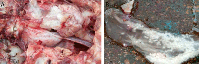
Figure 1: (A) A cyst settled in the left cerebral hemisphere, (B) A cyst after protruded from the brain.
Conclusion and Recommendations
Coenurosis is a disease of the brain and spinal cord caused by the intermediate stage of Taenia multiceps which inhabits the intestine of dogs, cats and wild carnivores. Clinically the disease occurs in sheep and Goats. The best way of confirming the case was through macroscopic visualization of the cyst from the brain of clinical animals as the victim has no way to recover once infected. The present case investigation concludes for the presence of a clearly visualized large sized C.cerebralis. Since the location of cyst especially in the brain determines the direction of circling movement due to the effect of pressure on the vital organ. The presence of free ranging dogs and wild carnivores like foxes and hyena can exacerbate the spread of the parasite over a wide grazing land. Based on the above conclusion the following recommendations will be forwarded:
• Whole villages should join forces to regularly (three months interval) deworm all dogs using praziquantel or other effective drugs for cestodes
• Burn or bury heads of small ruminants after slaughtering but not provide this part for dogs unless and otherwise the owner is not confident enough for the effectiveness of deworming.
References
- Vink WD, Lopes Pereira CM, Nota A, De Balogh KKIM. Prevalence of Coenurosis in goats in tete province, Mozambique. France. 1997; 72-99.
- Misra SS, Behl SM. The nervous system. In: Ruminant Surgery. Tyagi, R.P.S. and Singh, J. (Edts.). (1 Edn.) CSB, New st Delhi, India. 2015; 384-391.
- Radostits OM, Blood DC, Gay CC. A text books of cattle, sheep and goats, Veterinary Medicine. 6th Edition. Bailliere Tindall pub. London. England. pp: 492.1994.
- Abera S, Wubit T, Nejash A. Cerebral coenurosis in small ruminants: A review. J. Anim. Sci. Adv. 2016; 6: 1595-1608.
- Rahman MM, Sultana S, Hassan MZ,Rahma M.M. Surgical management of gid disease in goat at Rangpur district of Bangladesh. Asian J Med Biol Res. 2017; 3: 109-113.
- Sharma DK, ChauhanPPS.Coenurosis status in Afro-Asian region: a review. Small Ruminant Research, 2006; 64: 197-202.
- Fromsa A, Jobre Y. Infection prevalence of hydatidosis (Echinococcusgranulosus) in domestic animals in Ethiopia: a synthesis report of previous surveys. Ethiopia Veterinary Journal. 2011; 15: 11-33.
- Worku E, Muluneh H, Getahun E, Abaysew A. Prevalence major metacestodes of ruminant slaughtered at Elfora export abattoir and public health importance. College of Veterinary Medicine and Agriculture, Addis Ababa University, Ethiopia. 2019.
- Alemu Y, Merkel R. Sheep and goats’ production, Handbook for Ethiopia 2.2008.
- Acha PN, Szyfres B. Helminthiases: Cestodiases, Text book of zoonoses and communicable diseases common to man and animals, third edition, third volume, pan American health organization. 2003; 162-163.
- Komnenou A, Dessiris A, Giadinis N. Surgical treatment of coenurosis (gid) in sheep. Veterinary Record. 2000; 147: 242-244.
- Scala A, Varcasia A. Updates on morphobiology, epidemiology and molecular characterization of coenurosis in sheep. Parassitologia. 2006; 48: 61-3.