Research Article
Austin Virol and Retrovirology. 2015;2(1): 1009.
Pediatric Respiratory Severity Score (PRESS) for Respiratory Tract Infections in Children
Yumiko Miyaji1,2,5*, Kazuko Sugai³, Asako Nozawa², Miho Kobayashi4, Shoichi Niwa4, Hiroyuki Tsukagoshi4, Kunihisa Kozawa4, Masahiro Noda5, Hirokazu Kimura5 and Masaaki Mori2
1Department of Pediatrics, National Hospital Organization Yokohama Medical Center, Japan
2Department of Pediatrics, Yokohama City University Graduate School of Medicine, Japan
3Department of Pediatrics, National Hospital Organization Fukuyama Medical Center, Japan
4Department of Health Science, Gunma Prefectural Institute of Public Health and Environmental Sciences, Japan
5Infectious Disease Surveillance Center, National Institute of Infectious Diseases, Japan
*Corresponding author: Yumiko Miyaji, Department of Pediatrics, National Hospital Organization Yokohama Medical Center3-60-2 Harajuku, Totsuka-ku, Yokohama, Kanagawa, Japan
Received: April 29, 2015; Accepted: July 07, 2015; Published: July 10, 2015
Abstract
Background: Respiratory tract infections are common diseases in children. It is crucial therefore to evaluate the severity of the condition during the initial bedside assessment in the emergency department so that further examinations and hospital treatment can be conducted as appropriate. However, there are few such scoring systems for acute respiratory infection in childhood.
Objective: To evaluate a new simple bedside scoring system for the rapid assessment of pediatric respiratory infections in emergency settings.
Methods: We established a respiratory scoring system, namely “Pediatric Respiratory Severity Score (PRESS)”, and examined its utility for assessing severity in 202 children who visited our hospital due to respiratory symptoms between January 2010 and November 2011. The PRESS assessed tachypnea, wheezing, retraction (accessory muscle use), SpO2, and feeding difficulties, with each component given a score of 0 or 1, and total scores were classified as mild (0–1), moderate (2–3), or severe (4–5). In addition, we performed RT-PCR techniques to detect respiratory viruses from nasal swabs and the detected viruses were evaluated in relation to severity.
Results: According to the PRESS scores, the hospitalization rate was significantly higher in the moderate and severe groups than in the mild group. Oxygen therapy was longer in severe cases compared with other cases. There were no significant differences in the viral detection rate between the severity groups.
Conclusion: The PRESS scoring system is useful for the initial assessment of respiratory tract infections in children to identify the need for hospitalization and further examination in emergency settings.
Keywords: Severity score; Child; Respiratory infection; Virus; Triage
Abbreviations
PRESS: Pediatric Respiratory Severity Score; ARI: Acute Respiratory Illness; RSV: Respiratory Syncytial Virus; HRV: Human Rhinovirus; HPIV: Human Parainfluenza Virus; HMPV: Human Metapneumovirus; HEV: Human Enterovirus; HBoV: Human Bocavirus; AdV: Adenovirus
Introduction
Various viruses cause acute respiratory illness in children, including Respiratory Syncytial Virus (RSV), Human Rhinovirus (HRV), Human Parainfluenza Virus (HPIV), and Human Metapneumovirus (HMPV). Various symptoms (e.g., respiratory rate, wheezing, cyanosis, and use of the accessory respiratory muscles) may reflect the severity of respiratory disease and these clinical findings could be important for early diagnosis and treatment. Moreover, it is crucial to treat acute respiratory infection appropriately to avoid the risk of respiratory failure, which is sometimes fatal in children. Severe cases must be triaged and treated immediately; therefore, respiratory condition should be assessed at first contact, in a similar way that the APGAR scoring system is used to assess neonates [1]. In 2006, the American Academy of Pediatrics highlighted the importance of assessing severity for selecting the appropriate outpatient or inhospital treatment in Diagnosis and Management of Bronchiolitis, a guideline for acute bronchiolitis in infants [2]. Although some studies have evaluated the severity of bronchial asthma and acute respiratory infections in children, few suggest simple methods of bedside assessment [3-10].
To address this issue, we established a scoring system of the severity of respiratory infections in children, which we named the Pediatric Respiratory Severity Score (PRESS), and evaluated its utility for assessing the severity of infection caused by various pathogens and for deciding the necessity of further clinical examinations and the treatment to start. We focused on the severity of the respiratory symptoms because the supportive care required by the patients is not available at home and only available at the hospital. In addition, we examined the relationship between the severity of illness and pathogens profiles.
Materials and Methods
Subjects and samples
This study was carried out at the Department of Pediatrics, National Hospital Organization Yokohama Medical Center, an urban emergency hospital in Japan, between January 2010 and November 2011. We enrolled 202 children who visited the outpatient clinic or emergency department because of acute respiratory symptoms. Nasopharyngeal swabs were collected after written informed consent was obtained from the children’s parents. The study protocol was approved by the Ethics Committee on Human Research of the National Hospital Organization Yokohama Medical Center.
We collected nasal fluid samples using Advanced Flocked Swabs and Universal Transport Medium (Copan Innovation, Brescia, Italy), and stored the samples at -80 °C until used for viral detection. White blood cell counts (normal range: 7,000-11,000/μL in child) and C-reactive protein (CRP, normal range for children :< 1.2mg/dL) were measured at the first visit. We collected samples for bacterial culture before antibiotic therapy was initiated. We collected clinical data, radiographic evidence, and laboratory data from hospital charts.
PRESS score
We collected data on five components using the PRESS, namely, respiratory rate, wheezing, accessory muscle use, SpO2, and feeding difficulties (Table 1). Accessory muscle use was defined as visible retraction of one or more of the sternomastoid/suprasternal, intercostal, and subcostal muscles. Wheezing was defined by auscultation performed by experienced pediatricians. SpO2 was evaluated as above or below 95%. Feeding difficulties were assessed using information provided by the parents. Each component was given 0 or 1 point and the PRESS total score was classified as mild (0–1 points), moderate (2–3 points), or severe (4–5 points). Respiratory rate was evaluated based on the American Heart Association guidelines (Table 1) [11].
    Score Component
    Operational definition
    Scoring


    Respiratory rate
    Respiratory rate at rest, on    room air*
    0 or 1


    Wheezing
    High-pitch expiratory sound heard by auscultation
    0 or 1


    Accessory muscle use
    Any visible use of accessory muscles
    0 or 1


    SpO2
    Oxygen saturation <95% on room air
    0 or 1


    Feeding difficulties
    Refusing feedings
    0 or 1


    Sum of five components


    PRESS score
    0-1: mild 2-3: moderate 4-5:    severe
    0-5


    Criteria of tachypnea*
    Month
    Respiratory rate


    <12
    60<
    1


    12=, <35
    40<
    1


    36=, <156
    30<
    1


    156=
    20<
    1


    *Respiratory rate evaluated according to American Heart    Association guideline.
      PRESS, Pediatric Respiratory Severity Score
Table 1: PRESS Scoring System.
Virus detection
Nasopharyngeal swab samples were centrifuged at 3000 × g at 4°C for 15 min for viral DNA/RNA extraction, PCR and RT-PCR, and sequence analysis, and the supernatants were used as described previously [12,13]. RT-PCR was used for RNA virus detection including RSV, HRV, HMPV, HPIV, HEV, and influenza virus, while PCR was used for DNA virus detection such as Adenovirus (AdV) and Human Bocavirus (HBoV). Viral nucleic acid was extracted using QIAampMinElute Virus Spin Kit (Qiagen, Valencia, CA). The reverse transcription reaction mixture was incubated with random hexamers at 37°C for 15 min, followed by incubation at 85°C for 5 s using the PrimeScript RT reagent Kit (Takara Bio, Shiga, Japan) and amplification by thermal cycling. The PCR procedures for amplification of different viral genomes, including RSV [14], Human Rhinovirus (HRV)/Human Enterovirus (HEV) [15,16], HMPV [17], HPIV type1-4 [18], influenza virus subtypes A–C [19], Adenovirus (AdV) [20], and Human Bocavirus (HBoV) [21] were performed as described previously. Negative control (no virus) and positive controls using the prototype or clinical strains were also examined in each test. Viral RNA/DNA extraction was conducted in a room physically separate from that used for performing PCR to avoid carry-over and cross-contamination. Positive and negative controls were included in all PCR assays. PCR products were determined by electrophoresis on 3% agarose gels. Purification of DNA fragments and nucleotide sequence determination procedures were performed as described previously.
Statistical analysis
Data were analyzed using SPSS software (SPSS for Windows, Version 19.0). All data are expressed as mean ± Standard Deviation (SD). We performed Scheffe tests for multiple comparisons between three groups when there were significant differences found in the ANOVA tests. Statistical significance was set at p< 0.05.
Results
Of the 202 children enrolled, 123 (60.9%) were boys and 79 (39.1%) were girls, aged 27.6 ± 31.8 months (mean ± SD, 0–13 years old), and 128 in total were admitted to hospital. Patient characteristics are shown in Table 2. According to our respiratory severity scoring system PRESS, 99 (49.0%) participants were classified with mild infection, 70 (34.7%) with moderate, and 33 (16.3%) with severe (Table 2). The history of wheezing rate in moderate/severe cases was higher than in mild cases. The hospitalization rate was 32.3% in mild cases, 91.4% in moderate cases, and 97.0% in severe cases, with significant differences between the hospitalization rates of mild and moderate cases, and between mild and severe cases. There was no significant difference between moderate and severe cases (Table 2). The blood test results were as follows: WBC, 1.21 x 10³ ± 6.1 x 10²/μL in mild cases; 1.02 x 10³ ± 4.0 x10²/μL in moderate cases; and 1.23 x 10³ ± 5.3 x10²/μL in severe cases. CRP was 2.8 ± 3.0 mg/dL in mild cases, 1.59 ± 2.1 mg/dL in moderate cases and1.9 ± 2.6 mg/dL in severe cases. No significant difference among the data was found in the present cases (Table 2). Oxygen therapy was longer in severe cases compared with mild and moderate cases. Duration of fever in severe cases was significantly shorter compared with moderate and severe cases.
    All patients n=202

    Severity


    mild n=99
    moderate n=70
    severe n=33


    Age (months) †

    27.6±31.8
    29.8±32.9
    23.0±29.7
    30.7±32.7


    Male/female
    123/79
    51/48
    45/25
    6/27


    History of wheezing (%)
    123 (60.9)
    50 (50.5)*
    50 (71.4)*
    23 (69.7)*


    Hospitalization (%)
    128 (63.4)
    32 (32.3)*
    64 (91.4)*
    32 (97.0)*


    Virus detection (%)
    119 (58.9)
    53 (53.5)
    48 (68.6)
    18 (54.5)


    White blood cells (/μL) †

    11360±5200
    12100±6080
    10200±4040
    12300±5300


    CRP (mg/dL) †
    2.1±2.6
    2.8±3.0
    1.59±2.1
    1.9±2.6


    Duration (days) †


    Fever
    2.6±2.7
    3.6±2.7*
    2.3±2.8*
    1.8±1.9*


    Hospitalization
    7.7±2.7
    7.1±3.4
    7.8±2.8
    8.0±1.5


    Oxygen therapy
    1.6±2.0
    0.2±0.8**
    1.5±1.8**
    3.7±1.8**


    PRESS score    (%)


    Respiratory    rate
    42 (20.8)
    0 (0)**
    18 (25.7)**
    24 (72.7)**


    Wheezing
    143 (70.8)
    45 (45.5)*
    65 (92.9)*
    33 (100)*


    Accessory muscle use
    53 (26.2)
    0 (0)**
    21 (30.0)**
    32 (87.0)**


    Cyanosis
    54 (26.7)
    1 (1.0)**
    23 (32.9)**
    30 (90.9)**


    Feeding difficulties
    78 (38.6)
    6 (6.0)*
    47 (67.1)*
    25 (75.8)*


    †Mean±SD; *p <0.05, mild versus    moderate, severe; **p <0.05, mild versus    moderate versus severe Percentages indicate the number of cases in    each category/total patients in each severity group.
      PRESS, Pediatric Respiratory Severity Score
Table 2: Patient characteristics.
Of the 202 patients, 82 were aged 0–11 months, 102 were 12–71 months, and 18 were 72–180 months, and 41.4%, 72.6%, and 83.3%, respectively, had a history of wheezing (Table 3). In patients aged 0–11 months, 36 (43.9%) cases were classified as mild, 34 (41.5%) as moderate and 12 (14.6%) as severe. In the 12–71 months age group, 57 (55.9%) cases were classified as mild, 30(29.4%) as moderate and 17 (16.7%) as severe. In the group aged 72–180 months, 8 (44.4%) cases were classified as mild, 6 (33.3%) as moderate and 4 (22.2%) as severe. There was no significant difference in the percentage of severity cases between each groups (p<0.05) (Table 3).
    All patients n=202

    Age (months)


    0-11 n=82
    12-71 n=102
    72-180 n=18


    Age (months)†
    27.6±31.8
    5.7±3.4
    30.5±16.0
    111.0±25.4


    Male/female
    123/79
    54/21
    59/62
    10/8


    History of wheezing (%)
    123 (60.9)
    34 (41.4)
    74 (72.6)
    15 (83.3)


    Hospitalization (%)
    128 (63.4)
    57 (69.5)
    60 (58.8)
    11 (61.1)


    Virus detection (%)
    119 (58.9)
    53 (64.6)
    57 (55.9)
    9 (50.0)


    White blood cells (/μL)†
    11360±5200
    10740±4970
    12000±5330
    11200±5670


    CRP (mg/dL)†
    2.1±2.6
    2.2±2.7
    2.1±2.5
    0.7±0.6


    Duration (days)†


    Fever
    2.6±2.7
    2.1±2.4*,**
    3.4±2.8*,**
    2.0±2.9*,**


    Hospitalization
    7.7±2.7
    8.2±3.5
    7.2±1.8
    7.0±1.4


    Oxygen therapy
    1.6±2.0
    1.2±1.8
    2.0±2.0
    2.5±2.2


    PRESS score†


    Respiratory rate
    0.4±0.5
    0.2±0.4*
    0.6±0.5*
    0.5±0.5


    Wheezing
    0.9±0.4
    0.8±0.4
    0.9±0.3
    1.0±0.0


    Accessory muscle use
    0.5±0.5
    0.4±0.5
    0.5±0.5
    0.6±0.5


    Cyanosis
    0.5±0.5
    0.4±0.5
    0.6±0.5
    0.6±0.5


    Feeding difficulties
    0.6±0.5
    0.6±0.5
    0.6±0.5
    0.7±0.5


    Total
    1.8±1.5
    1.9±1.4
    1.7±1.6
    2.2±1.6


    Severity


    Mild
    82(40.6)
    36(43.9)
    57(55.9)
    8(44.4)


    Moderate
    102(50.5)
    34(41.5)
    30(29.4)
    6(33.3)


    Severe
    18(8.9)
    12(14.6)
    17(16.7)
    4(22.2)


    †Mean±SD; *: p <0.05, 0-11 months versus 12-71 months; **: p <0.05, 0-11 months versus 72-180    months Percentages indicate the number of cases in    each category/total patients in each age group.
      PRESS, Pediatric Respiratory Severity Score
Table 3: Characteristics and PRESS scores of patients.
Mean severity scores were 1.9±1.4, 1.7±1.6, and 2.2±1.6 in patients aged 0–11, 12–71, and 72–180 months, respectively. There was no significantly different in severity scores between each groups (p<0.05) (Table 3).
Virus profiles and severity scores
Viral pathogens were detected in 62.4% of all patients. According to severity grouping, viruses were detected in 53.5% of mild cases, 71.4% of moderate cases, and 69.7% of severe cases. There were no significant differences in the rate of virus detection among the groups. According to age group, the viral detection rate was 64.6% in those aged 0–11 months, 59.8% in those aged 12–71 months, and 66.7% in those aged 71–180 months, with no significant differences found between the groups (Table 4).
All patients n=202
Severity
Age (months)
Mild n=99
moderate n=70
Severe n=33
0-11 n=82
12-71 n=102
72-180 n=18
Virus detection (%)
RSV aloneNo. of patients (%)
126 (62.4)
39 (19.3)53 (53.5)
17 (17.2)50 (71.4) 17 (24.3)
23(69.7) 5(15.2)
53 (64.6)
17 (20.7)61 (59.8)
19 (18.6)12 (66.7) 3(16.7)
Score†
1.9±1.3***
0.7±0.5**
2.5±0.5**
4.2±0.5**
2.4±1.2
1.5±1.4***
1.7±1.2
HRV alone
No. of patients (%)
53 (26.2)
25 (25.2)
19 (27.1)
9 (27.3)
21 (25.6)
26 (25.5)
6 (33.3)
Score†
1.9±1.6***
0.4±0.5**
2.5±0.5**
4.6±0.5**
1.8±1.5
1.8±1.7***
2.5±1.9
HMPV alone
No. of patients (%)
1 (0.5)
1 (1.0)
0
0
1 (1.2)
0
0
Score†
1.0±0.0***
1.0±0.0
-
-
1.0±0.0
-
-
HPIV alone
No. of patients (%)
9 (4.5)
3 (3.0)
5 (7.1)
1 (3.0)
4 (4.9)
5 (4.9)
0
Score†
2.0±1.4***
0.3±0.6*
2.6±0.6*
4.0±0.0
1.8±2.1
2.2±0.8***
-
HBoV alone
No. of patients (%)
2 (1.0)
0
1 (1.4)
1 (3.0)
1 (1.2)
1 (1.0)
0
Score†
3.5±0.7
-
3.0±0.0
4.0±0.0
3.0±0.0
4.0±0.0
-
AdV alone
No. of patients (%)
3 (1.5)
3 (3.0)
0
0
1 (1.2)
2 (2.0)
0
Score†
0.3±0.6***
0.3±0.6
-
-
0.0±0.0
0.5±0.7***
-
HEV alone
No. of patients (%)
7 (3.5)
0
2 (2.9)
5 (15.2)
0
4 (3.9)
3 (16.7)
Score†
4.3±1.0***
-
3±0.0
4.8±0.5
-
4.8±0.5***
3.7±1.2
co-detection
No. of patients (%)
12 (5.9)
4 (4.0)
6 (8.6)
2 (11.1)
8 (9.8)
4 (3.9)
0
Score†
2.1±1.5***
0.5±0.6**
2.3±0.5**
4.5±0.7*
2.4±1.3
1.5±1.9***
-
†Mean±SD; *p<0.05, mild versus moderate; **p<0.05, mild versus moderate versus severe; ***p<0.05, HEV versus each virus Percentages indicate the number of cases in each category/total patients in each severity or age group.
PRESS: Pediatric Respiratory Severity Score; RSV: Respiratory Syncytial Virus; HRV: Human Rhinovirus; HPIV: Human Parainfluenza Virus; HMPV: Human Metapneumovirus; HEV: Human Enterovirus; HBoV: Human Bocavirus; AdV: Adenovirus
Table 4: PRESS scores of viruses detected.
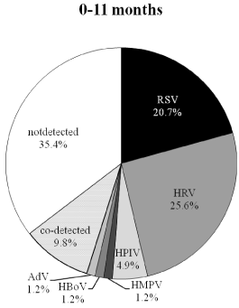
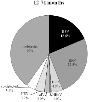
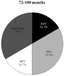
Next, HRV (25.6%), RSV (20.7%), and HPIV (4.9%) were detected in the group aged 0–11 months, HRV (25.5%), RSV (18.6%), and PIV (4.9%) in the 12–71 months group, and HRV (33.3%) and RSV (16.7%) in the 72–180 months group (Figures 1, 2 and 3). There was no significant difference in severity scores between each groups, with the exception of Human Enterovirus (HEV) (Table 4). Bacterial respiratory infections were diagnosed when laboratory reports showed white blood cell counts >10,000 /μL and C-reactive protein levels >5.0 mg/dL. According to our criteria, only 13 (6.4%) patients were diagnosed with bacterial infection. We performed nasopharyngeal cultures for 121 patients and detected Moraxella catarrhalis in 12 (9.9%) cases, Streptococcus pneumoniae in 16 (13.2%), and Haemophilus influenza in 7 (5.8%); a combination was detected in 61 cases (50.4%).

Figure 1: Viruses detected in patients aged 0-11 months.

Figure 2: Viruses detected in patients aged 12-71 months.

Figure 3: Viruses detected in patients aged 72-180 months.
Discussion
Severity score and clinical course
Respiratory tract infections in childhood can readily lead to respiratory distress and sometimes to severe dyspnea, which requires further examinations and hospitalization. An objective bedside assessment of the respiratory symptoms would enable such examinations and treatment to be commenced quickly, to avoid the condition worsening. In emergency settings particularly, medical staff must quickly assess a patient’s respiratory condition and general health to decide management. The World Health Organization has suggested that infected children exhibiting drowsiness, feeding difficulties, vomiting, convulsion, and dyspnea should be hospitalized quickly [22]. To avoid inappropriate antibiotic use, Ishiwada et.al reported a scoring system that differentiates between pediatric bacterial pneumonia and atypical pneumonia [23]. In the present study, we evaluated the effectiveness of a simple scoring system based on respiratory symptoms for assessing the need for further examinations and hospitalization.
Guidelines for the Management of Respiratory Infectious Diseases in Children in Japan 2011 proposed the following criteria for hospitalization in community-acquired pneumonia cases: moderate or severe cases determined according to the guidelines, under 1 year old, difficulty in taking oral medications, poor response to oral antibiotics, underlying disease, dehydration, and the patient’s doctor opting for hospitalization [24]. The guidelines define the criteria for severity assessment of pediatric community-acquired pneumonia as auscultation, respiratory rate, condition of respiratory assistance muscles, cyanosis, and radiographic data. Recent studies have described previous scoring systems for pediatric respiratory infections [3-10] and clarified that respiratory rate, wheezing, retraction, and SpO2 are all useful criteria for the assessment of respiratory status. In children, feeding difficulties are a very important sign of illness, so we adopted these former and latter components for our PRESS scoring system. We did not adopt heart rate and blood pressure because these parameters are difficult to evaluate in a crying child.
In our study, respiratory rate, retraction scores and Oxygen demand were significantly different between mild, moderate and severe cases, and wheezing and feeding difficulty scores differed significantly between mild and moderate/severe cases. Accordingly, these components are useful for assessing respiratory status. CRP 12100±6080 in mild,
There were no significant differences between the groups for criteria such as WBC and CRP (p>0.05), and duration of fever in severe cases was significantly shorter compared with moderate and severe cases. It suggested that clinical respiratory symptoms and feeding difficulties are the more useful assessment criteria.
In this study, the hospitalization rate in moderate/severe cases was significantly higher than in mild cases (p<0.001). In addition, the duration of oxygen therapy was significantly longer in severe cases compared with mild and moderate cases (p<0.001). The PRESS score can help predict the duration of in-hospital treatment and the clinical course.
Severity score and viral infection
Previous reports have clarified that respiratory viruses can cause not only acute respiratory infections but also infant wheezy illnesses, bronchial asthma, and exacerbation of asthma [25-27]. Friedlander et al. reported that viral infections are the main cause of exacerbations in 80% of child asthma patients and in 50% of adult asthma patients, and HRV contributed strongly to these cases at all ages [28]. Johnston et al. reported that respiratory viral infections contribute to asthma exacerbations in 80–85% of school-aged children with bronchial asthma and viruses such as HRV, enterovirus, corona virus, influenza virus, parainfluenza virus, and RSV were detected in these children [29]. Hamano et al. reported bacterial infection, including M. pneumonia and Chlamydia pneumonia, at a rate of 49.6%, viral pneumonia at 43.1%, and mixed infection (bacteria and virus) at 25.8% in 2008 after a comprehensive analysis of pediatric communityacquired pneumonia cases, using real-time PCR techniques [30,31]. The main cause of acute respiratory infections in children may be viral infection. Thus, we also compared the severity with the detected viruses. In the present study, there were no significant differences between each group for detection rate of viruses. HRV, RSV, and HPIV were detected in almost all patients, and both HRV and RSV were continuous. We detected viral pathogens in 60% of cases; therefore, it is clear that viral infections are very common and are major causative agents in pediatric respiratory infections, as reported previously [32]. There were no significant differences of severity scores between each virus, with the exception of (HEV). In a recent report, HEV infections are very common and are associated with more severe diseases compared with other common viruses such as RSV [33]. There is a possibility that HEV was included in our examination. HRV was the main causative pathogen for exacerbations of asthma in our school-aged children. To validate this, however, larger studies may be needed because the number of patients in the present cohort was relatively small.
The mechanism of respiratory distress caused by these viruses has not been clarified. The WHO Pneumonia Fact Sheet, 2013, reported that it is difficult to determine the causative agents in cases of bacterial and viral infections [34]. Ishiwada et al. suggested criteria to help discriminate between bacterial and community-acquired pneumonia and defined community-acquired pneumonia as follows: age >6 years, no prediagnosed diseases, no prescribed beta-lactam antibiotics in the last week, good general health, no wet cough, no crackles on auscultation, infiltration area on chest radiograph, <10,000/μL of white blood cells, and <4.0 mg/dL of C-reactive protein [23]. We defined bacterial infection as WBC >10,000/μL and C-reactive protein >5.0 mg/dL. According to these criteria, 13 of our patients (6.4%) were diagnosed with bacterial infection. On the other hand, major bacterial pathogens such as Moraxella catarrhalis, S. pneumoniae, and H. influenza were detected in nasopharyngeal cultures from 59.9% of patients.
Conclusion
The PRESS, with its simple components of respiratory rate, wheezing, retraction, SpO2, and feeding difficulties, may be useful and applicable to triage and assessment of respiratory status by medical staff at the initial bedside examinations. It would likely be useful in prehospital settings.
Patient Consent
Nasopharyngeal swabs were collected after written informed consent was obtained from the parents of subjects.
Acknowledgement
This work was supported by a Grant-in-Aid from the Japan Society for Promotion of Science and for Research on Emerging and Reemerging Infectious Diseases from the Ministry of Health, Labour and Welfare. We also thank the medical staff of the Department of Pediatrics, National Hospital Organization Medical Center, and technical support team of Gunma Prefectural Institute of Public Health and Environmental Sciences.
References
- APGAR V. A proposal for a new method of evaluation of the newborn infant. Curr Res Anesth Analg. 1953; 32: 260-267.
- American Academy of Pediatrics Subcommittee on Diagnosis and Management of Bronchiolitis. Diagnosis and management of bronchiolitis. Pediatrics. 2006; 118: 1774-1793.
- Carroll CL, Sekaran AK, Lerer TJ, Schramm CM. A modified pulmonary index score with predictive value for pediatric asthma exacerbations. Ann Allergy Asthma Immunol. 2005; 94: 355-359.
- Gorelick MH, Stevens MW, Schultz TR, Scribano PV. Performance of a novel clinical score, the Pediatric Asthma Severity Score (PASS), in the evaluation of acute asthma. Academic emergency medicine. Acad Emerg Med. 2004; 11: 10-18.
- Chalut DS, Ducharme FM, Davis GM. The Preschool Respiratory Assessment Measure (PRAM): a responsive index of acute asthma severity. J Pediatr. 2000; 137: 762-768.
- Arnold DH, Gebretsadik T, Abramo TJ, Moons KG, Sheller JR, Hartert TV. The RAD score: a simple acute asthma severity score compares favorably to more complex scores. Ann Allergy Asthma Immunol. 2011; 107: 22-28.
- Reed C, Madhi SA, Klugman KP, Kuwanda L, Ortiz JR, Finelli L, et al. Development of the Respiratory Index of Severity in Children (RISC) score among young children with respiratory infections in South Africa. PLoS One. 2012; 7: e27793.
- Tal A, Bavilski C, Yohai D, Bearman JE, Gorodischer R, Moses SW. Dexamethasone and salbutamol in the treatment of acute wheezing in infants. Pediatrics. 1983; 71: 13-18.
- Nariai A. Usefulness of a clinical scoring system to estimate a degree of seriousness in infants younger than 24 months with respiratory syncytial virus bronchiolitis. Nihon SyoniKokyukiShikkan Gakkai Zasshi. 2008; 19: 3-10.
- Fujitsuka A. Lower respiratory infection with respiratory virus associates to bronchial asthma in children. Allerugie. 2011; 60: 1393.
- ECC Committee, Subcommittees and Task Forces of the American Heart Association. 2005 American Heart Association Guidelines for Cardiopulmonary Resuscitation and Emergency Cardiovascular Care. Circulation. 2005; 112: IV1-203.
- Fujitsuka A, Tsukagoshi H, Arakawa M, Goto-Sugai K, Ryo A, Okayama Y, et al. A molecular epidemiological study of respiratory viruses detected in Japanese children with acute wheezing illness. BMC Infect Dis. 2011; 11: 168.
- Mizuta K, Hirata A, Suto A, Aoki Y, Ahiko T, Itagaki T, et al. Phylogenetic and cluster analysis of human rhinovirus species A (HRV-A) isolated from children with acute respiratory infections in Yamagata, Japan. Virus Res. 2010; 147: 265-274.
- Parveen S, Sullender WM, Fowler K, Lefkowitz EJ, Kapoor SK, Broor S. Genetic variability in the G protein gene of group A and B respiratory syncytial viruses from India. J Clin Microbiol. 2006; 44: 3055-3064.
- Ishiko H, Shimada Y, Yonaha M, Hashimoto O, Hayashi A, Sakae K, et al. Molecular diagnosis of human enteroviruses by phylogeny-based classification by use of the VP4 sequence. J Infect Dis. 2002; 185: 744-754.
- Olive DM, Al-Mufti S, Al-Mulla W, Khan MA, Pasca A, Stanway G, et al. Detection and differentiation of picornaviruses in clinical samples following genomic amplification. J Gen Virol. 1990; 71 : 2141-2147.
- Takao S, Shimozono H, Kashiwa H, Matsubara K, Sakano T, Ikeda M, et al. [The first report of an epidemic of human metapneumovirus infection in Japan: clinical and epidemiological study]. Kansenshogaku Zasshi. 2004; 78: 129-137.
- Bellau-Pujol S, Vabret A, Legrand L, Dina J, Gouarin S, Petitjean-Lecherbonnier J, et al. Development of three multiplex RT-PCR assays for the detection of 12 respiratory RNA viruses. J Virol Methods. 2005; 126: 53-63.
- Nicoll A, Ammon A, Amato Gauci A, Ciancio B, Zucs P, Devaux I, et al. Experience and lessons from surveillance and studies of the 2009 pandemic in Europe. Public Health. 2010; 124: 14-23.
- Miura-Ochiai R, Shimada Y, Konno T, Yamazaki S, Aoki K, Ohno S, et al. Quantitative detection and rapid identification of human adenoviruses. J Clin Microbiol. 2007; 45: 958-967.
- Allander T, Tammi MT, Eriksson M, Bjerkner A, Tiveljung-Lindell A, Andersson B. Cloning of a human parvovirus by molecular screening of respiratory tract samples. Proc Natl Acad Sci U S A. 2005; 102: 12891-12896.
- World Health Organization. Programme for the control of acute respiratory infections: technical bases for the WHO recommendations on the management of pneumonia in children at first-level health facilities. WHO/ARI/91.20.
- Ishiwada N, Nagai F, Tanaka J, Arima M, Abe K, Fukasawa C. Scoring system to distinguish between bacterial pneumonia and atypical pneumonia in children. The Journal of Pediatric Infectious Disease and Immunology. 2010; 22: 343-348.
- Uehara S, Sunakawa K, Eguchi H, Ouchi K, Okada K, Kurosaki T, et al. Japanese Guidelines for the Management of Respiratory Infectious Diseases in Children 2007 with focus on pneumonia. Pediatr Int. 2011; 53: 264-276.
- Martinez FD, Wright AL, Taussig LM, Holberg CJ, Halonen M, Morgan WJ. Asthma and wheezing in the first six years of life. The Group Health Medical Associates. N Engl J Med. 1995; 332: 133-138.
- Martinez FD, Stern DA, Wright AL, Taussig LM, Halonen M. Differential immune responses to acute lower respiratory illness in early life and subsequent development of persistent wheezing and asthma. J Allergy Clin Immunol. 1998; 102: 915-920.
- Busse WW, Lemanske RF Jr, Gern JE. Role of viral respiratory infections in asthma and asthma exacerbations. Lancet. 2010; 376: 826-834.
- Friedlander SL, Busse WW. The role of rhinovirus in asthma exacerbations. J Allergy Clin Immunol. 2005; 116: 267-273.
- Johnston SL, Pattemore PK, Sanderson G, Smith S, Lampe F, Josephs L, et al. Community study of role of viral infections in exacerbations of asthma in 9-11 year old children. BMJ. 1995; 310: 1225-1229.
- Hamano-Hasegawa K, Morozumi M, Nakayama E, Chiba N, Murayama SY, Takayanagi R, et al. Comprehensive detection of causative pathogens using real-time PCR to diagnose pediatric community-acquired pneumonia. J Infect Chemother. 2008; 14: 424-432.
- Ling CL, McHugh TD. Rapid detection of atypical respiratory bacterial pathogens by real-time PCR. Methods Mol Biol. 2013; 943: 125-133.
- Marcone DN, Ellis A, Videla C, Ekstrom J, Ricarte C, Carballal G, et al. Viral etiology of acute respiratory infections in hospitalized and outpatient children in Buenos Aires, Argentina. Infectious Disease Journal. 2013; 32: 105-110.
- Asner SA, Petrich A, Hamid JS, Mertz D, Richardson SE, Smieja M. Clinical severity of rhinovirus/enterovirus compared to other respiratory viruses in children. Influenza Other Respir Viruses. 2015; 8; 436-442.
- World Health Organization. Pneumonia Fact sheet N°331 2013.