Research Article
Austin Virol and Retrovirology. 2015; 2(1): 1012.
Evaluation of Reversion to Virulence on a Modified Live Highly Pathogenic Porcine Reproductive and Respiratory Syndrome Vaccine Strain in Pigs
Mingqi Xia, Wei Wang and Hua Wu*
Sinovet Biotechnology Co., Ltd., China
*Corresponding author: Hua Wu, Sinovet Biotechnology Co., Ltd., Beijing 100085, China
Received: August 20, 2015; Accepted: September 29, 2015; Published: October 01, 2015
Editorial
Highly Pathogenic Porcine Reproductive and Respiratory Syndrome Virus (HP-PRRSV) has spread most provinces of China. An attenuated vaccine strain, TJM-F92, was obtained by passaging HP-PRRSV strain TJ on Marc-145 cells for 92 passages and showed a continuous 120 amino acids deletion in nsp2 gene. The purpose of this study was to evaluate the virulence of TJM-F92 vaccine strain by serial passages in pigs and to analyze the genetic changes of the nsp2 region.TJM-F92 vaccine virus was passaged continuously five times in pigs. At the end of each passage, pigs were euthanized and necropsied, and tissue samples were collected and examined. The lungs were harvested for histopathological examination. The Nsp2 gene of isolates from the fifth passage (P5) was sequenced and compared to the Nsp2 gene sequences of TJM-F92 vaccine virus and its parent strain TJ. All pigs showed no clinical symptoms related to PRRS each passage. The viremia in pigs from P1 to P5 showed no significantly difference. No gross-lung lesions were observed in pigs from P1 to P5. Sequence analysis showed the continuous 120 aa deletion in nsp2 in TJM-F92 vaccine virus existed persistently in vivo. In conclusion, the TJM-F92 stain did not convert its pathogenicity to pigs after continuously passaging in pigs.
Keywords: Porcine reproductive and respiratory syndrome, Cytopathic Effect, Nsp2 gene, ELISA
Introduction
Porcine Reproductive and Respiratory Syndrome (PRRS) is characterized by respiratory distress in piglets and reproductive failure in sows [1-3]. The disease was first detected in North America in 1987 and in Europe in 1990 [4], and since then, the disease was identified quickly in many countries throughout the world. Since 2006, Highly Pathogenic PRRS (HP-PRRS) has spread to most provinces of China and its neighboring countries [5-7]. PRRS has caused significant economic problems to the swine industry worldwide [8].
The causative agent, PRRS Virus (PRRSV), is an envelope, singlestranded, positive sense RNA virus possessing a 15 kb genome that contains ten Open Reading Frames (ORFs) [9-13], which belongs to the family Arteriviridae, together with Lactate Dehydrogenase Elevating Virus (LDV) of mice, Equine Arteritis Virus (EAV) and Simian Hemorrhagic Fever Virus (SHFV) [9]. The nsp2 gene is the most variable region in the PRRSV genome. The highly pathogenic PRRSV isolated in China was found to contain a discontinuous 30 amino acids deletion in nsp2 gene [5,14-17].
Vaccination is the most effective and practical method in control of PRRS. Both inactivated and Modified Live Virus (MLV) vaccines have been used in gilts, sows, and growing pigs for the control of PRRSV [18]. The commercially available inactivated vaccines are generally safe to use, but do not provide sufficient protection [19-21]. MLV vaccines have shown efficacy in reducing disease occurrence and severity in growing pigs [22-25]. However, there are some safety concerns, as the vaccine may spread and revert to virulence [19, 26- 28].
A live-attenuated vaccine strain, TJM-F92 [29], was obtained by passaging virulent PRRSV strain TJ on Marc-145 cells (for 92 passages). In this study, the TJM-F92 vaccine virus was passaged five times in pigs to evaluate the virulence of the vaccine strain by serial passages in pigs and to analyze the nsp2 genetic changes.
Materials and Methods
Experimental animals
Thirty-four 5-6-weeks-old healthy weaned pigs free of PRRS virus and antibody were used in this study. All animals were transported to the facilities with a Biological Safety Level 3 (BSL3) at Jilin Teyan Biological Technology Limited Liability Company. The study was approved by Sinovet Animal ethics Committee.
Virus and inoculation
The PRRSV TJM-F92 vaccine strain was obtained by passaged 92from HP-PRRSV TJ strain (GenBankaccession no. EU860248) on Marc-145 cells cultures. Thirty-four pigs were divided randomly into 5 treatment groups and 5 control groups. Table 1 describes the group setting and animal numbers in each group for each passage. Briefly, 5 pigs were used in passage 1 and 5 (P1 and P5) and 3 pigs were used in passage 2 to 4 (P2 to P4) for in vivo virus inoculation groups. In each passage, there were 3 pigs in control group, served as both mockinfected negative controls and environmental sentinels.
    Passage
    Group
    Number of pigs
    Inocula
    Volume (ml/pig)
    Temperature (d.p.i)
    Clinical signs (d.p.i)
    Serum sample (d.p.i)
    Necropsy (d.p.i)


    P1
    Treatment
    5
    PRRSV TJM-F92
    2ml
    -1→14
    -1→14
    -1→14
    14


    Control
    3
    PBS
    2ml
    -1→14
    -1→14
    -1→14
    14


    P2
    Treatment
    3
    Mixed Serum from P1a
    5ml
    -1→14
    -1→14
    -1→14
    14


    Control
    3
    PBS
    5ml
    -1→14
    -1→14
    -1→14
    14


    P3
    Treatment
    3
    Mixed Serum from P2a
    5ml
    -1→14
    -1→14
    -1→14
    14


    Control
    3
    PBS
    5ml
    -1→14
    -1→14
    -1→14
    14


    P4
    Treatment
    3
    Mixed Serum from P3a
    5ml
    -1→14
    -1→14
    -1→14
    14


    Control
    3
    PBS
    5ml
    -1→14
    -1→14
    -1→14
    14


    P5
    Treatment
    5
    Mixed Serum from P4a
    5ml
    -1→21
    -1→21
    -1→21
    21


    Control
    3
    PBS
    5ml
    -1→21
    -1→21
    -1→21
    21


    aPigs in P2 to P5 were inoculated with    mixed PRRSV-positive serum which on the high level of viremia obtained from    pigs in the previous passage.
Table 1: Experimental design and study grouping.
In the first passage (P1), 5 pigs were inoculated intramuscularly with PRRSV TJM-F92 at a titer of 105.7 50% Tissue Culture Infective Doses (TCID50) per ml, 2 ml per pig. After inoculation, the serum samples were collected daily from the inoculated pigs and PRRSV was isolated from the serum. Then, the positive serum samples which showed high level of viremia were pooled and used for the second passage (P2) inoculation. 3 pigs were inoculated in P2, with 5 ml of serum per pig. Similarly, the serum samples from P2 were used to inoculate the pigs in P3; The serum samples from P3 were used to inoculate the pigs in P4; and finally, the serum samples from P4 were used to inoculate the pigs in P5. The control pigs (3 pigs) in each passage were inoculated with same amount of PBS.
Clinical assessment
Pigs were observed for clinical signs on days -2, -1 and 0 prior to inoculation, and days 1 through 14(in P1 to P4) or days 1 through 21 (in P5) d.p.i. Clinical signs monitored included depression, appetite, sneeze, cough and respiration, etc. Rectal temperatures were collected daily at the same time schedule.
Biological samples
Blood samples were collected daily from all pigs for virus isolation (Table 1). The serum was harvested by centrifugation of the sample at 3000 rpm for 10 min and stored at -80°C. The virus was isolated on Marc-145 cells cultures. The serum samples from day 0, day7 and day14 were also used for serology. The positive serum samples in P5 treatment group were used for Nsp2 gene sequencing. At 14(in P1 to P4) or 21 (in P5) d.p.i., pigs were euthanized and necropsied. Tissues from the lung, spleen, kidney, liver, heart, tonsil, and lymph nodes were collected and examined. Furthermore, the lungs were used for histopathology by the Inner Mongolia Agricultural University.
Virus isolation
MonolayerMarc-145 cells in 96-well culture plates were used for virus isolation. Fifty microliters (50μl) of each serum sample was added to two wells of a 96-well culture plate containing 48 hours old confluent Marc-145 cells monolayers. The plates were incubated for one hour at 37°C in a humidified 5% CO2 incubator for absorption and then rinsed once with PBS. Two hundred microliters (200μl) of MEM with 8 % fetal bovine serum was added to each well; then, the plates were incubated at 37°C in a humidified 5% CO2 incubator for 4-5 days and were observed daily for Cytopathic Effect (CPE). If CPE were not evident, the cells were fixed with aqueous 80% acetone solution, and the samples were identified as PRRSV positive by immunostaining with PRRSV-positive antiserum.
Virus titration
A microtitration infectivity assay was performed to assess the virus titer as well as the levels of PRRSV in serum samples collected from pigs in P1 through P5. Samples were serially diluted 10-fold (10-1 to 10-5) in a culture medium. One hundred microliters (100μl) of each dilution was added to 8 wells of a 96-well microtitration plate containing 48 hours old confluent Marc-145 cell monolayers. Inoculated cells were incubated at 37°C in a humidified 5% CO2 incubator for 4-5 days. Cytopathic Effect (CPE) was observed daily; virus titers were determined by the Spearman Karber method and reported as log10 TCID50 per milliliter (ml).
Serology
The serum samples from day 0, day7 and day14 were also used for serology. A commercial Enzyme-Linked Immunosorbent Assay (ELISA) kit (IDEXX Laboratories, Inc.,) for the detection of antibody specific for PRRSV was used by following the directions supplied by the manufacturer. According to the manufacturer, a sample was considered positive for antibodies to PRRSV if the sample-to-positive ratio was = 0.4. At the same time, serum neutralization was used for the detection of neutralizing antibody.
Gross pathology and histopathology of the lung tissue
At 14(in P1 to P4) or 21 (in P5) d.p.i., pigs were euthanized and necropsied. Complete necropsies were performed on all pigs, and all organ systems were examined. All lungs were examined in a blind fashion and given a subjective score for severity of gross lung lesions. The evaluation used an established scoring system that estimated the percentage of the lung affected by pneumonia [30,31]. Lung samples were collected and fixed in 10 % neutral buffered formalin, and processed for histopathological examination. The sections were examined under a light microscope and given a score between 0–4 for severity of interstitial pneumonia as described previously [30,31].
RT-PCR and sequencing
The virus from the positive serum samples in P5 was used to identify the Nsp2 genome nucleotide and amino acid sequences. Viral RNA for RT-PCR amplification and sequencing was extracted using the QIA amp viral RNA kit (Qiagen), and used to generate cDNA using random primers and SuperScriptTM reverse transcriptase (Invitrogen).
The cDNA was then used in PCR amplifications using primers specific for viral genes as described in Table 2. The PCR reaction conditions were: 3μl of cDNA, 2.5μl of 10×Ex TaqPCR buffer, 2μl (2.5mM) of each dNTP, 0.5μl (10pmol) of each primer, 0.25μl (5U/ μl) of Ex Taq (TaKaRa), adjusted to a final volume of 25μl with doubly distilled water. The cycling protocol included an initial denaturation at 95°C for 5 min, followed by 35 cycles of denaturation at 94°C for 1 min, annealing at 49-55°C (Table 2) for 1 min, extension at 72°C for 1 min, and finally the products were polished by incubation at 72°C for 10 min. PCR products were analyzed by electrophoresis on a 1% agarose gel containing 0.5mg/ml ethidium bromide. The bands were observed and photographed under ultraviolet light.
    Primer
    Sequence(5’-3’)
    Positiona
    Size(bp)
    Anneal temperature    (oC)


    S1 F
    TGACCGACACACATGGACCTAT
    1142-1163
    988
    49


    S1 R
    GTTGCGCCACGGAGGTACTGAT
    2108-2129


    S2 F
    CCGCTACTACGTGGACTGTTTC
    1937-1958
    1103
    53


    S2 R
    CGCCTCCAGGATACCCATGTTC
    3018-3039


    S3 F
    GAGCCGATGACACCTAT
    2815-2831
    1050
    53.4


    S3 R
    CGGAGAATAACCACTGT
    3848-3864


    S4 F
    AGGCTCTTAGACCAACTG
    3766-3783
    1605
    55


    S4 R
    GACGAGACCAGCAATGTT
    5353-5370


    aPosition relative to the published    sequence of the PRRSV TJ strain (GenBank accession no: EU860248).
Table 2: Primers used for amplification of PRRSV Nsp2 gene.
The PCR products were purified using an Agarose Gel DNA Purification Kit (TaKaRa) and then cloned into PMD 18-T vector (TaKaRa) to generate recombinant clones. When the recombinant clones were identified as positive clones, they were purified using the QIAquick® gel extraction kit (QIAGEN) and then sequenced by Invitrogen (Shanghai, China).
Data analysis
The gene sequences of Nsp2 were aligned using the sequences analysis software Vector NTI Advance 11.5.1.
Results
Clinical observations, viremia, and lesions in the lung
Following inoculation with cell culture-derived virus of TJMF92 strain or the positive blend serum samples which were on the high level of viremia from the previous pig passage, the pigs in the experimental group and the negative-control group all showed no clinical signs of illness at any time during the experiment. Rectal temperatures were all no more than 40.5°C (Table 3).
    Passage
    No. of piglets whose samples virus was isolated/total no.a


    Day 0
    Day 1
    Day 2
    Day 3
    Day 4
    Day 5
    Day 6
    Day 7
    Day 8
    Day 9
    Day 10
    Day 11
    Day 12
    Day 13
    Day 14


    P1
    0/5
    0/5
    0/5
    1/5
    4/5
    5/5
    5/5
    5/5
    5/5
    5/5
    5/5
    1/5
    0/5
    0/5
    0/5


    P2
    0/3
    0/3
    0/3
    1/3
    3/3
    3/3
    3/3
    3/3
    3/3
    3/3
    2/3
    1/3
    0/3
    0/3
    0/3


    P3
    0/3
    0/3
    0/3
    0/3
    1/3
    2/3
    2/3
    2/3
    2/3
    2/3
    2/3
    0/3
    0/3
    0/3
    0/3


    P4
    0/3
    0/3
    0/3
    0/3
    2/3
    3/3
    3/3
    3/3
    3/3
    3/3
    3/3
    2/3
    1/3
    0/3
    0/3


    P5
    0/5
    0/5
    0/5
    1/5
    4/5
    5/5
    5/5
    5/5
    5/5
    4/5
    3/5
    2/5
    1/5
    0/5
    0/5


    aViruses were isolated on Marc-145    cells.
Table 3: PRRSV isolation from piglets.
All of the five pigs in P1 became viremic after inoculation with TJM-F92, and the pigs in P2, P4 and P5 all became viremic following inoculation with the positive pooled-serum samples, whereas two of the three pigs in P3 became viremic (Table 4). The duration of viremia was from day 3 to day 12, and the days of peak viremia were around day 5 to day 9. Therefore, the serum samples from day 5 to day 9 were pooled as the inocula for the next passage. The virus titer in the pooled-serum samples was around 102.5TCID50per ml (Table 5).
    Group
    Animal ID
    Days after inoculation


    0
    1
    2
    3
    4
    5
    6
    7
    8
    9
    10
    11
    12
    13
    14


    Treatment from P1
    No.2589
    /
    /
    /
    /
    1.75
    2.0
    2.25
    2.13
    2.13
    2.0
    1.75
    /
    /
    /
    /


    No.2576
    /
    /
    /
    /
    1.75
    2.0
    2.13
    2.5
    2.6
    2.38
    2.0
    1.88
    /
    /
    /


    No.2591
    /
    /
    /
    /
    1.88
    2.13
    2.0
    2.38
    2.25
    2.13
    1.75
    /
    /
    /
    /


    No.2496
    /
    /
    /
    /
    /
    2.0
    2.25
    2.38
    2.25
    2.0
    1.75
    /
    /
    /
    /


    No.2500
    /
    /
    /
    1.88
    2.0
    2.25
    2.13
    2.38
    2.0
    2.13
    1.75
    /
    /
    /
    /


    Treatment from P2
    No.2481
    /
    /
    /
    /
    1.88
    2.0
    2.25
    2.5
    2.38
    2.13
    2.0
    1.75
    /
    /
    /


    No.2407
    /
    /
    /
    /
    1.75
    2.0
    2.5
    2.38
    2.25
    1.88
    /
    /
    /
    /
    /


    No.2495
    /
    /
    /
    /
    1.63
    1.88
    2.5
    2.63
    2.63
    2.25
    1.75
    /
    /
    /
    /


    Treatment from P3
    No.2460
    /
    /
    /
    /
    1.75
    1.88
    2.13
    2.63
    2.5
    2.13
    1.88
    /
    /
    /
    /


    No.2422
    /
    /
    /
    /
    /
    2.0
    2.5
    2.75
    2.63
    2.25
    1.75
    /
    /
    /
    /


    No.2464
    /
    /
    /
    /
    /
    /
    /
    /
    /
    /
    /
    /
    /
    /
    /


    Treatment from P4
    No.2451
    /
    /
    /
    /
    1.88
    2.25
    2.5
    2.63
    2.5
    2.13
    1.88
    1.75
    /
    /
    /


    No.2498
    /
    /
    /
    /
    2.0
    2.38
    2.38
    2.5
    2.25
    2.13
    1.88
    /
    /
    /
    /


    No.2488
    /
    /
    /
    /
    /
    2.0
    2.38
    2.38
    2.5
    2.25
    2.0
    1.75
    /
    /
    /


    Treatment from P5
    No.2431
    /
    /
    /
    /
    1.75
    2.0
    2.25
    2.25
    2.0
    /
    /
    /
    /
    /
    /


    No.2433
    /
    /
    /
    /
    2.0
    2.25
    2.5
    2.75
    2.5
    2.38
    2.0
    1.88
    /
    /
    /


    No.2437
    /
    /
    /
    /
    1.88
    2.0
    2.13
    2.38
    2.13
    1.88
    /
    /
    /
    /
    /


    No.2424
    /
    /
    /
    /
    /
    1.75
    2.13
    2.38
    2.25
    2.0
    1.88
    /
    /
    /
    /


    No.2466
    /
    /
    /
    /
    /
    2.0
    2.38
    2.75
    2.5
    2.63
    2.25
    2.13
    1.75
    /
    /
Table 4: Viremia titers.
    ORFs
    Encoded protein
    positiona
    TJ
    TJM-F92
    P5


    ORF1a
    nsp2
    8
    P
    T
    T


    25
    E
    K
    K


    26
    T
    I
    I


    182
    I
    I
    T


    262
    G
    D
    D


    298
    A
    T
    T


    313
    L
    P
    P


    324
    K
    E
    E


    355
    V
    A
    V


    397
    K
    E
    E


    450
    G
    D
    D


    485
    P
    L
    L


    488
    L
    F
    F


    554
    F
    S
    S


    557
    A
    T
    T


    568
    G
    G
    V


    572
    V
    A
    A


    574
    E
    G
    G


    598-717
    S-I
    Deletion
    Deletion


    771
    M
    I
    I


    773
    A
    V
    V


    809
    G
    E
    E
Table 5:  Amino acid mutations occurred in Nsp2 from P1 to P5. The revertant
amino acids are in bold.
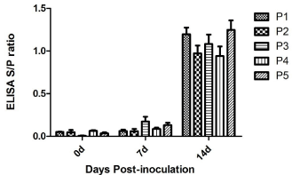
ELISA-detectable antibody responses in successive passages (P2 to P5) showed that the inoculation of pigs with serum resulted in transmission of the infection. All pigs were negative(S/P<0.4) for PRRSV serum antibodies at the time of inoculation (5 to 6 weeks of age). Pigs from P2 to P5 all sero converted to anti-PRRSV at 14 d.p.i., and showed similar antibody responses (Figure 1). The detection of neutralizing antibody in day0, day7 and day14 serum samples showed that all pigs were negative for PRRSV neutralizing antibody.

    Figure 1:  Antibody response to PRRSV infection. Values represent means
and standard deviations of ELISA S/P ratios over all passages (P1 to P5).
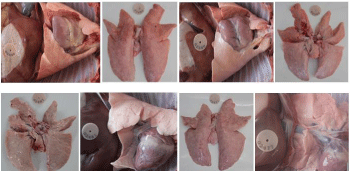
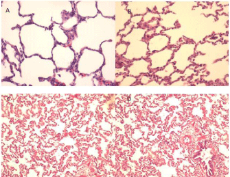
No gross-lung lesions were observed in pigs from P1 to P5 (Figure 2). Microscopically, the lungs from inoculated pigs exhibited no obvious pneumonia (Figure 3), with no significant differences observed in the pneumonia scores between the experimental pigs and the control pigs from P1 to P5.

    Figure 2:  Lung was observed for gross lesions in the pigs from P1 to P5. No
gross lung lesions and consolidations were found in whole process of the
study. Pictures A, B, C and D are lungs from inoculated pigs in P1; E is the
lung from inoculated pig in P2; F is the lung from inoculated pig in P4; G and
H are lungs from inoculated and control pigs in P5, respectively.

    Figure 3:  Photomicrographs of hematoxylin and eosin (H&E)-stained lungs
from inoculated and control pigs. The alveolar structure was normal and
clear, with a few lymphocytes observed around the bronchi and vessels, both
from the inoculated and control pigs. No interstitial pneumonia lesion was
observed. A. lung from inoculated pig in P1; B. lung from control pig in P1; C.
lung from inoculated pig in P5; D. lung from control pig in P5.
All pigs that served as negative controls and environmental sentinels remained free of PRRSV. These results provided evidence that biosecurity procedures effectively prevented the inadvertent transmission of PRRSV among pigs.
Amino acid mutations and deletion during TJM-F92 passaging in pigs
The Nsp2 gene of viruses from P5 was sequenced. There were three mutations in nucleotides found by comparing to PRRSV TJM F92 strain. Two mutations were conversion mutations (From one pyrimidine to another pyrimidine, T545&rarC545 and C1064→T1064), one was mutation (substitutions between purines and pyrimidines, G1703→T1703).
Based on the deduced acids, three amino acids were mutated during the process of five passages in pigs (Table 6); and the mutation rate is 0.36% (3/830). The 2 of 3 mutations occurred newly in this animal passage study and they were different from both TJM F92 strain and TJ strain (I182→T182 and G568→V568). The third mutation occurred as a reverse mutation (A355→V355). A continuous 120 amino acid deletion was identified between PRRSV strain TJ and its derived vaccine strain TJM-F92 [29]. After five passages in pigs sequence analysis of P5 viruses showed that the continuous 120 amino acids deletion still existed.
    Passage
    Group
    Days post    immunization (dpi)


    0d
    2d
    3d
    4d
    5d
    6d
    7d
    8d
    9d
    10d
    11d
    12d
    13d
    14d
    15d
    16d
    17d
    18d
    19d
    20d
    21d


    P1
    Treatment
    39.5
    39.6
    39.6
    39.5
    39.7
    39.5
    39.5
    39.5
    39.6
    39.5
    39.4
    39.7
    39.4
    39.5
    39.6
    /
    /
    /
    /
    /
    /


    Control
    39.6
    39.4
    39.6
    39.6
    39.7
    39.6
    39.6
    39.6
    39.6
    39.6
    39.6
    39.6
    39.5
    39.7
    39.5
    /
    /
    /
    /
    /
    /


    P2
    Treatment
    39.4
    39.6
    39.6
    39.4
    39.4
    39.3
    39.4
    39.4
    39.3
    39.5
    39.5
    39.7
    39.7
    39.6
    39.7
    /
    /
    /
    /
    /
    /


    Control
    39.7
    39.6
    39.6
    39.6
    39.3
    39.2
    39.2
    39.3
    39.6
    39.3
    39.6
    39.3
    39.3
    39.7
    39.7
    /
    /
    /
    /
    /
    /


    P3
    Treatment
    39.6
    39.4
    39.4
    39.6
    39.6
    39.7
    39.5
    39.7
    39.7
    39.6
    39.6
    39.4
    39.6
    39.8
    39.4
    /
    /
    /
    /
    /
    /


    Control
    39.4
    39.4
    39.3
    39.3
    39.5
    39.6
    39.6
    39.4
    39.6
    39.7
    39.5
    39.4
    39.2
    39.6
    39.5
    /
    /
    /
    /
    /
    /


    P4
    Treatment
    39.5
    39.5
    39.2
    39.4
    39.6
    39.3
    39.6
    39.6
    39.3
    39.5
    39.4
    39.5
    39.4
    39.5
    39.5
    /
    /
    /
    /
    /
    /


    Control
    39.6
    39.3
    39.6
    39.4
    39.6
    39.6
    39.4
    39.5
    39.2
    39.5
    39.5
    39.3
    39.5
    39.3
    39.6
    /
    /
    /
    /
    /
    /


    P5
    Treatment
    39.6
    39.6
    39.7
    39.7
    39.7
    39.7
    39.7
    39.6
    39.7
    39.5
    39.6
    39.5
    39.7
    39.6
    39.4
    39.5
    39.5
    39.5
    39.5
    39.5
    39.5


    Control
    39.6
    39.5
    39.6
    39.5
    39.2
    39.5
    39.5
    39.7
    39.7
    39.7
    39.9
    39.4
    39.4
    39.5
    39.4
    39.6
    39.7
    39.5
    39.6
    39.5
    39.5
Table 6: Average temperature from P1 to P5.
Discussion
Most live veterinary viral vaccines induce mild infections with live organisms derived from non-target hosts or attenuated through passage in different cell line cultures or chicken embryos. These vaccines can replicate and induce both cellular and humoral immunity and generally do not require an adjuvant to be effective [32]. However, they can pose a risk of residual virulence and reversion to pathogenic wild types as well as provide a potential source of environmental contamination. This was highlighted during a program to control Porcine Respiratory and Reproductive Syndrome (PRRS) in Denmark [33,34]. This disease first emerged in North America in the late 1980s and spread quickly in Europe in the early 1990s. The two main types of PRRS virus, European and North American, are only 55 to 80% identical at the nucleotide level [33] and cause distinguishable serological responses. Following vaccination with the live, attenuated North American PRRS vaccine against the European PRRS virus type present in Denmark in 1996, the vaccine virus reverted and spread within vaccinated herds as well as from vaccinated to non-vaccinated herds, leaving both virus types in the Danish pig population [34]. Despite such drawbacks of live viral vaccines, they have played a major role in successful disease control and eradication in the world.
The virus inoculum used in P1 of the animal experiment was the PRRSV TJM-F92 vaccine strain, which was the 92nd passage virus (F92) of TJ on Marc-145 cells. It had been passaged continuously in Marc-145 cells and cloned by plaque every 5-10 passages, and has been approved as a safe PRRSV vaccine strain. From P2 to P5, the use of PRRSV-positive serum to pass virus directly from pig to pig was meant to circumvent the selection pressures arising from the in vitro isolation and propagation of viruses. Pigs from P1 to P5 showed no clinical symptoms of PRRS in current study, which was different from those reported by Chang et al., where all pigs (from P1 to P7) in the experimental group exhibited mild to moderate clinical signs such as lethargy and anorexia, occasionally with dyspnea, following inoculation with cell culture-derived VR-2332 virus or with tissue homogenate filtrates from inoculated animals [35]. Taken together, these results demonstrate that the TJM-F92 strain does not show obvious pathogenicity in host animals during the five passages in the present experimental conditions. The TJM-F92 strain has had a wide range of applications in China since 2010 and no PRRSassociated reproductive and respiratory problems were observed in the vaccinated piglets and the TJM-F92 vaccine can offer effective protection against PRRSV infection [25].
In P3, one of the three pigs inoculated with the positive blend serum samples which were on the high level of viremia from P2 did not develop any evidence of virus replication in pig, and did not seroconvert. This result was similar to those observed in previous studies [36]. In one such experiment, two of six pigs apparently did not become infected following intramuscular injection with 2×10⁶ TCID50 NADC-8 PRRSV, and the other four pigs did become infected and seroconverted [36]. This phenomenon showed that the replication level of highly attenuated PRRSV vaccine virus in vivo decreased.
Porcine Reproductive and Respiratory Syndrome Virus (PRRSV) is a single-stranded RNA virus [37]. The mode of replication of the virus makes it prone to high rates of mutation and recombination [38]. More pathogenic variants have been described in the past, a high degree of diversity in the virus population has recently been reported in the United States [39], China [40], Spain [41,42], and eastern Europe [43].
The nsp2 is recognized as the most variable region in the genome of PRRSV, which endure a number of mutations, insertions and deletions [16,44]. The highly pathogenic PRRSV that has spread to most provinces of China since 2006 was found to contain a discontinuous 30 amino acid deletion in nsp2; this deletion is considered to be a potential determining factor that causes the fatal clinical symptoms [5]. The TJ strain was sequenced and analyzed, and a comparison to PRRSV prototype the VR-2332 strain identified a discontinuous 30 aa deletion in the nsp2 region with deletions of 1 and 29 aa, respectively, corresponding to the amino acid positions 481 and 533-561, the same as the HP-PRRSV strains JXA1.
A 120 amino acids (628-747, corresponding to VR-2332) deletion in nsp2, found in passage 19 of the TJ strain, was still present in passage 92 of the TJM strain [29]. Recent studies have characterized the phenomena of amino acid insertions and deletions in nsp2 from virulent and vaccine strains(15, 16,44).Kim constructed a mutant with 131 aa deletion (657-787, corresponding to VR-2332) within a relatively well conserved region of nsp2, and the gross and microhistopathology showed that this construct was less virulent in pigs [45]. By alignment, we found that there was 90 amino acids identity between the 131 amino acid deletion in the mutant and the 120 amino acids deletion in TJM. Therefore, we hypothesize that the 120amino acids deletion in nsp2 has a similar function [29].
The Nsp2 gene of viruses from P5 was sequenced and compared to that of PRRSV TJM F92 strain. Three nucleotide mutations were found, and two mutations were conversion mutations, and the other one was a transversion mutation. Based on the deduced acids, three amino acids were found mutated during the process of five passages in pigs and the mutation rate is 0.36% (3/830) comparing with TJM F92 strain. Whereas the mutation rate of TJM F92 strain is 2.28% (19/830) during its attenuation process from PRRSV TJ strain. After five passages in pigs, sequence analysis of P5 viruses also showed that the continuous 120 amino acids deletion still existed, suggesting the stable characteristic of the deletion in vivo.
Conclusion
The study results showed that the 120 amino acid deletion in nsp2 is stable and the vaccine virus did not show reversion to virulence. As the TJM-F92 strain has a continuous 120 amino acids deletion in nsp2, it also has the potential DIVA function by differentiation of antibody responses induced by the vaccine (no antibodies generated to deleted genes) from those induced during infection with the wildtype virus.
Acknowledgement
The authors especially thank Dr. WenzhiXue and BinBin Wu for helping with the manuscript preparation.
References
- Terpstra C, Wensvoort G, Pol JM. Experimental reproduction of porcine epidemic abortion and respiratory syndrome (mystery swine disease) by infection with Lelystad virus: Koch's postulates fulfilled. Vet Q. 1991; 13: 131-136.
- Rossow KD, Bautista EM, Goyal SM, Molitor TW, Murtaugh MP, Morrison RB, et al. Experimental porcine reproductive and respiratory syndrome virus infection in one-, four-, and 10-week-old pigs. J Vet Diagn Invest. 1994; 6: 3-12.
- Christianson WT, Collins JE, Benfield DA, Harris L, Gorcyca DE, Chladek DW, et al. Experimental reproduction of swine infertility and respiratory syndrome in pregnant sows. Am J Vet Res. 1992; 53: 485-488.
- Wensvoort G, Terpstra C, Pol JM, ter Laak EA, Bloemraad M, de Kluyver EP, et al. Mystery swine disease in The Netherlands: the isolation of Lelystad virus. Vet Q. 1991; 13: 121-130.
- Tian K, Yu X, Zhao T, Feng Y, Cao Z, Wang C, et al. Emergence of fatal PRRSV variants: unparalleled outbreaks of atypical PRRS in China and molecular dissection of the unique hallmark. PLoS One. 2007; 2: e526.
- Feng Y, Zhao T, Nguyen T, Inui K, Ma Y, Nguyen TH, et al. Porcine respiratory and reproductive syndrome virus variants, Vietnam and China, 2007. Emerg Infect Dis. 2008; 14: 1774-1776.
- Ni J, Yang S, Bounlom D, Yu X, Zhou Z, Song J, et al. Emergence and pathogenicity of highly pathogenic Porcine reproductive and respiratory syndrome virus in Vientiane, Lao People's Democratic Republic. J Vet Diagn Invest. 2012; 24: 349-354.
- Neumann EJ, Kliebenstein JB, Johnson CD, Mabry JW, Bush EJ, Seitzinger AH, et al. Assessment of the economic impact of porcine reproductive and respiratory syndrome on swine production in the United States. J Am Vet Med Assoc. 2005; 227: 385-392.
- Dea S, Gagnon CA, Mardassi H, Pirzadeh B, Rogan D. Current knowledge on the structural proteins of porcine reproductive and respiratory syndrome (PRRS) virus: comparison of the North American and European isolates. Arch Virol. 2000; 145: 659-688.
- Johnson CR, Griggs TF, Gnanandarajah J, Murtaugh MP. Novel structural protein in porcine reproductive and respiratory syndrome virus encoded by an alternative ORF5 present in all arteriviruses. J Gen Virol. 2011; 92: 1107-1116.
- Firth AE, Zevenhoven-Dobbe JC, Wills NM, Go YY, Balasuriya UB, Atkins JF, et al. Discovery of a small arterivirus gene that overlaps the GP5 coding sequence and is important for virus production. J Gen Virol. 2011; 92: 1097-1106.
- Allende R, Lewis TL, Lu Z, Rock DL, Kutish GF, Ali A, et al. North American and European porcine reproductive and respiratory syndrome viruses differ in non-structural protein coding regions. J Gen Virol 1999; 80: 307-315.
- Fang Y, Snijder EJ. The PRRSV replicase: exploring the multifunctionality of an intriguing set of nonstructural proteins. Virus Res. 2010; 154: 61-76.
- Yuan S, Mickelson D, Murtaugh MP, Faaberg KS. Complete genome comparison of porcine reproductive and respiratory syndrome virus parental and attenuated strains. Virus Res. 2001; 79: 189-200.
- Han J, Wang Y, Faaberg KS. Complete genome analysis of RFLP 184 isolates of porcine reproductive and respiratory syndrome virus. Virus Res. 2006; 122: 175-182.
- Shen S, Kwang J, Liu W, Liu DX. Determination of the complete nucleotide sequence of a vaccine strain of porcine reproductive and respiratory syndrome virus and identification of the Nsp2 gene with a unique insertion. Arch Virol. 2000; 145: 871-883.
- Tong GZ, Zhou YJ, Hao XF, Tian ZJ, An TQ, Qiu HJ. Highly pathogenic porcine reproductive and respiratory syndrome, China. Emerg Infect Dis. 2007; 13: 1434-1436.
- Corzo CA, Mondaca E, Wayne S, Torremorell M, Dee S, Davies P, et al. Control and elimination of porcine reproductive and respiratory syndrome virus. Virus Res. 2010; 154: 185-192.
- Nielsen TL, Nielsen J, Have P, Baekbo P, Hoff-Jørgensen R, Bøtner A. Examination of virus shedding in semen from vaccinated and from previously infected boars after experimental challenge with porcine reproductive and respiratory syndrome virus. Vet Microbiol. 1997; 54: 101-112.
- Nilubol D, Platt KB, Halbur PG, Torremorell M, Harris DL. The effect of a killed Porcine Reproductive and Respiratory Syndrome Virus (PRRSV) vaccine treatment on virus shedding in previously PRRSV infected pigs. Vet Microbiol. 2004; 102: 11-18.
- Zuckermann FA, Garcia EA, Luque ID, Christopher-Hennings J, Doster A, Brito M, et al. Assessment of the efficacy of commercial Porcine Reproductive and Respiratory Syndrome Virus (PRRSV) vaccines based on measurement of serologic response, frequency of gamma-IFN-producing cells and virological parameters of protection upon challenge. Vet Microbiol. 2007; 123: 69-85.https://www.ncbi.nlm.nih.gov/pubmed/12513927
- Foss DL, Zilliox MJ, Meier W, Zuckermann F, Murtaugh MP. Adjuvant danger signals increase the immune response to porcine reproductive and respiratory syndrome virus. Viral Immunol. 2002; 15: 557-566.
- Meier WA, Galeota J, Osorio FA, Husmann RJ, Schnitzlein WM, Zuckermann FA. Gradual development of the interferon-gamma response of swine to porcine reproductive and respiratory syndrome virus infection or vaccination. Virology. 2003; 309: 18-31.
- Charerntantanakul W, Platt R, Johnson W, Roof M, Vaughn E, Roth JA. Immune responses and protection by vaccine and various vaccine adjuvant candidates to virulent porcine reproductive and respiratory syndrome virus. Vet Immunol Immunopathol. 2006; 109: 99-115.
- Leng X, Li Z, Xia M, He Y, Wu H. Evaluation of the efficacy of an attenuated live vaccine against highly pathogenic porcine reproductive and respiratory syndrome virus in young pigs. Clin Vaccine Immunol 2012; 19: 1199-1206.
- Dewey CE, Wilson S, Buck P, Leyenaar JK. The reproductive performance of sows after PRRS vaccination depends on stage of gestation. Prev Vet Med. 1999; 40: 233-241.
- Mengeling WL, Vorwald AC, Lager KM, Clouser DF, Wesley RD. Identification and clinical assessment of suspected vaccine-related field strains of porcine reproductive and respiratory syndrome virus. Am J Vet Res. 1999; 60: 334-340.
- Nielsen HS, Oleksiewicz MB, Forsberg R, Stadejek T, Bøtner A, Storgaard T. Reversion of a live porcine reproductive and respiratory syndrome virus vaccine investigated by parallel mutations. J Gen Virol. 2001; 82: 1263-1272.
- Leng X, Li Z, Xia M, Li X, Wang F, Wang W, et al. Mutations in the genome of the highly pathogenic porcine reproductive and respiratory syndrome virus potentially related to attenuation. Vet Microbiol. 2012; 157: 50-60.
- Halbur PG, Paul PS, Frey ML, Landgraf J, Eernisse K, Meng XJ, et al. Comparison of the pathogenicity of two US porcine reproductive and respiratory syndrome virus isolates with that of the Lelystad virus. Vet Pathol. 1995; 32: 648-660.
- Halbur PG, Paul PS, Meng XJ, Lum MA, Andrews JJ, Rathje JA. Comparative pathogenicity of nine US porcine reproductive and respiratory syndrome virus (PRRSV) isolates in a five-week-old cesarean-derived, colostrum-deprived pig model. J Vet Diagn Invest. 1996; 8: 11-20.
- Meeusen EN, Walker J, Peters A, Pastoret PP, Jungersen G. Current status of veterinary vaccines. Clin Microbiol Rev. 2007; 20: 489-510.
- Murtaugh MP, Elam MR, Kakach LT. Comparison of the structural protein coding sequences of the VR-2332 and Lelystad virus strains of the PRRS virus. Arch Virol. 1995; 140: 1451-1460.
- Mortensen S, Stryhn H, Søgaard R, Boklund A, Stärk KD, Christensen J, et al. Risk factors for infection of sow herds with porcine reproductive and respiratory syndrome (PRRS) virus. Prev Vet Med. 2002; 53: 83-101.
- Chang CC, Yoon KJ, Zimmerman JJ, Harmon KM, Dixon PM, Dvorak CM, et al. Evolution of porcine reproductive and respiratory syndrome virus during sequential passages in pigs. J Virol. 2002; 76: 4750-4763.
- Grebennikova TV, Clouser DF, Vorwald AC, Musienko MI, Mengeling WL, Lager KM, et al. Genomic characterization of virulent, attenuated, and revertant passages of a North American porcine reproductive and respiratory syndrome virus strain. Virology. 2004; 321: 383-390.
- Rossow KD. Porcine reproductive and respiratory syndrome. Vet Pathol. 1998; 35: 1-20.
- Gorbalenya AE, Enjuanes L, Ziebuhr J, Snijder EJ. Nidovirales: evolving the largest RNA virus genome. Virus Res. 2006; 117: 17-37.
- Fang Y, Schneider P, Zhang WP, Faaberg KS, Nelson EA, Rowland RR. Diversity and evolution of a newly emerged North American Type 1 porcine arterivirus: analysis of isolates collected between 1999 and 2004. Arch Virol. 2007; 152: 1009-1017.
- Li Y, Wang X, Jiang P, Wang X, Chen W, Wang X, et al. Genetic variation analysis of porcine reproductive and respiratory syndrome virus isolated in China from 2002 to 2007 based on ORF5. Vet Microbiol. 2009; 138: 150-155.
- Mateu E, Díaz I, Darwich L, Casal J, Martín M, Pujols J. Evolution of ORF5 of Spanish porcine reproductive and respiratory syndrome virus strains from 1991 to 2005. Virus Res. 2006; 115: 198-206.
- Prieto C, Vázquez A, Núñez JI, Alvarez E, Simarro I, Castro JM. Influence of time on the genetic heterogeneity of Spanish porcine reproductive and respiratory syndrome virus isolates. Vet J. 2009; 180: 363-370.
- Stadejek T, Oleksiewicz MB, Scherbakov AV, Timina AM, Krabbe JS, Chabros K, et al. Definition of subtypes in the European genotype of porcine reproductive and respiratory syndrome virus: nucleocapsid characteristics and geographical distribution in Europe. Arch Virol. 2008; 153: 1479-1488.D
- Fang Y, Kim DY, Ropp S, Steen P, Christopher-Hennings J, Nelson EA, et al. Heterogeneity in Nsp2 of European-like porcine reproductive and respiratory syndrome viruses isolated in the United States. Virus Res. 2004; 100: 229-235.
- Kim DY, Kaiser TJ, Horlen K, Keith ML, Taylor LP, Jolie R, et al. Insertion and deletion in a non-essential region of the nonstructural protein 2 (nsp2) of porcine reproductive and respiratory syndrome (PRRS) virus: effects on virulence and immunogenicity. Virus Genes 2009; 38: 118-128.