Abstract
Yoga has become a popular form of exercise for improving core strength and stability in individuals with Low Back Pain (LBP). Researchers have quantified muscle activation during core stabilization exercises, believing that exercises which require greater activation will benefit that needing improved core stabilization. Minimal attention has been directed toward muscle activation during yoga. The purpose of this study was to determine and compare the relative activation of core muscles during yoga to traditional back exercises. Surface Electro Myo Graphy (EMG) was used to quantify the relative activation of the Rectus Abdominis (RA), Abdominal Obliques (AO), Lumbar Extensors (LE), and Gluteus Maximus (GMX) during four yoga poses (Chair, High Plank, Upward-Facing Dog, and Dominant-Side Warrior 1). Data were expressed as 100% of a Maximum Voluntary Isometric Contraction (100% MVIC). Separate analyses of variance with repeated measures were used to compare muscle activity across each exercise. The sequentially rejective Bonferroni test was used for post hoc testing. EMG activity during the High Plank was moderate (28% MVIC) and high (44% MVIC) for the RA and AO, respectively. The AO had moderate (27% MVIC) activity during the Upward-Facing Dog. EMG activity during the Chair was moderate (38% MVIC) only for the LE. GMX activity was low (< 21% MVIC) during all the exercises. These findings suggest that certain yoga poses may be useful for improving core endurance and strength. Clinicians may use these data when developing and implementing an evidence-based core exercise program for individuals with LBP who prefer a yoga treatment strategy.
Keywords: Complementary medicine; Surface electromyography; Core stabilization
Introduction
Classified as a Complementary and Alternative Medicine (CAM) mind-body therapy, the practice of yoga is growing in popularity in the United States. As such, yoga is being adopted in conventional physical therapy practice as an integral therapy [1]. The National Center for Complementary and Alternative Medicine defines CAM as a group of diverse medical and healthcare systems practices not presently considered part of conventional healthcare. CAM is commonly used as an adjunct intervention with conventional medicine and represents the synthesis of conventional practice and evidenced-based complementary medicine [2]. National survey trends of adult users of CAM illustrate that approximately 33.2% of adults aged 18 years and over have used one form of CAM in the last 12 months [1,2].
Yoga is a popular CAM intervention, with 34% of adults in 2016 likely to practice yoga within the next 12-month period. More than 15% (36.7 million) of adults in the US consistently practice yoga, an increase of over 16 million since 2012. Most importantly, 74% of those who engage in yoga are relatively new to the practice, becoming involved within the last five years. Significance is the emerging and growing use of yoga across ethnic dimensions. Non-Hispanic adults showed a 30% increase in use of yoga from 2002 to 2012; significant increases in use by Hispanic (5.1%) and African American (5.6%) adults has been reported as well [1].
Yoga is defined as “a combination of breathing exercises, physical postures, and meditation used to calm the nervous system and balance the body, mind, and spirit” [1]. Approximately 55% of physical therapists regularly use yoga as a common form of alternative strength training [3], which may reflect its adaptability for addressing various musculoskeletal problems [4]. The incorporation of lowintensity forms of exercise can be varied and scaled to age, medical complexity, and preference across all age and ethnic groups. The use of yoga suggests a positive relationship to the alleviation of chronic Low Back Pain (LBP) [5] and supports the premise that yoga brings balance and health to the physical, mental, emotional, and spiritual dimensions of an individual [6].
Yoga incorporates core strengthening and stabilization exercises considered important for the treatment of non-specific LBP [5]. This core comprises the lumbar spine, pelvis, and hip, as well as the ligaments and muscles that control their movement [7]. Core stabilization refers to the ability of the core musculature to control the position and motion of the trunk over the pelvis and lower extremities during movement [8]. The importance of core stabilization has led to substantial evidence quantifying the relative Electro Myo Graphy (EMG) activity of core muscles during therapeutic exercise [9-14]. These studies were based on the premise that exercises which require greater EMG activity will result in increased muscle strength [15].
To date, limited data exist regarding the relative EMG activity of core muscles during yoga poses. Ni and colleagues are the only researchers who have examined such EMG activity during yoga practice. They reported differences in muscle activation during various poses performed by trained yoga practitioners [16]. They also found that muscle activation varied per practitioner skill level [17]. A limitation of these studies was the use of experienced subjects. With respect to physical therapy practice, patients who use yoga for rehabilitation purposes most likely have limited, if any, prior experience. Because skill level can influence muscle activity, additional studies are needed to examine EMG activity in untrained individuals. Therefore, the primary purpose of this study was to determine the relative EMG activity of core muscles during yoga poses in individuals with minimal yoga experience. The secondary purpose was to compare core activation during yoga to back exercises commonly prescribed for individuals with LBP. Our study was based on the null hypothesis that no differences would exist in core muscle activation during the chosen yoga poses and when compared to traditional back exercises.
Methods
Subjects
Thirty individuals with minimal experience with yoga participated (15 females, mean age 24.7 + 2.1 y, mass 71.6 + 13.0 kg, height 1.7 + 0.1 m). A convenience sample was recruited from the greater Central Savannah River Area. Inclusion criteria included healthy subjects between the age of 18 and 40 years with less than four weeks of yoga experience. Additionally, subjects had no history of spine or upper/ lower extremity surgery. None had incurred any significant spine or lower extremity injury in the past two years. The investigators explained the benefits and risks of this study to all participants, who then signed an informed consent document approved by the Georgia Regents University (now Augusta University) Institutional Review Board.
Procedures
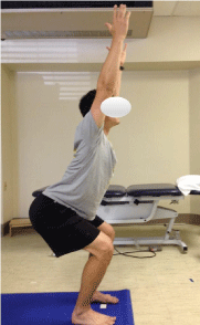
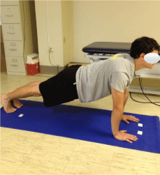
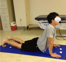
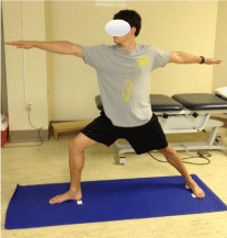
Following the informed consent process, all subjects completed a warm-up session that consisted of gentle stretching exercises for the trunk extensors and rotators, hamstrings, quadriceps, and calf muscles. Subjects performed each stretch three times with a 15-second hold. Next, an investigator instructed each subject in the following yoga poses: Chair, High Plank (Plank), Upward-Facing Dog (Dog), and Dominant-Side Warrior 1 (Warrior) [16]. Based on our clinical experience, we chose these poses because they emulated those typically used for the rehabilitation of individuals with pathologies like LBP [18]. For the Chair pose (Figure 1), subjects were instructed to stand and flex the knees 45 degrees (as if to sit in a chair) while keeping their backs straight, upper extremities overhead, and palms facing inward. Stance width was standardized using a “fist-width” measure (Figure 2). For this purpose, subjects placed both hands in a closed-fist position with a side-by-side orientation. An investigator measured the distance from the most ulnar side of the head of the 5th metacarpal on each hand to the nearest 1/10th of a centimeter. This exercise was chosen to emulate a static position to facilitate core stabilization during a stand-to-sit transfer. For the Plank (Figure 3), subjects assumed a full push-up position. This exercise was chosen since it has been prescribed to improve trunk endurance. For the Dog, subjects assumed a prone position and then extended their spines (Figure 4). This exercise was chosen because of its similarity to the McKenzie extension exercise. For the Warrior (Figure 5), subjects lunged toward the same side as the dominant hand. Care was taken to ensure that subjects kept their shoulders, trunks, and non-dominantside lower extremities facing forward. Stance width was standardized as the distance equal to the lower limb length of the dominant hand side. Subjects lunged to the position where the tibia on the dominant hand side was vertical to the floor. This exercise was chosen to facilitate core stabilization during a frontal plane movement.

Figure 1: The chair pose.

Figure 2: The “first-width” measure.

Figure 3: The high plank pose.

Figure 4: The upward-facing dog pose.

Figure 5: The dominant-side warrior 1 pose.
Next, subject’s skin was prepared for surface EMG electrodes by shaving (if needed) and cleaning the skin with isopropyl alcohol over the Rectus Abdominis (RA), Abdominal Obliques (AO), Lumbar Extensors (LE), and Gluteus Maximus (GMX). Trigno™ wireless sensors (Delsys®, Boston, MA) were placed parallel over the muscle belly of each muscle in a standardized manner [19,20]. Electrode placement was confirmed by observing electrical signals on an oscilloscope as an investigator applied muscle resistance in accordance with common manual muscle testing techniques [21]. Following electrode placement, subjects performed three Maximum Voluntary Isometric Contractions (MVIC) for each muscle to normalize raw EMG data. All MVICs were conducted in accordance with the “make” test [22], where subjects generated force over a two-second period and held the MVIC for five seconds. Subjects completed one practice trial and three test trials [23]. Test positions for each muscle were used in accordance with previously described methods [24]. Subjects received strong verbal encouragement during each trial and rested 30 seconds between trials.
For testing, subjects held each pose for a 20-second period and EMG activity during the last 15 seconds was used for analysis [16]. They also rested at least one minute between poses to minimize fatigue. The order of testing was counterbalanced to reduce the possibility of order bias [23]. For all exercises, subjects were instructed to breathe normally and simultaneously contract their abdominal and gluteal muscles. They also received verbal feedback, if needed, to maintain the proper form. After testing, subjects were instructed to refrain from any physical activity, other than normal walking, for a 24-hour period to minimize the potential for muscle soreness.
EMG instrumentation and analysis
A four-channel wireless EMG system (Delsys™, Natick, MA) collected all EMG data, which were sampled at 2000 Hz and bandpass filtered between 20 and 450 Hz. Unit specifications also included a common mode rejection ratio greater than 80 dB. All data were Root-Mean-Squared (RMS) over a 30-ms moving window [24]. For the MVICs, a computer algorithm determined the maximum RMS amplitude over a moving 500-ms average window across the MVICs [25]. The window with the highest amplitude was used to normalize all data as a percentage of MVIC (% MVIC). The average amplitude of EMG data during the last 15 seconds of individual yoga poses was expressed as %MVIC and used for statistical analysis.
Statistical analysis
Separate analyses of variance with repeated measures were used to determine differences in average muscle amplitudes across exercises. All statistical analyses were conducted using IBM SPSS version 24.0 (IBM SPSS, Inc., Armonk, NY) with a 0.05 level of significance. The level of significance was adjusted using the sequentially rejective Bonferroni test to account for multiple pair wise comparisons, thus protecting against possible type I error [26].
Results
EMG activity for the ranged from 9.3% to 28.2% MVIC. A main effect existed (P<0.0001), and the post-hoc analysis showed that subjects generated significantly greater RA activity during the Plank (P<0.0001) compared to the other exercises. EMG activity for the AO ranged from 17.5% to 44.5% MVIC. A main effect existed (P<0.0001), and the post-hoc analysis showed that subjects generated significantly greater AO activity during the Plank (P<0.0001) compared to the Chair, Dog, and Warrior poses. Subjects also generated greater AO activity during the Dog compared to the Chair (P<0.0001) and Warrior (P<0.0001). EMG activity for the LE ranged from 10.8% to 38.6% MVIC. A main effect existed (P<0.0001), and the posthoc analysis showed that subjects generated significantly greater LE activity during the Chair (P<0.0001) compared to the Plank, Dog, and Warrior. EMG activity for the GMX ranged from 14.3% to 20.4% MVIC; no main effect existed (P = 0.09). The table summarizes all EMG data (Table 1).
    Yoga Pose


    Muscle
    Chair
    High Plank
    Upward-Facing Dog
    Dominant-Side Warrior 1


    Rectus Abdominisa
    10.6 (7.5)
    28.2 (15.8)
    12.7 (7.6)
    9.3 (6.7)


    Abdominal Obliquesb,c
    18.9 (8.5)
    44.5 (20.4)
    27.3 (12.4)
    17.5 (7.7)


    Lumbar Extensorsd
    38.6 (13.7)
    13.7 (8.5)
    12.5 (7.9)
    10.8 (7.7)


    Gluteus Maximuse
    15.6 (11.2)
    14.3 (8.7)
    20.4 (14.9)
    18.9 (13.7)


      aSignificantly    greater rectus abdominis activation during the High Plank compared to all    other exercises; P<0.0001
      bSignificantly    greater abdominal oblique activation during the High Plank compared to the    Chair, Upward-Facing Dog, and Dominant-Side Warrior 1; P<0.0001
      cSignificantly    greater abdominal oblique activation during the Upward-Facing Dog compared to    the Chair and Dominant-Side Warrior 1; P<0.0001
      dSignificantly    greater lumbar extensor activation during the Chair compared to all other    exercises; P<0.0001
      eSimilar    gluteus maximus activation during all exercises; P>0.05
Table 1: Mean + (standard deviation) of individual muscle activity during the yoga poses expressed as 100% of a Maximum Voluntary Isometric Contraction (MVIC).
Discussion
The purpose of this study was to determine the relative EMG activity of core muscles during yoga poses in untrained individuals. Historically, researchers have measured the relative activation based on a % of MVIC during therapeutic exercises and concluded that those requiring greater EMG activity would result in greater strength gains [9,11,27]. To improve interpretation, the following activation ranges were used: 0-20% MVIC (low); 21-40% MVIC (moderate); 41-60% MVIC (high); and greater than 60% MVIC (very high) [15]. Exercises that generate moderate EMG activity have been thought to improve muscle endurance, whereas those requiring high and very high EMG activity may produce greater strength gains. Understanding these differences can assist clinicians in determining the expected gains from each yoga pose.
Rectus abdominis
EMG activity for the RA was low for all poses except the Plank, which had moderate activation (28.2% MVIC). This activation level was similar (27.0% MVIC) to that of yoga practitioners during this pose [16]. Cholewicki and VanVliet [28] found that a combination of low level EMG activity (i.e., less than 30% MVIC) from multiple trunk muscles, not high level activity from a single muscle, contributed most to lumbar spine stability. This combined lower level activity highlights the importance of core muscle endurance, and not necessarily strength, for core stability. Therefore, regardless of yoga skill level, individuals can perform the Plank to improve RA endurance [15]. However, clinicians would need to prescribe more demanding exercises than the Plank to obtain RA-strengthening effects.
Abdominal obliques
Compared to the RA, subjects generated relatively greater AO EMG activity during all poses. Subjects, on average, generated moderate activity (27.3% MVIC) during the Dog and high activity (44.5% MVIC) during the Plank. Ni et al. [16] reported much higher AO activity (78.0% MVIC and 66.0% MVIC for the Plank and Dog, respectively) for yoga practitioners. This difference could have reflected the ability of skilled individuals to activate the AO better than novices. An interesting pattern of activation between the RA and AO during the Plank and Dog also existed between studies. Subjects in both studies generated at least 1.5 times greater AO activity than RA activity during these exercises. This result suggests that the AO may have a greater stabilizing effect than the RA during these poses. Clinicians may use these data when prescribing a progressive yoga program for untrained individuals. The untrained individual with AO weakness may initially benefit from the Dog to improve muscle endurance and then transition to the Plank for greater strengthening effects. Care must be taken for individuals with pathologies like LBP, however, as trunk extension during the Dog can increase lumbar spine loading [16].
Lumbar extensors
EMG activity for the LE was low for all poses except the Chair, which had moderate activation (38.6% MVIC). This finding was not unexpected as LE activation is needed to control the amount of trunk flexion required during this pose. While LE activity for our subjects during the Chair was similar to that of yoga practitioners (32.0% MVIC), differences existed with the Dog [16]. Our subjects generated low LE activity (12.5% MVIC) during the Dog compared to yoga practitioners (34.0% MVIC) [16]. A possible explanation may be the different strategies used to maintain trunk extension during this pose. Experienced individuals can maintain trunk extension via LE activation, whereas inexperienced individuals may push through the upper extremities (e.g., extending the shoulders and elbows). Additional studies are needed to make this determination, however. Based on our findings, untrained individuals may benefit from the Chair, but not the Dog, to improve trunk extensor endurance. Like the RA during the Plank, clinicians would need to prescribe more demanding exercises than the Chair to obtain LE-strengthening effects.
Gluteus maximus
EMG activity for the GMX was low across all poses, ranging from 14.3% to 20.4% MVIC. This finding suggests that the poses would provide minimal benefit for individuals needing improved GMX endurance and strength. However, yoga practitioners have generated high GMX activity during the Dog (41.0% MVIC) and Warrior (40.0% MVIC) poses [16]. While we instructed our subjects to activate the GMX during all poses, individuals with experience may have learned to engage this muscle better than untrained individuals. However, the design of this study did not allow us to make this determination. Relatively low GMX activation during all the poses suggests that they will not benefit individuals in need of improved GMX function.
Comparison of activation during yoga to traditional back exercises
The secondary purpose of this study was to compare EMG activity during yoga poses to traditional back exercises. The most investigated exercise similar to those used in the current study is the Plank. EMG activity of the RA from prior studies [9,10,13,29] has ranged from 18% to 36% MVIC, while that of the AO has ranged from 30% to 47% MVIC. These ranges agree with our data. Regarding the LE, EMG activity during traditional back extension exercises has ranged from 65% to 80% MVIC [13,30]. Differences in LE activity, especially for the Dog in the current study, are most likely due to subjects in other studies performing the back extension exercises without upper extremity support. As discussed in the LE section, individuals who perform the Dog will most likely need instruction to activate the LE instead of relying solely on upper extremity support (i.e., primarily pushing upward using the upper extremities).
Based on our clinical experience, we considered the alternating arm/leg raise in a quadruped position as a commonly prescribed core exercise [9]. Results from prior investigations have reported trunk extensor EMG activity ranging from 32%to 46% MVIC [31- 33]. Subjects in the current study generated LE activity equal to 38% MVIC during the Chair, which suggests that the Chair pose may be viable for individuals who need improved LE function but cannot assume a quadruped position. In summary, many of the yoga poses required similar levels of EMG activation as commonly prescribed back exercises.
Limitations
This study has certain limitations. First, it only included healthy individuals, which prevents generalization to those with musculoskeletal or neurological problems. Second, signal crosstalk from adjacent muscles is a possibility with the use of surface EMG. However, we minimized this possibility by applying electrodes in a standardized manner [19,20]. We did not assess activity from other core muscles like the transversus abdominis or multifidi. These muscles are best assessed using fine-wire EMG, while the extent of their activation during each pose is currently unknown.
Conclusion
Emerging evidence supports the use of yoga as an effective alternative or complement to physical therapy for the treatment of non-specific LBP [34,35]. Interventions like tai chi and yoga have resulted in improved function and reduced pain over general exercise in individuals with chronic LBP [34]. Yoga can also be as effective as traditional stretching exercises for the trunk and lower extremity muscles [36]. It has no more risk of adverse effects than traditional back exercises [37], indicating that yoga may be a safe alternative treatment approach. Finally, a specifically designed yoga intervention that addresses impairments associated with chronic, non-specific LBP can be as effective as physical therapy in improving function and alleviating pain [38]. Emerging research on the efficacy of yoga has provided valuable evidence for the physical therapy practitioner choosing to integrate yoga into a plan of care. Factors such as patient preference, cost, or ease of a self-directed home exercise program can support yoga as a viable treatment strategy. Clinicians may use data from the current study when developing and implementing an evidence-based yoga intervention designed to improve the endurance and strength of core muscles.
References
- Clarke TC, Black LI, Stussman BJ, Barnes PM, Nahin RL. Trends in the use of complementary health approaches among adults: United States. 2002- 2012. Natl Health Stat Report. 2015; 79: 1-16.
- Barnes PM, Bloom B, Nahin RL. Complementary and alternative medicine use among adults and children: United States. 2007. Natl Health Stat Report. 2008; 12: 1-23.
- Tapley H, Dotson M, Hallila D, Haley McCrory, Kendall Moss, Kris Neelon, et al. Participation in strength training activities among US physical therapists: A nationwide survey. Int J Ther Rehabil. 2015; 22: 79-85.
- Yoga Journal & Yoga Alliance. Yoga in America study. 2016.
- Patil NJ, Nagarathna R, Tekur P, Patil DN, Nagendra HR, Subramanya P. Designing, validation, and feasibility of integrated yoga therapy module for chronic low back pain. Int J Yoga. 2015; 8: 103-108.
- Ross A, Thomas S. The health benefits of yoga and exercise: a review of comparison studies. J Altern Complement Med. 2010; 16: 3-12.
- Willson JD, Dougherty CP, Ireland ML, Davis IM. Core stability and its relationship to lower extremity function and injury. J Am Acad Orthop Surg. 2005; 13: 316-325.
- Kibler WB, Press J, Sciascia A. The role of core stability in athletic function. Sports Med. 2006; 36: 189-198.
- Ekstrom RA, Donatelli RA, Carp KC. Electromyographic analysis of core trunk, hip, and thigh muscles during 9 rehabilitation exercises. J Orthop Sports Phys Ther. 2007; 37: 754-762.
- Czaprowski D, Afeltowicz A, Gebicka A, Pawowska P, Kedra A, Barrios C, et al. Abdominal muscle EMG-activity during bridge exercises on stable and unstable surfaces. Phys Ther Sport. 2014; 15: 162-168.
- Youdas JW, Boor MM, Darfler AL, Koenig MK, Mills KM, Hollman JH. Surface electromyographic analysis of core trunk and hip muscles during selected rehabilitation exercises in the side-bridge to neutral spine position. Sports Health. 2014; 6: 416-421.
- Escamilla RF, Lewis C, Bell D, Bramblet G, Daffron J, Lambert S, et al. Core muscle activation during Swiss ball and traditional abdominal exercises. J Orthop Sports Phys Ther. 2010; 40: 265-276.
- Maeo S, Takahashi T, Takai Y, Kanehisa H. Trunk muscle activities during abdominal bracing: comparison among muscles and exercises. J Sports Sci Med. 2013; 12: 467-474.
- Atkins SJ, Bentley I, Brooks D, Burrows MP, Hurst HT, Sinclair JK. Electromyographic response of global abdominal stabilizers in response to stable- and unstable-base isometric exercise. J Strength Cond Res. 2015; 29: 1609-1615.
- Reiman MP, Bolgla LA, Loudon JK. A literature review of studies evaluating gluteus maximus and gluteus medius activation during rehabilitation exercises. Physiother Theory Pract. 2012; 28: 257-268.
- Ni M, Mooney K, Harriell K, Balachandran A, Signorile J. Core muscle function during specific yoga poses. Complement Ther Med. 2014; 22: 235-243.
- Ni M, Mooney K, Balachandran A, Richards L, Harriell K, Signorile JF. Muscle utilization patterns vary by skill levels of the practitioners across specific yoga poses (asanas). Complement Ther Med. 2014; 22: 662-669.
- Hoogenboom BJ, Keisel K. Core stabilization training. Clinical Orthopedic Rehabilitation. 3rd edn. Philadelphia: Elsevier. 2011; 467-481.
- Criswell E. Cram’s Introduction to Surface Electromyography. 2nd edn. Sudbury, MA: Jones and Bartlett Publishers. 2011.
- Rainoldi A, Melchiorri G, Caruso I. A method for positioning electrodes during surface EMG recordings in lower limb muscles. J Neurosci Methods. 2004; 134: 37-43.
- Kendall FP, McCreary EK, Provance P, Rodgers MM, Romani WA. Muscles: Testing and Function with, Posture and Pain. 5th edn. Baltimore, MD: Lippincott Williams & Wilkins. 2010.
- Bohannon RW. Reference values for extremity muscle strength obtained by hand-held dynamometry from adults aged 20 to 79 years. Arch Phys Med Rehabil. 1997; 78: 26-32.
- Bolgla LA, Uhl TL. Reliability of electromyographic normalization methods for evaluating the hip musculature. J Electromyogr Kinesiol. 2007; 17: 102-111.
- Bolgla LA, Cruz MF, Roberts LH, Buice AM, Pou TS. Relative electromyographic activity in trunk, hip, and knee muscles during unilateral weight bearing exercises: Implications for rehabilitation. Physiother Theory Pract. 2016; 32: 130-138.
- Bamman MM, Ingram SG, Caruso JF, Greenisen MC. Evaluation of surface electromyography during maximal voluntary contraction. J Strength Cond Res. 1997; 11: 68-72.
- Holm S. A simple sequentially rejective multiple test procedure. Scand J Stat. 1979; 6: 65-70.
- Boren K, Conrey C, Le Coguic J, Paprocki L, Voight M, Robinson TK. Electromyographic analysis of gluteus medius and gluteus maximus during rehabilitation exercises. Int J Sports Phys Ther. 2011; 6: 206-223.
- Cholewicki J, VanVliet JJ. Relative contribution of trunk muscles to the stability of the lumbar spine during isometric exertions. Clin Biomech (Bristol, Avon). 2002; 17: 99-105.
- Snarr RL, Esco MR. Electromyographical comparison of plank variations performed with and without instability devices. J Strength Cond Res. 2014; 28: 3298-3305.
- Kim SM, Yoo WG. Comparison of trunk and hip muscle activity during different degrees of lumbar and hip extension. J Phys Ther Sci. 2015; 27: 2717-2718.
- Souza GM, Baker LL, Powers CM. Electromyographic activity of selected trunk muscles during dynamic spine stabilization exercises. Arch Phys Med Rehabil. 2001; 82: 1551-1557.
- Kelly M, Jacobs D, Wooten ME, Edeer AO. Comparison of electromyographic activities of lumbar iliocostalis and lumbar multifidus muscles during stabilization exercises in prone, quadruped, and sitting positions. J Phys Ther Sci. 2016; 28: 2950-2954.
- Yoon TL, Cynn HS, Choi SA, Choi WJ, Jeong HJ, Lee JH, et al. Trunk muscle activation during different quadruped stabilization exercises in individuals with chronic low back pain. Physiother Res Int. 2015; 20: 126-132.
- Chou R, Deyo R, Friedly J, Skelly A, Hashimoto R, Weimer M, et al. Nonpharmacologic therapies for low back pain: a systematic review for an American College of Physicians clinical practice guideline. Ann Intern Med. 2017; 166: 493-505.
- Goode AP, Coeytaux RR, McDuffie J, Duan-Porter W, Sharma P, Mennella H, et al. An evidence map of yoga for low back pain. Complement Ther Med. 2016; 25: 170-177.
- Sherman KJ, Cherkin DC, Wellman RD, Cook AJ, Hawkes RJ, Delaney K, et al. A randomized trial comparing yoga, stretching, and a self-care book for chronic low back pain. Arch Intern Med. 2011; 171: 2019-2026.
- Wieland LS, Skoetz N, Pilkington K, Vempati R, D’Adamo CR, Berman BM. Yoga treatment for chronic non-specific low back pain. Cochrane Database Syst Rev. 2017; 1.
- Saper RB, Lemaster C, Delitto A, Sherman KJ, Herman PM, Sadikova E, et al. Yoga, physical therapy, or education for chronic low back pain: a randomized noninferiority trial. Ann Intern Med. 2017; 167: 85-94.
