
Case Report
Austin J Anesthesia and Analgesia. 2016; 4(1): 1044.
Air Q® LMA Assisted Fiberoptic Tracheal Tube Intubation in Cervical Spine Fracture: A Rescue Technique
Haleem S¹*, Athar M², Ansari MM³, Mahmood A² and Fatima N²
¹Department of Anaesthesiology and Critical Care, Jawaharlal Nehru Medical College, India
²Department of Anaesthesiology, Jawaharlal Nehru Medical College, India
³Department of Surgery, Jawaharlal Nehru Medical College, India
*Corresponding author: Shahla Haleem, Department of Anaesthesiology and Critical Care, Jawaharlal Nehru Medical College, AMU, Aligarh, India
Received: April 21, 2016; Accepted: May 14, 2016; Published: May 18, 2016
Abstract
Cervical spine fracture presents a complex challenge for the anesthetist in performing endotracheal intubation especially when cervical collar is used. Conventional Laryngoscopy and intubation usually pose difficulty in such fracture patients requiring Manual-In-Line-Stabilization (MILS) or fixed cervical. Herein, we presents a clinical study of 4 cases intubated for cervical spine fracture where an improvised technique of endotracheal tube guided fiberoptic intubation through Air Q® as a rescue measure is described. The unique use of tracheal tube curvature as a guide to the fiberscope through Air Q® was utilized in obliterated laryngeal axis in case of cervical spine (C1-C2) fracture with minimal atlanto-occipital extension. This technique can effectively deal with the patients of fixed cervical spine, improving the success rate and decreasing the chances of failed intubation.
Keywords: Air Q® LMA; Cervical spine fracture; Difficult intubation; Endotracheal tube guided fiberoptic intubation; Tracheal tube curvature
Introduction
Failed or difficult tracheal intubation is an infrequent but an important cause of mortality and morbidity. Incidents of difficult airways that are neither easily identified, nor predicted constitute the single largest class of injury in the American Society of Anesthesiology Closed Claims Study [1]. Tracheal intubation is considered to be the “gold standard” for securing the airway [2] but often it is extremely difficult to provide immediate and/or emergent airway control especially in cases of obliterated airway axis. Conventional Laryngoscopy and intubation has been rarely successful under ideal conditions of Manual-In-Line Stabilization (MILS) or fixed cervical spine (cervical collar) in patients of cervical injury. Fiber-optic endotracheal intubation is useful and usually performed technique of airway management in these cases, but it has a limited role in emergency situations and with the novice health care providers.
Recently, modified supraglottic airway devices tailored to facilitate tracheal intubation, utilized as a conduit to tracheal intubation in the emergency pathway; termed as an Intubating Laryngeal Airways (ILA). Fastrach Intubating LMA, I-Gel, LMA Supreme, AMBU LMA and Air Q ILA are some of the most commonly used intubating supraglottic airway devices. Air-Q® intubating laryngeal airway (Cookgas LLC, Mercury Medical) designed primarily for blind tracheal intubation.
Herein, we describe a novel rescue technique of fiberoptic tracheal tube intubation using Air Q® in cervical spine fracture. The unique role of Air Q® as a health professional’s life saving measures to guide the twin assembly of tracheal tube and fiberscope in obliterated laryngeal axis in C1-C2 fracture with minimal atlanto-occipital extension.
Case Report
A 13 year old, 35 kg male with Atlanto Axial Dislocation with Basilar Invagination having compression myelopathy and inability to walk with bilateral hyper-reflexia since 2 and ½ months, was posted for C1-C2 fusion with dislocation and joint manipulation. He was kept on Modified Garden Well Cervical Traction with hard cervical collar. His X-ray cervical spine (AP/Lateral view) shows exaggerated cervical lordosis with an increase in C2-C3 intervertebral disc space, posterior subluxation of C2 over C3 and lateral subluxation of atlanto occipital joint. His Non Contrast Computerized Tomography (NCCT) cervical spine shows evidence of reduced Clivus canal angle, and the tip of the odontoid process lying approximately 10 mm above the chamberlains. CT image morphology demonstrated features of cord compression and lateral subluxation of atlanto-occipital joint (Figures 1a&1b).
Fiberoptic intubation was planned under sedation; airway was topicalized with nebulization and spray of lignocaine. Premedication was done with glycopyrrolate 0.01 mg-kg, ondansetron 0.08 mg-kg, midazolam 0.03 mg-kg and fentanyl 1μgm-kg intravenously. After preoxygenation with 100% O2 for 3 minutes, propofol 1 mg-kg was given intravenously and repeated in incremental doses to ease fiberoptic intubation. Fiberoptic laryngoscopy was attempted, but found to be grade-4 (Table 1) due to hard cervical collar, large floppy epiglottis, and atlanto-occipital assimilation. Jaw thrust was not possible as the patient was on cervical collar. The lingual traction was applied hoping for some improvement, but even that did not make a difference. After checking the adequacy of bag mask ventilation injection rocuronium was given to ease the fiberscope advancement beyond epiglottis but failed. Finally, as a rescue measure Air Q® laryngeal airway was inserted and adequate ventilation was assured with square wave pattern on capnograph. Because, the endotracheal intubation was a mandatory requirement of the surgical procedure, the fiberscope was advanced through the Air Q® again, but it was obstructed and could not be advanced further. We wanted to explore the site of airway obstruction to the fiberscope and laryngeal alignment in relation to the distorted laryngeal anatomy secondary to cervical fracture and hence, blind tracheal intubation through Air Q® was not attempted.
We, passed the appropriate size lubricated, cuffed tracheal tube preloaded with fiberscope through Air Q®. Here the fiberscope was utilized as a lighted stylet and preloaded Endotracheal Tube (ETT) was railroaded over the tip of fiberscope till the bevel end of tracheal tube. We took the advantage of the natural curvature of tracheal tube. The twin assembly (scope & tube) was advanced through Air Q®, and endotracheal tube passed easily under vision through vocal cord in first attempt (Figures 1d,1e,1f,&1g ). Correct placement of tracheal intubation was further confirmed by EtCO2 tracing. Air Q® was removed and ETT was secured. The surgery was completed successfully with proper fixation of cervical spine (Figure 1c) and patient regained full motor power thereafter. The analogous technique was utilized in four similar cases of cervical spine fracture (Table1), where we took the advantage of the natural curvature of ETT to guide fiber scope with the assistance of Air Q® as a conduit to tracheal tube.
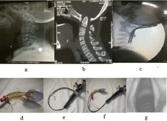
    Figure 1:  1a and b: Showing X-ray cervical spine and CT morphology before
surgery; c: X-ray cervical spine after surgery; d-g: shows the technique in a
situation of obliterated laryngeal axis as described. Step1: Proper Placement
& confirmation of ventilation and oxygenation via the Air Q® as a rescue
device. Step 2: preloaded Endotracheal Tube (ETT) was railroaded over the
tip of fiberscope till the bevel end of tracheal tube (Figure 1e &1f). Step 3:
The twin assembly (scope & tube) were advanced through the Air Q®, and
the endotracheal tube passed easily under vision through vocal cord in the
first attempt (Figure 1f &1g). Step4: Correct placement of tracheal intubation
was further confirmed by EtCO2 tracing. Air Q® was removed and ETT was
secured.
Discussion
Sometime, preplanned anticipated difficult airway may create a crisis [3]. Difficult airway should always be considered in patients presenting for surgery of the cervical spine. Laryngeal Mask Airways (LMA) is very useful in these situations especially the Intubating Laryngeal Mask Airway (I-LMA). The use of Air Q®-ILA guided tracheal intubation is found to have high success rate [4] particularly in patients with limited cervical spine mobility or minimal atlantooccipital extension [5,6].
The unique features in Air Q® (Figure 1d) LMA are the elevation ramp in airway outlet, which better approximates the airway anatomy for easy insertion; facilitates intubation and directs the tracheal tube towards the laryngeal inlet. Keyhole-shaped airway outlet elevates the epiglottis to provide unobstructed access to laryngeal inlet. Recessed mask tip approximates the shape of posterior pharynx which helps in easier positioning and improved airway alignment. An auxiliary hole improves airway flow in the event of partial airway obstruction by the epiglottis [7,8].
However, this technique further requires the guidance of tracheal tube curvature for intubation under direct vision by fiberscope. The technique reported here to is to create awareness among the anesthesiologist to modify the techniques of intubation according to clinical scenario. A similar technique was used in four other cases of cervical spine fracture with cervical (Table 1) without any difficulty in a safe and effective manner. Therefore, this technique can effectively deal with the patients of fixed cervical spine, improving the success rate and decreasing the chances of failed intubation.
![]()
    variable
    Case No.    1
    Case No.    2
    Case No.    3
    Case    No.4


    Age
    45
    35
    45
    60


    Sex
    M
    M
    F
    M


    Weight    (Kg)
    62
    70
    50
    65


    Height    (cm)
    162.5
    170
    130
    160


    Diagnosis
    C6-C7    subluxation
    C5-C6
    C5-C6
    C5-C6


    Size of    air-Q
    3.5
    3.5
    2.5
    3.5


    Number    of attempts for placement
    1
    1
    1
    2


    Time for    successful placement (sec)
    25
    20
    22
    35


    Fibreoptic    grade of view
    5
    2
    2
    4


    Fiberoptic    grade of view: 1, larynx only seen; 2, larynx and epiglottis posterior    surface seen; 3, larynx and epiglottis tip of anterior surface seen, < 50%    visual obstruction of epiglottis to larynx; 4, epiglottis downfolded and its    anterior surface seen, > 50% visual obstruction of epiglottis to larynx;    5, epiglottis downfolded and larynx cannot be seen directly.
Table 1:  Fiberoptic Tracheal Tube Intubation Using Air Q® In Cervical Spine
Fracture.
Acknowledgment
The authors acknowledge the contribution of Mr. Mohammad Asif (Aanesthesia Technician) who has made substantial contributions to the case by arranging and preparing the equipments and timely assistance.
References
- APAGBI Paediatric Airway Guidelines.
- Karim YM, Swanson DE. Comparison of blind tracheal intubation through the intubating laryngeal mask airway (LMA Fastrach) and the Air-Q. Anaesthesia. 2011; 66: 185-190.
- Girgis KK, Youssef MMI, and ElZayyat NS. Comparison of the air-Q intubating laryngeal airway and the cobra perilaryngeal airway as conduits for fiber opticguided intubation in pediatric patients. Saudi J Anaesth. 2014; 8: 470-476.
- Samir EM, Sakr SA. The air-Q as a conduit for fiberoptic aided tracheal intubation in adult patients undergoing cervical spine fixation: A prospective randomized study. Egyptian Journal of Anaesthesia. 2012; 28: 133-137.
- Air-Q: Quick Tips Reference Guide.
- Jagannathan N, Kozlowski RJ, Sohn LE, Langen KE, Roth AG, Mukherji II, et al. A clinical evaluation of the intubating laryngeal airway as a conduit for tracheal intubation in children. Anesth Analg. 2011; 112: 176-182.
- Jagannathan N, Roth AG, Sohn LE, Pak TY, Amin S, Suresh S. The new air-Q intubating laryngeal airway for tracheal intubation in children with anticipated difficult airway: a case series. Pediatric Anesthesia. 2009; 19: 618-622.
- Sohn LE, Jagannathan N, Sequera-Ramos L, Sawardekar A, Schaldenbrand K, De Oliveira GS. A randomised comparison of free-handed vs air-Q assisted fibreoptic-guided tracheal intubation in children < 2 years of age. Anaesthesia. 2014; 69: 723-728.