
Case Report
Austin Neurol. 2016; 1(1): 1005.
Successful Thrombolysis of a Stroke Patient beyond the AHA Guidelines
Rana SS¹, Saini N¹, Goel D²* and Rohatgi S²
¹Resident, Max Institute of Neurosciences Dehradun (MIND), India
²Consultant Neurology, Max Institute of Neurosciences Dehradun (MIND), India
*Corresponding author: Deepak Goel, Principal Consultant Neurology, Max Institute of Neurosciences Dehradun (MIND), State Uttarakhand, India
Received: August 09, 2016; Accepted: August 31, 2016; Published: September 02, 2016
Abstract
Intravenous thrombolysis is approved and a proven treatment for acute ischemic stroke in the window period of 4.5 hours. The therapeutic benefit is not extended to patients beyond the window period as they are excluded in the protocol for thrombolysis given by AHA. We report a case of successful IV thrombolysis in a 43-year-old female presented with acute onset right sided weakness and slurring of speech, 5.5 hours post onset of symptoms. She also had concurrent DVT and Pulmonary embolism. IV tissue plasminogen activator was administered 14 hours post onset of symptoms for PE. The patient’s neurological and respiratory status both improved following thrombolysis without any complications.
Keywords: Acute stroke; Thrombolysis; Guidelines
Case Report
A 43 years old right handed female, presented at around 2:11pm on 27th February, 2016 with history of acute onset, right sided weakness and slurring of speech at around 8am while climbing a hill. She felt that she was breathless, followed by light headedness and fell on the ground without any loss of consciousness. She had weakness of right side of the body and was unable to move her right upper and lower limb. She was a diagnosed case of bronchial asthma on/off medication. There was no history of diabetes mellitus, hypertension, or oral contraceptive intake. She had given history of previous 2 spontaneous abortions at less than 10 weeks of gestation.
On examination she was pale, tachypnoeic, and her extremities were cool and cyanotic with a respiratory rate of 32 per minute, a normal volume pulse of 140 beats per minute, and a blood pressure of 136/70mm Hg. She was maintaining a Spo2 of 70% on room air and 94% at 15 l of O2. On Central nervous examination, her speech was incomprehensible, right 7th upper motor neuron facial palsy and right sided hemiplegia with power of 2/5. Her NIHSS score on arrival was 15. Her MRI brain showed acute infarction in Left middle cerebral artery distribution (Figure 1). Full blood count and routine chemistry were unremarkable. Hemoglobin level was 7.4gm/dl and blood picture was suggestive of chronic hypo chromic anemia.
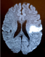
    Figure 1:  AXIAL MRI BRAIN DWI sequence showing restricted diffusion in
the left inferior frontal gyrus.
ECG showed a sinus tachycardia with p pulmonale and right bundle branch block. Chest X-ray was normal. A MRI brain scan demonstrated left fronto-parietal cerebral infarction. Bedside echocardiogram was remarkable for raised PA pressure, dilated RA and RV, mild tricuspid regurgitation and pulmonary arterial hypertension suggestive of pulmonary embolism. A CT pulmonary angiogram demonstrated multiple defects in bilateral pulmonary trunks (Figure 2). CT peripheral angiogram showed thrombus in the left femoral vein (Figure 3).
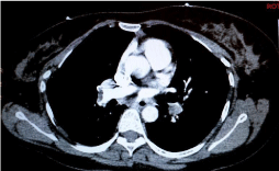
    Figure 2:  AXIAL CT PULMONARY ANGIOGRAM illustrating acute thrombus
involvement in bilateral main branches of pulmonary trunk extending into the
lobar and segmental branches.
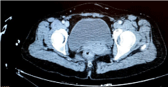
    Figure 3:  AXIAL CT PERIPHERAL ANGIOGRAPHY done to look for DVT
showing thrombus in left external iliac extending up to the common femoral
vein, superficial femoral and popliteal vein.
The patient was already beyond the window period for thrombolysis. In view of systemic venous, pulmonary arterial and systemic arterial embolism, a possibility of Anti-Phospholipid (APLA) Syndrome was kept, especially, in view of recurrent abortions in the past.
A contrast ECHO followed by Trans-esophageal echocardiography was done. There was no evidence of intramural thrombosis and no direct communication between the right and left side of the heart was demonstrated. Serum samples for thrombophilia profile, serum homocysteine, Prothrombin III, protein C, protein S, factor V Leiden, Anti-Nuclear Antibody (ANA), APLA and Anti-cardiolipin antibody ACLA were sent.
She was started on heparin infusion at the rate of 500 units/hour along with supportive treatment as she was beyond the window period for alteplase infusion. Same day evening, she became tachypnoeic, tachycardic, and de-saturated to spo2 of 80%. Having explained clearly all life threatening risks, thrombolysis with Alteplase was decided upon. She was administered 100mg alteplase according to the standard protocol of pulmonary embolism at 9:25pm of 27th February, 2016. The procedure was well tolerated. Her hemodynamic status improved markedly. And above all, clinically there was reversal of aphasia with power in the right side improving to 4/5 of MRC grade. Her NIHSS score 2 hours post thrombolysis was 3. The morning of 28th February she was extubated. Repeat NCCT brain scan showed no evidence of hemorrhage.
She continued to progress well without any deterioration over the next several days. Low hemoglobin was corrected by blood transfusion. Later, she was taken up for Inferior Vena Cava filter for secondary prevention. She got discharged in stable condition after 1 week on oral anticoagulants. One month later, on follow-up she was having mild upper limb weakness for fine movement. Her blood test for anti-cardiolipin antibody and other factors was negative.
Discussion
This case of young stroke with pulmonary embolism of unknown etiology was successfully treated with thrombolysis beyond window period. Etiology of stroke has shown a impact on outcome after thrombolysis as suggested by Anticoli, et al. 2016 [1]. In our patient, the etiology of stroke remains cryptogenic in spite of all investigations including transesophageal ECHO (to rule out PFO) being normal. Screening for thrombophilia also did not reveal any abnormality. She was not having any risk factors including diabetes, hypertension, hyperlipidemia or any factor for atherosclerosis. In young people, the substance abuse may represent a vascular risk factor for ischemic stroke. In cocaine users, the probability of stroke is up to 14 times greater than that in non-drug users (cardioembolism (43%) respect to large- artery atherosclerosis (18%) and small-vessel occlusion (21%) [2]. The patient and the family members denied any drug or substance abuse. In India cocaine abuse is very uncommon as compared to other countries and especially in women from rural background. Hence urine for toxicology panel is not routinely done.
The most important and effective treatment to improve the neurological outcome in a stroke patient is clot bursting therapy (Thrombolysis). However, this evidence based treatment is of limited utility, due to narrow window period and logistic problems pertaining to available health care system in India. An estimated, only about 7% of stroke patients reach hospitals, and out of them, mere 3% get the required thrombolysis in India. Our current guidelines, for thrombolytic therapy in acute stroke, have been under revision since May 2009. Again in March 2013, AHA/ASA guidelines were revised to expand the window from 3 to 4.5 hours [3-7]. However, this revision is not FDA approved.
Attempts to expand the window period, beyond 3 hours, were tried by many groups, only, after effectiveness of thrombolysis was proved, with immediate reversal of disabling stroke [8]. The major risk of delayed thrombolysis was the risk of intracranial bleeding that may be fatal sometime. The NINDS investigators reported a time to treatment interaction in a subgroup analysis. Treatment with rtPA started within 90 minutes of symptom onset was favorable as compared to patients who were thrombolysed between 90 to 180 minutes. A subsequent pooled analysis of all large multicenter, placebo controlled trials for acute stroke confirmed a time effect, but the upper limit of the treatment window may be as late as 5-6 hours [9].
Our patient was thrombolysed with high dose of alteplase, with the primary target being fatal pulmonary embolism, and not stroke, after 10 hours of stroke onset. Rapid reversal of neurological deficit was a surprise for us but was true. There are a number of authors who also have reported the use of thrombolytic therapy beyond the recommended period and have had success in their endeavor. Coster, et al. reported successful thrombolysis, 10 and 11 hours after onset of stroke in two young patients but with intra-arterial rout [10]. Naidoo P, et al. reported successful thrombolysis in a HIV positive patient who presented with 4 day old stroke and massive pulmonary embolism [11].
ASA/AHA guidelines also proposed a contraindication for thrombolysis, that a patient should not have had received heparin or anticoagulation in the last 48 hours. Our patient had been given heparin before alteplase, as thrombolysis was not planned till that time. Thus, this is a case of successful intravenous thrombolysis; 10 hours post stroke symptoms, having received heparin in the last 48 hours, with high dose of alteplase. Though a single case cannot be taken as an example to thrombolysis stroke patients beyond guidelines, but this case tells us that there are many hidden facts, that need to be explored, for effective thrombolysis in acute stroke. A large number of patients can therefore get the benefit out of it. Several phase 2 and one phase 3 trials has used multimodal CT or MRI to identify select 3- to 9-hour post onset patients who still harbor substantial salvageable tissue and are likely to benefit from late intravenous treatment [12,13]. This strategy appears highly promising but is not yet validated by an unambiguously positive phase 3 trials. Ideally perfusion images to be done before thrombolysis given beyond window period but it was not done primarily for ischemic stroke in our case. As is evident from the case above, it was initiated for pulmonary embolism. Hence DW-PW mismatch was not assessed in this case.
To summarize our case, I hereby present that in a young female, with probability of hyper-coagulation state, a delayed thrombolysis done for pulmonary embolism had the unintended benefit of resolution of the ischemic stroke as well. As I have already mentioned, as per the AHA/ASA guidelines the thrombotic treatment window period for acute stroke is still in evolution [14] and we are yet to identify the factors that were responsible for such an outcome. Therefore, we submit that delayed thrombolysis may be considered in young patients and extensive research should be conducted.
References
- Anticoli S, Bravi MC, Perillo G, Siniscalchi A, Pozzessere C, Pezzella FR, et al. Effect of cardioembolic etiology on Intravenous Thrombolysis Efficacy for Acute Inschemic Stroke. Curr Neurovasc Res. 2016; 13: 193-198.
- Siniscalchi A, Bonci A, Mercuri NB, De Siena A, De Sarro G, Malferrari G, et al. Cocaine dependence and stroke: pathogenesis and management. Curr Neurovasc Res. 2015; 12: 163-172.
- Diedler J, Ahmed N, Sykora M, Uyttenboogaart M, Overgaard K, Luijckx GJ, et al. Safety of intravenous thrombolysis for acute ischemic stroke in patients receiving antiplatelet therapy at stroke onset. Stroke. 2010; 41: 288-294.
- Donnan GA, Hommel M, Davis SM, McNeil JJ. Streptokinase in acute ischaemic stroke. Steering Committees of the ASK and MAST-E trials. Australian Streptokinase Trial. Lancet. 1995; 346: 56.
- Alexandrov AV, Molina CA, Grotta JC, Garami Z, Ford SR, Alvarez-Sabin J, et al. Ultrasound-enhanced systemic thrombolysis for acute ischemic stroke. N Engl J Med. 2004; 351: 2170-2178.
- Tsivgoulis G, Eggers J, Ribo M, Perren F, Saqqur M, Rubiera M, et al. Safety and efficacy of ultrasound-enhanced thrombolysis: a comprehensive review and meta-analysis of randomized and nonrandomized studies. Stroke. 2010; 41: 280-287.
- Jauch EC, Saver JL, Adams HP Jr, Bruno A, Connors JJ, Demaerschalk BM, et al. Guidelines for the early management of patients with acute ischemic stroke: a guideline for healthcare professionals from the American Heart Association/American Stroke Association. Stroke. 2013; 44: 870-947.
- Gregory WA. Expanding the window of thrombolytic therapy in acute stroke. The potential role of MRI for patient selection. Stroke. 1999; 30: 2230-2237.
- Hacke W, Donnan G, Fieschi C, Kaste M, Von Kummer R, Broderick JP, et al. ATLANTIS Trials Investigators, ECASS Trials Investigators, NINDS rt-PA Study Group Investigators. Association of outcome with early stroke treatment: pooled analysis of ATLANTIS, ECASS, and NINDS rt-PA stroke trials. Lancet. 2004; 363: 768–774.
- Coster S, Van Dijk LC, Treurniet FE, Van Overhagen H, Van Woerkom TC. Successful intra-arterial thrombolysis beyond the accepted 6-hour time window in two young patients. J Neurol Sci. 2010; 288: 182-185.
- Naidoo P, Hift R. Massive pulmonary Thromboembolism and stroke. Epub. 2011.
- Ducrocq X, Bracard S, Taillandier L, Anxionnat R, Lacour JC, Guillemin F, et al. Comparison of intravenous and intra-arterial urokinase thrombolysis for acute ischaemic stroke. J Neuroradiol. 2005; 32: 26-32.
- Berlis A, Lutsep H, Barnwell S, Norbash A, Wechsler L, Jungreis CA, et al. Mechanical thrombolysis in acute ischemic stroke with endovascular photoacoustic recanalization. Stroke. 2004; 35: 1112-1116.
- Stemer A, Lyden P. “Evolution of the thrombolytic treatment window for acute ischemic stroke”. Current Neurol Neurosci Rep. 2010; 10: 29-33.