
Research Article
Austin J Cerebrovasc Dis & Stroke. 2014;1(1): 1005.
Complex Visual Hallucinations Following Stroke: Epileptic origin or a Deafferentation Phenomenon?
Emre Kumral*, Arzu Uluakay and Ilknur Dönmez
Department of Neurology, Ege University Medical School Hospital, Turkey
*Corresponding author: Emre Kumral, Department of Neurology, Stroke Unit, Ege University Medical School, Bornova, Izmir 35100, Turkey
Received: July 07, 2014; Accepted: July 14, 2014; Published: July 16, 2014
Abstract
Objective: Complex and recurrent visual hallucinations are uncommon disorder in patients with visual pathway pathologic defects.
Patients: To describe 8 patients that were diagnosed as having Charles Bonnet syndrome (CBS) after having experienced complex visual hallucinations following stroke involving occipital lobe. Two of these patients also had epileptic seizures.
Results: Of 9560 patients with stroke in our Stroke Registry, 8 (0.08%) patients experienced a CBS following acute stroke. They had various complex visual hallucinations in their defective visual field. Hallucinations persisted for more than one month in half of the patients, and during hallucinations no electrographic seizures were recorded through a 24 hour video electroencephalographic monitoring.
Conclusion: Charles Bonnet syndrome (CBS) may develop in patients with stroke involving occipital lobe following ischemic or hemorrhagic events. Charles Bonnet syndrome (CBS) associated with unilateral or bilateral medial occipital lesion and epilepsy may coexist and reflects the abnormal functioning of an integrated dorsal occipitoparietal processing system and ventral occipitotemporal processing neuronal network.
Keywords: Charles Bonnet syndrome; Occipital lobe infarction; Hemorrhage; Visual hallucinations
Introduction
Charles Bonnet syndrome (CBS) is a disorder with visual field deficit and complex visual vivid, complex recurrent visual hallucinations in conscious patients. It has been described firstly by Charles Bonnet in 1760 [1]. Previously, Charles Bonnet syndrome has been found in association with variable pathologic conditions of the eyes and central visual pathways, and occipital lobe [2]. Occipital lobe lesion is an important cause in visual field deficit and elementary, simple hallucinations, whereas complex hallucinations of elaborate, complex images of objects including animals, people or landscape related to damage to the occipitotemporal and occipitoparietal visual association neocortex [3]. We describe herein 8 patients who experienced complex visual hallucinations following stroke with unilateral or bilateral lesions of the occipital lobe.
Methods
Between 2000 and 2012, 9560 patients with first-ever stroke were admitted to the Neurology Department of the Ege University Hospital and prospectively entered in our Ege Stroke Registry [4]. A total of 2390 patients with MRI-proven lesions restricted to the posterior circulation were identified. Among them, 217 patients had occipital lobe lesion. Patients with old hemorrhagic lesions, old infarcts on imaging, or simultaneous acute unilateral or multiple lesions were excluded. Patients with Charles Bonnet syndrome (CBS) having complex, non-stereotyped multimodal hallucinations following a vascular lesion in the occipital lobe differentiated from other possible causes of visual hallucinations in the elderly or the patients with delirium, seizure, psychiatric symptoms, alcohol or drug use, Parkinson’s disease, and dementia. The mental examination including time, place orientation was performed in all patients, and Mini-Mental State Examination (MMSE) was done by a clinical neuropsychologist in our department. Standard laboratory tests were performed in all patients. MRI was performed within 48 hours of admission by 1.5T or 3T scanners (Siemens Sonata, Siemens Medical Solutions, and Erlangen, Germany). MRI scanners consisted of axial T1- and T2-weighted spin-echo and T2 fluid-attenuated inversion recovery (FLAIR) and diffusion-weighted imaging (DWI). Video- EEG monitoring was performed in all patients to determine the cause of the visual hallucinations and to check if there were recurrent electrical discharges. The study was approved by the Ethic Committee of University Hospital, and all patients or their designated relatives provided informed consent.
Results
In our Stroke Registry, 9560 patients enrolled consecutively within first week of stroke in our Aegean region. Among them there were 8 patients with CBS (4 males and 4 females) with a mean of 65+12 years-old (range 45-82 years old). All patients are right-handed. Acute visual loss was seen in all patients in addition to headache in 4 patients and dizziness in 6 patients. In 24 hours they experienced different type of visual hallucinations as appearance of scenes with moving animals, known faces, trees, gestures, colors, with varied degrees of clarity depending on the stimuli in their visual fields (Table 1). They were able to give detailed descriptions of the hallucinations. Patient 2 and 7 reported images and sensation of ants walking on different parts of their bodies. No auditory or olfactory components were present and images were not related to their personal memories. They had no history of seizure, psychiatric symptoms, alcohol or drug use, Parkinson’s disease, Parkinsonism and dementia before admission. The mental-status examination revealed preserved orientation and consciousness. Mini-Mental State Examination score of patients were within normal limits. The only abnormality revealed by the neurological evaluations was a homonymous hemianopsia in 7 patients and tubular vision in one patient (no 7). The neuro-ophthalmologic examination revealed no other finding. The routine laboratory examination including serum electrolytes, glucose, liver and kidney functions were normal. In the first day of admission, Patient 2 and 7 had seizures, which were characterized by flexed posture of both upper extremities and secondary generalization. Brain magnetic resonance imaging (MRI) and diffusion MRI showed acute ischemic lesion in the posterior cerebral artery territory in seven patients, and one had a hemorrhagic lesion involving medial occipital region. There was no other lesion involving midbrain or other territories. Six patients had right sided lesion, one had left sided lesion and one had bilateral lesions. Lesions in the occipital lobe were mainly affected descendens gyrus and medial occipital gyrus (Figure 1). Patients 1, 5 and 7 had an interictal electroencephalogram (EEG) that showed regional spikes with most occurring in the temporo-occipital area. Hallucinations did not disappear completely and they persisted in the visual field over one month in half of patients (Patients 1,2,5,7). The hallucinations occurred when the patients were fully awake. They were not accompanied by loss of consciousness or abnormal behavior and persisted throughout the whole day. Antiepileptic drug was started in patients 2 and 7 having seizures, and EEG showed spikes and waves at T8 and C4 in patient 7. Electrographic seizures were not recorded when he was experiencing hallucinations. During follow-up, visual hallucinations ceased in all patients in 3 months without a specific treatment, and only visual field deficits remained without change as sequelae.
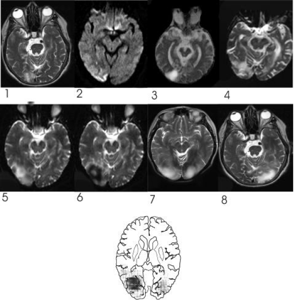
Figure 1 : T2-weighted or diffusion MRIs demonstrated unilateral ischemic lesion in territory of posterior cerebral artery in 6 patients (nos.1, 2, 3, 4, 5, 8) and bilateral lesions in one patient (no. 7) and hemorrhagic lesion on the right occipital lobe in one patient (no. 6). Bottom schematic figure shows overlapping lesions affecting mainly gyrus descendens and medial occipital gyrus.
![]()
    Patient/    Age/Sex
    Pathology
    Lesion    localization
    Hallucination    characteristics
    EEG


    1. 60/M
    Ischemic    lesion
    Right    occipital lobe
    Green and    yellow colors, geometrical shapes
    Spikes and    waves at T4


    2. 69/M
    Ischemic    lesion
    Right medial    occipital lobe
    Faces, gestures, dressing
      and    movements of animate items
    Normal


    3. 77/M
    Ischemic    lesion
    Right medial    occipital lobe
    Faces,    landscapes, miniaturized persons doing various activities
    Normal


    4. 56/F
    Ischemic lesion
    Right medial    occipital lobe
    Shape, color, and size    changes and pygmy characters, vegetables, and small animals
    Normal


    5. 82/F
    Ischemic    lesion
    Right medial    occipital lobe
    Formed    unknown face hallucinations
    Spikes and    waves at T3


    6. 60/M
    Hemorrhagic
      lesion
    Right    occipital lobe
    Geometrical    shapes, coloured figures
    Normal


    7. 72/F
    Bilateral    ischemic lesions
    Bilaateral    occipital lobe
    Images of a    strange child, geometrical shapes, and and a human with colored eyes,    touching of ants
    Spikes and    waves at T8 and C4


    8. 45/F
    Ischemic    lesion
    Left medial    occipital lobe
    Formed    visual hallucinations
    Normal
Table 1: Characteristics of current and previous patients.
Discussion
In our Registry we found 8 patients who experienced complex visual hallucinations with acute stroke involving the occipital lobe. Our patients had typical CBS according to the diagnostic criteria of Gold and Rabins [5]. The main characteristics of hallucinations were complex visual hallucinations of geometrical figures and known faces, bizarre human movements and gestures, which occurred with the eyes open and do not appeared to be just illusions triggered by low lighting levels. Following impairment or loss of vision, hallucinations developed after a period of 24 hours, persisted without change of characteristics, and disappeared within following months in all patients. Pathophysiology of CBS is generally interpreted either as a release phenomenon (deafferentation) or it may be caused by sensory deprivation and “phantom vision,” or an abnormal focus activating neuronal network [6-8]. Its frequency in patients with structural damage such as infarction or vascular malformation has suggested that isolated lesions to the occipital lobe can lead to complex visual and somatosensory hallucinations. In our series, descendens gyrus and medial occipital gyrus were mainly damaged which carries information on spatial relations to the parietal lobe and object properties to the inferotemporal cortex [9]. Visual hallucinations in our patients may be developed by interruption of dorsal occipitoparietal processing system or ventral occipitotemporal processing stream that are related with perception of spatial functions and pattern discrimination and visual identification of objects [10,11].
Previous reports using SPECT, patients with optic nerve lesions or macular lesions [12,13] showed asymmetric hyperperfusions in the lateral temporal cortex, striatum, and thalamus, with visual hallucinations precipitated by excessive cortical compensation. Occipital hypoperfusion has also been suggested as a cause of CBS [13]. It is possible that clinicians may interpret visual hallucinations as part of the remaining visual aura in patients with occipital lobe epilepsy (OLE). However, a recent report found that complex hallucinations were never seen in patients with occipital lobe seizures [14]. We can exclude the possibility OLE in our patients because they had only complex hallucinations and interictal recordings did not demonstrate any epileptic activities. Although, intracranial recordings were not performed in our patients and small seizure foci could be missed during scalp-recorded EEG. It is obvious that any possibility of misdiagnosis can be prevented through a detailed medical history of the nature of the visual hallucinations and through video-EEG monitoring. The characteristics of our patients’ hallucinations were identical to those of CBS and they have persisted for more than one month, therefore we considered that they were unlikely caused by an epileptic activity. It is important to determine if the visual hallucinations are caused by persisting occipital lobe seizures or by newly developed lesion causing deafferentation of the occipital association areas.
References
- de Morsier G. [The Charles Bonnet syndrome: visual hallucinations in the aged without mental deficiency]. Ann Med Psychol (Paris). 1967; 2: 678-702.
- Beniczky S, Kéri S, Vörös E, Ungureán A, Benedek G, Janka Z, et al. Complex hallucinations following occipital lobe damage. Eur J Neurol. 2002; 9: 175-176.
- Kölmel HW. Complex visual hallucinations in the hemianopic field. J Neurol Neurosurg Psychiatry. 1985; 48: 29-38.
- Kumral E, Ozkaya B, Sagduyu A, Sirin H, Vardarli E, Pehlivan M, et al. The Ege Stroke Registry: a hospital-based study in the Aegean region, Izmir, Turkey. Analysis of 2,000 stroke patients. Cerebrovasc Dis. 1998; 8: 278-288.
- Gold K, Rabins PV. Isolated visual hallucinations and the Charles Bonnet syndrome: a review of the literature and presentation of six cases. Compr Psychiatry. 1989; 30: 90-98.
- Menon GJ, Rahman I, Menon SJ, Dutton GN. Complex visual hallucinations in the visually impaired: the Charles Bonnet Syndrome. Surv Ophthalmol. 2003; 48: 58-72.
- Siatkowski RM, Zimmer B, Rosenberg PR. The Charles Bonnet syndrome. Visual perceptive dysfunction in sensory deprivation. J Clin Neuroophthalmol. 1990; 10: 215-218.
- Schultz G, Melzack R. The Charles Bonnet syndrome: 'phantom visual images'. Perception. 1991; 20: 809-825.
- Ungerleider LG, Mishkin M. Two cortical visual systems. In: Analysis of visual behaviour. Ingle DJ, Goodale MA, Mansfield RJW, editors. MIT Press, Cambridge MA. 1982; 549-586.
- Nieuwenhuys R, Voogd J, Van Huijzen C. The human Central Nervous System. Springer-Verlag, New York. 2008; 604-605.
- Schultz G, Needham W, Taylor R, Shindell S, Melzack R. Properties of complex hallucinations associated with deficits in vision. Perception. 1996; 25: 715-726.
- Adachi N, Watanabe T, Matsuda H, Onuma T. Hyperperfusion in the lateral temporal cortex, the striatum and the thalamus during complex visual hallucinations: single photon emission computed tomography findings in patients with Charles Bonnet syndrome. Psychiatry Clin Neurosci. 2000; 54: 157-162.
- Kishi T, Uegaki J, Kitani M, Fujimoto A, Naganuma R. The usefulness of single photon emission computed tomography in Charles Bonnet syndrome: a case with occipital lobe involvement. Gen Hosp Psychiatry. 2000; 22: 132-135.
- Bien CG, Benninger FO, Urbach H, Schramm J, Kurthen M, Elger CE, et al. Localizing value of epileptic visual auras. Brain. 2000; 123: 244-253.