
Case Series
Austin J Cerebrovasc Dis & Stroke. 2015;2(1): 1034.
Difficulties in Making Decision on use of Intravenous Thrombolysis in Patients with Mild Ischemic Stroke Manifesting Isolate Cerebellar Syndrome
Piotr Sobolewski*
Department of Neurology and Stroke Unit of Holy Spirit Specialist Hospital, Poland
*Corresponding author: Piotr Sobolewski, Department of Neurology and Stroke Unit of Holy Spirit Specialist Hospital in Sandomierz, 13 Schinzla Str. 27-600 Sandomierz, Poland.
Received: March 25, 2015; Accepted: May 19, 2015; Published: May 30, 2015
Abstract
Background: Minor Stroke (MS) and Transient Ischemic Attack (TIA) collectively represent the largest group of cerebrovascular events. Many studies have demonstrated that after TIA or MS there is an approximately 10% risk of subsequent stroke within 90 days, including patients with symptoms from posterior circulation. The significant part of vertebrobasilar territory infarcts were in the cerebellum and in the posterior inferior cerebellar artery territory. In these cases the main symptoms are cerebellar syndrome ones. However, many of these patients have poor long-term outcome.
Aims: The aim of this review was discussing problem of use of rt-PA in patients with minor stroke, especially in the cases with ischemia in the posterior circulation and turning the attention to the patients with isolated cerebellar syndrome and patients with recurrent TIA in posterior circulation with milder symptoms.
Cases: To illustrate this difficult clinical problem three cases of patients with minor stoke in posterior circulation are presented. In two cases the procedures of the diagnosis and treatment were inappropriate and in one patient thrombolytic therapy were used and she had favorable long-term outcome.
Conclusion: Selected patients with isolated cerebellar syndrome in the course of cerebral ischemia should be treated with iv-thrombolysis. The use of MRI with DWI sequence can significantly help to make therapeutic decisions beneficial for patients.
Keywords: Mild stroke; Posterior circulation; iv-thrombolysis; rt-PA
Introduction
Minor Stroke (MS) and Transient Ischemic Attack (TIA) collectively represent the largest group of cerebrovascular events [1]. As endorsed by 2009 guidelines from the American Heart Association and American Stroke Association (AHA/ASA), TIA is defined as a transient episode of neurologic dysfunction caused by focal brain, spinal cord, or retinal ischemia, without acute infarction [2]. The term MS is often used for stroke patients with mild and nondisabling symptoms, however, there are use different definitions. Mild ischemia implies a stroke mechanism which involves a small volume of brain and recovery from previous more severe injury or that brain injury has been well compensated for by collateral perfusion [3]. The incidence of diagnosis of MS, according to definition, is from 18,9% to 54,9% of all patients with ischemic stroke [3].
Many recent studies have demonstrated that after TIA or MS there is an approximately 10% risk of subsequent stroke within 90 days [4-9]. These categories of stroke are also associated with high rates of short-term and long-term disability and death [10].
Among 407 New England Medical Center Posterior Circulation registry patients, 59% had strokes without transient ischemic attacks (TIAs), 24% had TIAs then strokes, and 16% had only TIAs [11]. Posterior circulation territory stroke accounts for 20% of all strokes [12]. Traditionally, posterior circulation stroke and TIA have been thought to have a lower recurrent stroke risk than other types of stroke. However, other studies and meta-analysis have shown that the risk is as high as that seen in anterior circulation stroke [13,14] and that there is a threefold increased risk of stroke after posterior circulation TIA or minor stroke in patients with symptomatic vertebrobasilar stenosis than in those without stenosis [14-16].
In the posterior stroke patients there are symptoms associated with cerebellum, brain stem and occipital cortex damage. A large single center series of 1000 patients reported that 7% of vertebrobasilar territory infarcts were in the cerebellum and 36% in the posterior inferior cerebellar artery territory [17]. In the course of stroke in such locations cerebellar syndrome symptoms, nausea and vomiting dominate [18]. In these cases, it is difficult to make the right decision on how to diagnose and how to treat patients is most difficult. Lack of deficits from the brain stem often puts to sleep alertness the clinician’s to make decision.
Posterior circulation stroke (POCI) is diagnosed on the basis of history and clinical examination, assisted by imaging. In the neurological evaluation of these patients the generally applicable the National Institutes of Health Stroke Scale (NIHSS) is not perfect [19]. The Israeli Vertebrobasilar Stroke Scale (IVBSS) may be more helpful [20].
Non-contrast Computed Tomography (CT) is still the mainstay of investigation for TIA and MS at most stroke centers. Magnetic Resonance Imaging (MRI) with Diffusion Weighted Imaging (DWI) is the brain imaging modality of choice for suspected damage of brain in the course of TIA or MS, especially in POCI stroke. MRI is superior to CT for demonstrating focal ischemic changed in small-volume lesions [21]. MRI is far more sensitive than CT in the diagnosis of acute ischemic stroke for all vascular territories, 80-95% sensitivity in the first 24 hours when DWI is used, versus 16% sensitivity with CT [21,22]. However, it is difficult to access MRI in many countries, especially in the hyperacute phase.
Prolonged Transcranial Doppler (TCD) with emboli detection is also useful in the assessment and risk stratification of TIA and MS patients, mainly in patients with unstable atherosclerotic plaque in the large vessels [23-25].
Patients with milder or resolving symptoms, especially in posterior circulation, are often mistakenly considered poor candidates for revascularization. Specialist assessment and intravenous administration of rt-PA are slower in patients with POCI stroke compared with those with anterior circulation stroke, because of delayed or missed diagnosis [26,27]. Among other things, for this reason up to 43% of patients with mild or resolving deficits are not offered iv-thrombolysis, and almost a third of these patients will die or be dependent at hospital discharge [28,29].
To illustrate this difficult clinical problem three cases of patients with minor stoke in posterior circulation are presented. In two cases, the procedures of the diagnosis and treatment were inappropriate and in one patient thrombolytic therapy were used and she had favorable long-term outcome.
Cases Presentation
Case 1
A 42-year-old Caucasian male with a history of hypertension and dyslipidemia was on a maintenance dose of 75mg per day of aspirin. The patient was admitted with vertigo, ataxia, nausea, vomiting and horizontal nystagmus with fast phase toward the right of 205-min duration (National Institutes of Health Stroke Scale, NIHSS - 2 pts; IVBSS - 4 pts.). On admission, his blood pressure (BP) was 180/95 mmHg, and results of laboratory tests were normal. In the baseline CT early ischemic changes (IC) were not found. The patient was not treated with intravenous thrombolysis. In the following hours of hospitalization the patient experienced right hemiparesis and dysarthria. After 24 hours, persistence of neurological symptoms was observed (NIHSS - 6 pts.; IVBSS – 14 pts.). In the controlled MRI, performed 48-h after admission, small IC was located in the right cerebellum lobe and in the pons (Figure 1 a, b, c.). Cardioembolic stroke was diagnosed and anticoagulant therapy was enabled. 3 months after stroke onset, the patient was functionally independent (NIHSS – 1 pts.; IVBSS – 6 pts.; modified Rankin Scale – 2 pts.). The patient remained cerebellar syndrome symptoms.
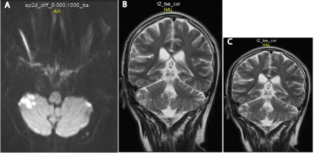
    Figure 1:  (A, B and C) Brain MRI scans of the 1st patient performed 48-h
after stroke onset.
Case 2
A 68-year-old Caucasian female with a history of hypertension, rheumatoid arthritis and chronic steroid therapy was admitted with vertigo, ataxia, nausea, vomiting and diplopia of 190-min duration (National Institutes of Health Stroke Scale, NIHSS- 1 pts.; IVBSS –12 pts). On admission, her blood pressure (BP) was 160/90 mm Hg, and results of laboratory tests were normal. In the baseline CT early IC were not found. Patients do not treat with intravenous thrombolysis. After 24 hours, persistence of cerebellar syndrome symptoms was observed (NIHSS - 6 pts.; IVBSS – 14 pts.). In the controlled MRI, performed at fourth day after admission, large IC was located in the left cerebellum lobe (Figure 2 a, b, c.). 3 months after stroke onset, the patient was functionally dependent (NIHSS – 1 pts.; IVBSS – 6 pts.; modified Rankin Scale – 3 pts.). Cerebellar syndrome symptoms remained in the patients and she walks using the walker.
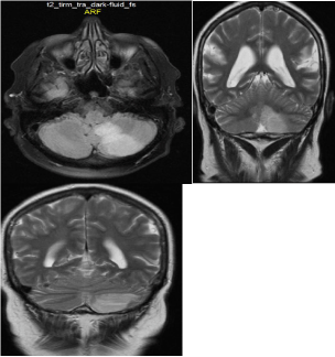
    Figure 2:  a,b,c: Brain MRI scans of the 2nd patient performed at 4th day after
stroke onset.
Case 3
A 48-year-old Caucasian female with a history of hypertension and smoking was admitted with vertigo, nausea, vomiting, dysarthria and mild disturbances of consciousness of 220-min duration (National Institutes of Health Stroke Scale, NIHSS - 2 pts.; IVBSS –12 pts). On admission, her blood pressure (BP) was 170/90 mm Hg, and results of laboratory tests were normal. In the baseline CT early ICs were not found; MRI-DWI showed early IC located in the pons (Figure 3 a. b. c.). At 330 min after symptom onset, she received 60 mg rt-PA (weight estimated at 66 kg) without complications. After 24 hours, patient was asymptomatic (NIHSS - 0 pts.; IVBSS – 0 pts.). Controlled CTs, performed 24-h and on 7th day after thrombolysis did not show any ICs or hemorrhagic transformation. 3 months after stroke onset, the patient was functionally independent (NIHSS – 0 pts.; IVBSS – 0 pts.; modified Rankin Scale – 0 pts.).
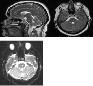
Figure 3: a,b,c: The baseline brain MRI scans of the 3rd patient.
Discussion
The results from randomized controlled trials and registers of patients in ischemic stroke showed that iv-thrombolysis with use of alteplase (recombinant tPA) improves functional outcome using the modified Rankin score (mRS) at three months [30-36]. However, many patients do not receive intravenous alteplase because of mild or improving stroke symptoms and the uncertain risk benefit ratio. A part of these patients have unfavorable outcomes and require further rehabilitation. About one third of acute stroke patients with rapid improvement of neurological deficit on arrival at the hospital develop severe subsequent deterioration [28,37]. Thus, withholding intravenous thrombolysis because of mild or improving symptoms may not always be justified.
In most randomized trials patients with POCI stroke were excluded from the study. There are only a few previously published studies evaluating safety and effectiveness of iv-thrombolysis in the posterior circulation strokes. In most of these studies rt-PA was administered in patients with basilar artery occlusion [38- 41]. A metaanalysis comparing intravenous versus intra-arterial thrombolytic treatment found that survival and outcome were roughly equal (24% vs. 22%of patients reached good outcomes) [39]. The previously published study by Förster et al. showed prolonged door to needle time in patients with posterior circulation stroke. This one-center study also showed that safety, mortality and incidence of intracerebral hemorrhage were similar in patients with stroke in anterior and posterior circulation [26]. In the Third International Stroke Trial (IST-3), a group of patients with POCI stroke treated with thrombolysis accounted 7% of all treated patients compared to 9% patients in a controlled group. There were no statistically significant differences between both groups regarding long-term outcome [34].
There are no studies evaluating safety and effectiveness of patients with milder or resolving symptoms in posterior circulation undergoing iv-thrombolysis. Planning and performing such studies would be very difficult.
I believe that discussing this clinical problem and presenting the negative cases, with respect to the clinical management, I turned the attention to the fact of too frequent exclusion of patients with isolated cerebellar syndrome and patients with recurrence TIA in posterior circulation with milder symptoms from iv-thrombolysis.
Conclusion
Selected patients with isolated cerebellar syndrome in the course of cerebral ischemia should be treated with iv-thrombolysis. The use of MRI with DWI sequence can significantly help to make a beneficial for patient therapeutic decision.
References
- Morgenstern LB, Lisabeth LD, Mecozzi AC, Smith MA, Longwell PJ, McFarling DA, et al. A population-based study of acute stroke and TIA diagnosis. Neurology. 2004; 62: 895-900.
- Easton JD, Saver JL, Albers GW, Alberts MJ, Chaturvedi S, Feldmann E, et al. Definition and evaluation of transient ischemic attack: a scientific statement for healthcare professionals from the American Heart Association/American Stroke Association Stroke Council; Council on Cardiovascular Surgery and Anesthesia; Council on Cardiovascular Radiology and Intervention; Council on Cardiovascular Nursing; and the Interdisciplinary Council on Peripheral Vascular Disease. The American Academy of Neurology affirms the value of this statement as an educational tool for neurologists. Stroke. 2009; 40: 2276-2293.
- Fischer U, Baumgartner A, Arnold M, Nedeltchev K, Gralla J, De Marchis GM, et al. What is a minor stroke? Stroke. 2010; 41: 661-666.
- Coull AJ, Lovett JK, Rothwell PM; Oxford Vascular Study. Population based study of early risk of stroke after transient ischaemic attack or minor stroke: implications for public education and organisation of services. BMJ. 2004; 328: 326.
- Hill MD, Yiannakoulias N, Jeerakathil T, Tu JV, Svenson LW, Schopflocher DP. The high risk of stroke immediately after transient ischemic attack: a population-based study. Neurology. 2004; 62: 2015-2020.
- Dennis MS, Bamford JM, Sandercock PA, Warlow CP. A comparison of risk factors and prognosis for transient ischemic attacks and minor ischemic strokes. The Oxfordshire Community Stroke Project. Stroke. 1989; 20: 1494-1499.
- Giles MF, Rothwell PM. Risk of stroke early after transient ischaemic attack: a systematic review and meta-analysis. Lancet Neurol. 2007; 6: 1063-1072.
- Wu CM, McLaughlin K, Lorenzetti DL, Hill MD, Manns BJ, Ghali WA. Early risk of stroke after transient ischemic attack: a systematic review and meta-analysis. Arch Intern Med. 2007; 167: 2417-2422.
- Johnston SC, Gress DR, Browner WS, Sidney S. Short-term prognosis after emergency department diagnosis of TIA. JAMA. 2000; 284: 2901-2906.
- Coutts SB, Hill MD, Simon JE, Sohn CH, Scott JN, Demchuk AM; VISION Study Group. Silent ischemia in minor stroke and TIA patients identified on MR imaging. Neurology. 2005; 65: 513-517.
- Caplan LR, Wityk RJ, Glass TA, Tapia J, Pazdera L, Chang HM, et al. New England medical center posterior circulation registry. Lancet Neurol. 2013; 12: 65-71.
- Bogousslavsky J, Van Melle G, Regli F. The Lausanne Stroke Registry: analysis of 1,000 consecutive patients with first stroke. Stroke. 1988; 19: 1083-1092.
- Flossmann E, Rothwell PM. Prognosis of vertebrobasilar transient ischaemic attack and minor stroke. Brain. 2003; 126: 1940-1954.
- Gulli G, Khan S, Markus HS. Vertebrobasilar stenosis predicts high early recurrent stroke risk in posterior circulation stroke and TIA. Stroke. 2009; 40: 2732-2737.
- Marquardt L, Kuker W, Chandratheva A, Geraghty O, Rothwell PM. Incidence and prognosis of > or = 50% symptomatic vertebral or basilar artery stenosis: prospective population-based study. Brain. 2009; 132: 982-988.
- Gulli G, Marquardt L, Rothwell PM, Markus HS. Stroke risk after posterior circulation stroke/transient ischemic attack and its relationship to site of vertebrobasilar stenosis: Pooled data analysis from prospective studies. Stroke. 2013; 44: 598-604.
- Bogousslavsky J, Van Melle G, Regli F. The Lausanne Stroke Registry: analysis of 1,000 consecutive patients with first stroke. Stroke. 1988; 19: 1083-1092.
- Searls DE, Pazdera L, Korbel E, Vysata O, Caplan LR. Symptoms and signs of posterior circulation ischemia in the new England medical center posterior circulation registry. Arch Neurol. 2012; 69: 346-351.
- Lyden P, Brott T, Tilley B, Welch KM, Mascha EJ, Levine S, et al. Improved reliability of the NIH Stroke Scale using video training. NINDS TPA Stroke Study Group. Stroke. 1994; 25: 2220-2226.
- Gur AY, Lampl Y, Gross B, Royter V, Shopin L, Bornstein NM. A new scale for assessing patients with vertebrobasilar stroke-the Israeli Vertebrobasilar Stroke Scale (IVBSS): inter-rater reliability and concurrent validity. Clin Neurol Neurosurg. 2007; 109: 317-322.
- Chalela JA, Kidwell CS, Nentwich LM, Luby M, Butman JA, Demchuk AM, et al. Magnetic resonance imaging and computed tomography in emergency assessment of patients with suspected acute stroke: a prospective comparison. Lancet. 2007; 369: 293-298.
- Edlow JA, Newman-Toker DE, Savitz SI. Diagnosis and initial management of cerebellar infarction. Lancet Neurol. 2008; 7: 951-964.
- Markus H. Monitoring embolism in real time. Circulation. 2000; 102: 826-828.
- Markus HS, MacKinnon A. Asymptomatic embolization detected by Doppler ultrasound predicts stroke risk in symptomatic carotid artery stenosis. Stroke. 2005; 36: 971-975.
- Valton L, Larrue V, le Traon AP, Massabuau P, Géraud G. Microembolic signals and risk of early recurrence in patients with stroke or transient ischemic attack. Stroke. 1998; 29: 2125-2128.
- Förster A, Gass A, Kern R, Griebe M, Hennerici MG, Szabo K. Thrombolysis in posterior circulation stroke: stroke subtypes and patterns, complications and outcome. Cerebrovasc Dis. 2011; 32: 349-353.
- Sarraj A, Medrek S, Albright K, Martin-Schild S, Bibars W, Vahidy F, et al. Posterior circulation stroke is associated with prolonged door-to-needle time. Int J Stroke. 2013.
- Barber PA, Zhang J, Demchuk AM, Hill MD, Buchan AM. Why are stroke patients excluded from TPA therapy? An analysis of patient eligibility. Neurology. 2001; 56: 1015-1020.
- Smith EE, Abdullah AR, Petkovska I, Rosenthal E, Koroshetz WJ, Schwamm LH. Poor outcomes in patients who do not receive intravenous tissue plasminogen activator because of mild or improving ischemic stroke. Stroke. 2005; 36: 2497-2499.
- [No authors listed]. Tissue plasminogen activator for acute ischemic stroke. The National Institute of Neurological Disorders and Stroke rt-PA Stroke Study Group. N Engl J Med. 1995; 333: 1581-1587.
- Hacke W, Kaste M, FieschiC, Toni D, Lesaffre E, von Kummer R, et al. Intravenous thrombolysis with recombinant tissue plasminogen activator for acute hemispheric stroke. The European Cooperative Acute Stroke Study (ECASS). JAMA. 1995; 274: 1017-1025.
- Hacke W, Kaste M, Fieschi C, von Kummer R, Davalos A, Meier D, et al. Randomised double-blind placebo-controlled trial of thrombolytic therapy with intravenous alteplase in acute ischaemic stroke (ECASS II). Lancet. 1998; 352: 1245-1251.
- Hacke W, Kaste M, Bluhmki E, Brozman M, Dávalos A, Guidetti D, rt al. Thrombolysis with alteplase 3 to 4.5 hours after acute ischemic stroke. N Engl J Med. 2008; 359: 1317-1329.
- IST-3 collaborative group, Sandercock P, Wardlaw JM, Lindley RI, Dennis M, Cohen G, et al. The benefits and harms of intravenous thrombolysis with recombinant tissue plasminogen activator within 6 h of acute ischaemic stroke (the third international stroke trial [IST-3]): a randomised controlled trial. Lancet. 2012; 379: 2352-2363.
- Wahlgren N, Ahmed N, Dávalos A, Ford GA, Grond M, Hacke W, et al; SITS-MOST investigators. Thrombolysis with alteplase for acute ischaemic stroke in the Safe Implementation of Thrombolysis in Stroke-Monitoring Study (SITS-MOST): an observational study. Lancet. 2007; 369: 275-282.
- Wahlgren N, Ahmed N, Errikson N, Aichner F, Bluhmki E, Dávalos A, et al; SITS-MOST Investigators. Multivariable Analysis of Outcome Predictors and Adjustment of Main Outcome Results to Baseline Data Profile in Randomized Controlled Trials: Safe Implementation of Thrombolysis in Stroke-Monitoring Study (SITS-MOST). Stroke.2008; 39: 3316-3322.
- Smith EE, Abdullah AR, Petkovska I, Rosenthal E, Koroshetz WJ, Schwamm LH, et al. Poor outcomes in patients who do not receive intravenous tissue plasminogen activator because of mild or improving ischemic stroke. Stroke. 2005; 36: 2497-2499.
- Lindsberg PJ, Soinne L, Tatlisumak T, Roine RO, Kallela M, Häppölä O, et al. Long-term outcome after intravenous thrombolysis of basilar artery occlusion. JAMA. 2004; 292: 1862-1866.
- Lindsberg PJ, Mattle HP. Therapy of basilar artery occlusion: a systematic analysis comparing intra-arterial and intravenous thrombolysis. Stroke. 2006; 37: 922-928.
- Strbian D, Sairanen T, Silvennoinen H, Salonen O, Kaste M, Lindsberg PJ. Thrombolysis of basilar artery occlusion: impact of baseline ischemia and time. Ann Neurol. 2013; 73: 688-694.
- Vergouwen MD, Algra A, Pfefferkorn T, Weimar C, Rueckert CM, Thijs V, et al. Basilar Artery International Cooperation Study (BASICS) Study Group. Time is brain (stem) in basilar artery occlusion. Stroke. 2012; 43: 3003-3006.