
Dermatology Clinic, University of Catania, Italy
*Corresponding author: Giuseppe Micali, Dermatology Clinic, University of Catania, A.O.U. Policlinico-Vittorio Emanuele, Via Santa Sofia, 78 - 95123, Catania, Italy
Received: August 06, 2014; Accepted: November 18, 2014; Published: November 20, 2014
Citation: Lacarrubba F, Verzì AE and Micali G. Dermatoscopy and Video Dermatoscopy in the Diagnosis and Therapeutic Monitoring of Plaque Psoriasis: A Review. Austin J Dermatolog. 2014;1(6): 1030. ISSN:2381-9189
Dermatoscopy and Video Dermatoscopy (VD) are two non-invasive techniques, which are widely used in dermatology in the differential diagnosis of pigmented skin lesions. These techniques have been demonstrated to be useful in a wide variety of skin disorders, including plaque-type psoriasis. In psoriatic skin, dermatoscopy (X10 magnification) can provide an overview of the vascular pattern, showing the presence of a typical feature consisting of uniformly distributed “dotted” or “pinpoint” red capillaries or “red dots”. VD, performed with magnifications ≥ X100, shows, in involved skin, the presence of dilated and twisted capillaries, with a “bushy” or “basket-like” appearance, which is regularly and homogeneously distributed throughout the entire lesion. These patterns were confirmed by several studies and correlate with the dilation of capillary vessels within the dermal papillae, one of the earliest detectable histological changes of psoriasis and typical in all stages of lesional development. In this paper, the role of dermatoscopy and VD in the diagnosis and therapeutic follow-up of plaque-type psoriasis will be reviewed and discussed.
Keywords: Psoriasis; Dermatoscopy; Video dermatoscopy; Vascular pattern; Diagnosis; Therapeutic monitoring
Psoriasis is a common inflammatory, chronic-relapsing, erythematous-desquamative dermatosis which affects about 3% of the overall population of the world [1]. The most common variant is psoriasis vulgaris or plaque-type; it appears as having red, roundish skin patches which are covered in silver/white scales. Psoriasis develops as a result of abnormal keratinocytes proliferation and differentiation, and is associated with: 1) local inflammation of the Th1/Th17-type response and 2) vascular modification. The latter represents one of the earliest detectable histological changes and is typical in all stages of lesional development. In early psoriatic lesions, vascular modifications include increased permeability, dilation of dermal capillaries and mild dermal edema. These features are followed by the development of dilated and slightly tortuous blood vessels and lymphatic capillaries within the elongated dermal papillae [2]. This phenomenon could be caused by the vasodilation and elongation of existing vessels, rather than by new vessel formation, because capillary density in lesional skin is similar to that of uninvolved skin. The endothelial cells in the extrapapillary portion of the venous limb undergo a division that leads to a venous configuration of the loop, with the intrapapillary venous limb being longer than the arterial limb [3]. Keratinocytes in lesional skin are a major source of pro-angiogenic cytokines in psoriasis, including the Vascular Endothelial Growth Factor (VEGF), the Endothelial Cell Stimulating Angiogenesis Factor (ESAF), the Platelet-Derived Endothelial Cell Growth Factor/Thymidine Phosphorylase (PDECGE/TP), the Tumor Necrosis Factor (TNF)-a, the Transforming Growth Factor (TGF)-a and the Platelet-Derived Epidermal Growth Factor (PDGF) [4-6]. Cutaneous microvascular modifications are essential for the maintenance of the clinical lesional state in psoriasis, because they provide cellular and tissue nutrition to hyperplastic keratinocytes and promote inflammatory cell migration [3].
Psoriatic vascular modifications are easily detectable by using two non-invasive techniques which are widely used in dermatology in the differential diagnosis of pigmented skin lesions: these are dermatoscopy and Video Dermatoscopy (VD). Dermatoscopy allows for an in vivo visualization of the morphological features of the skin, which are not discernible to the naked eye, and provides a magnified (X10) horizontal view of the skin structures up to the superficial/ medium dermis. Dermatoscopy is performed with a manual device (dermatoscope), which can be equipped with a non-polarized light source (allowing for a traditional contact technique with the use of immersion oil) and a cross-polarized one. Video Dermatoscopy (VD) is performed with a video-camera equipped with lenses, which provide higher magnification (i.e. up to X1000). Both dermatoscopy and VD have been demonstrated to be useful in the study of a wide variety of skin disorders, including inflammatory conditions such as psoriasis [7-9].
In the present article the role of dermatoscopy and VD in the diagnosis and therapeutic follow-up of plaque-type psoriasis will be reviewed and discussed.
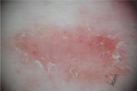
In psoriatic skin, dermatoscopy can provide an overview of the vascular pattern, showing the presence of a typical feature consisting of uniformly distributed “dotted” or “pinpoint” red capillaries or “red dots” (Figure 1). These dots represent the view from the top of the tortuous, ecstatic and elongated capillaries within the elongated dermal papillae [10].
The presence of red dots may also be observed in other inflammatory skin disorders (e.g. eczematous dermatitis, lichen planus, pityriasis rosea, pityriasis rubra pilaris and porokeratosis), but in these cases they are not uniformly distributed over the whole area. Moreover, different vascular arrangements and other dermoscopic features may provide useful hints for the correct diagnosis [11-17]. In a prospective study conducted on 83 patients with plaque psoriasis and 86 patients with dermatitis (n=41), lichen planus (n=25) and pityriasis rosea (n=20), each associated dermatoscopic pattern was determined and compared [12]. The combination of homogeneously distributed dotted vessels over a light red background associated with diffuse white scales allowed for a correct diagnosis of psoriasis with 88% of specificity and 84.9% of sensitivity. The authors of the study mentioned above suggested that, even though dotted vessels are common among all disorders, their arrangement and other associated criteria differ significantly among them. When dotted vessels appear in a patchy distribution together with yellow scales, they are indicative of dermatitis; when they appear in a peripheral arrangement with a yellowish background color, they are likely related to pityriasis rosea; and when they appear with a light red background color in combination with white crossing lines (Wickham striae), they are associated with lichen planus. In another retrospective observational study, 55 patients with scalp psoriasis and 41 patients with seborrheic dermatitis were enrolled to evaluate their distinctive dermatoscopic pattern. In scalp psoriasis, the main features discovered were red dots and globules (in 87% of patients), twisted red loops, and glomerular vessels. Instead, seborrheic dermatitis was associated with the presence of atypical red vessels (in 71% of patients), arborizing vessels, and featureless areas [17]. Finally, pityriasis rubra pilaris lesions showed round/oval yellowish areas surrounded by vessels of mixed morphology (i.e. linear and dotted) [13].
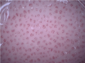
VD, performed with magnifications ≥ X100, works as a videocapillaroscope in the evaluation of psoriasis, showing the presence of dilated and twisted capillaries. These capillaries have a typically “bushy” or “basket-like” appearance, which is regularly and homogeneously extended to the entire lesion (Figure 2) [3]. This pattern was confirmed by several studies and correlates with the dilation of capillary vessels within the dermal papillae [18-23]. By using VD, one is able to assess the coursing of erythrocytes into the vascular lumen and to measure the diameter of each “bush”. This diameter generally ranges from 50 μm to 110 μm, which is much higher than the capillary loops of healthy skin (15-25 μm). At the edge of a psoriatic plaque, the capillary loops are elongated so as to form a “hairpin” shape, which is arranged in a parallel fashion to the skin surface. Importantly, VD examination should always be performed by gently applying a glass slide to the skin, in order to obtain a good visualization of the vascular structures. A limitation of the use of VD in psoriasis is represented by the presence of severe hyperkeratosis, which may hamper an optimal visualization of the capillaries. In this case, the typical vascular pattern should be searched in the erythematous areas of the lesion (e.g. periphery). Systems equipped with a 400-nm light source were employed to magnify vessel details and enhance vessel contrast compared to standard VD imaging [19].
VD appears to be a valid non-invasive aid in the diagnosis of psoriasis, particularly in cases where the clinical presentation is doubtful. This was demonstrated in hand, scalp and genital psoriasis, in which cases clinical diagnosis may sometimes be troublesome. In a study conducted on 32 patients with palmar and/or plantar erythematous scaly lesions, VD examination at X200 revealed a “bushy” aspect in 15 cases. This indicated a diagnosis of psoriasis which was later confirmed by histopathology. In the remaining 17 cases, in which there was no evidence of “bushy” capillaries, the histopathologic diagnosis was of spongiotic dermatitis [20]. Another study conducted on 90 subjects compared with one another capillary morphology in psoriasis, seborrheic dermatitis and normal skin of the scalp. Scalp psoriasis exhibited a “bushy” pattern, with a 73 +/- 17 μm diameter of capillary bushes. In contrast, scalp seborrheic dermatitis presented a multiform pattern, with mildly tortuous capillary loops and isolated dilated capillaries, but a substantial preservation of the local microangioarchitecture. The mean diameter of the capillary bushes was significantly lower (27 +/- 4 μm; p < 0.001) and similar to that of the scalp of healthy subjects (21 +/- 5 μm). The capillary loop density was similar among patients with psoriasis, seborrheic dermatitis, and healthy scalp skin (21). Genital psoriasis often represents a clinical dilemma especially in those cases in which other psoriatic lesions in the typical areas or elsewhere on the body are absent. In a study conducted on 12 patients with balanitis, the “bushy” pattern was observed (X100-X200) in only 6 patients with biopsy-proven psoriatic balanitis, while a non-specific vascular pattern consisting of dilated, linear, irregularly distributed capillaries, became apparent in the remaining cases of non-psoriatic balanitis (lichen sclerosus, Zoon balanitis and candidiasis) [22].
In addition to the results summarized above, VD was demonstrated to be diagnostically valid in clinically doubtful erythematous-desquamative lesions also in pediatric age. In an open comparative study, which was conducted on 24 children with clinical diagnosis of psoriasis, the presence of dilated capillaries with a “bushy” aspect, homogeneously distributed in all fields, was seen in all of the plaques considered. For an additional group of 36 children with other different erythematous-desquamative disorders, on the other hand, the VD findings were not specific, showing normal-looking capillaries, slightly dilated vessels, or a few isolated “bushes” [23].
The possibility to measure the diameter of “bushy” capillaries by VD resulted to be particularly useful in therapeutic monitoring. Several studies have demonstrated that the capillary diameter decreases after a positive response to various topical and systemic treatments (Figure 3) [24-33].
Regarding topical treatments, a study conducted by making use of VD (X200) evaluated the morphology of capillaries in psoriatic plaques both before and after treatment with tacalcitol ointment. Clinical evaluation was made after 3 and 6 weeks of therapy, with the following results: after 3 weeks, a reduction in erythema and scaling was noticed and, in some areas, the tortuosity of the capillaries decreased; after 6 weeks, the capillaries appeared to be less dilated and tortuous in the whole plaque and had lost the large and tortuous appearance of active psoriasis [25]. An additional study examined by means of VD 24 patients which were treated with mometasone fuoroate 0.1% once a day. At the end of the study, the diameters of the dilated and convoluted capillaries in the psoriatic plaque were significantly reduced (baseline, 69.2 μm; after 12 weeks, 29.3 μm; P<0.0001) in all subjects (26). Out of 24 patients, 12 showed clinical healing at the end of the treatment period, although the capillaroscopic picture returned to normal in 2 patients. Finally, in a study conducted on 30 patients, topical therapy with combined betamethasone dipropionate and calcipotriol induced a more marked decrease in erythema, infiltration and desquamation (P<0.001), and a significant reduction of the mean “bush” diameter (P<0.001) compared with betamethasone or calcipotriol alone [27].
With reference to the traditional systemic treatments of psoriasis, two studies evaluated the vascular pattern in psoriasis during treatment with oral cyclosporine. In the first study, 12 patients with psoriasis vulgaris were treated with a dose of 4 mg/kg/day over a period of 3 months with clinical and capillaroscopic (X300) assessments. A clinical resolution of the lesions and a reduction in the dimension of convoluted “basket-like” capillaries were observed in 70% of patients. The modification of the capillaroscopic aspects took place in a progressive manner in all patients with an average reduction of 64.8% in the “basket” diameter; however, none of these patients returned to a normal capillaroscopic pattern [28]. Similar results were observed in a different study, in which 20 patients with moderate-to-severe psoriasis were divided into two groups (A and B) based on PASI score. Patients belonging to Group A (PASI >16) were treated with 5 mg/Kg/day Cs for the first 4 weeks, and with 3 mg/ Kg/day Cs for an additional period of 4 weeks. Patients belonging to Group B (PASI 10-16) received 3 mg/Kg/day Cs for 8 weeks. At the end of the study, the normalization rate of the vascular pattern, which was assessed by VD at X150, appeared to be low in both groups (46% and 22% for Group A and Group B respectively), despite the fact that complete normalization by clinical and ultrasound assessment was observed in most cases [29].
With regard to biological treatments, the first study using VD was conducted in 2005 with infliximab on a 31-year-old man with active psoriasis. Two weeks after the first infusion, VD observation (X100-X200 magnification) showed some improvement of the capillary loops in the psoriatic lesions, which appeared as less tortuous and dilated and had an evident reduction in shape and size; after a second infusion, no “bushy” capillaries were detected in the same areas [30]. In another study, 18 patients with plaque-type psoriasis were treated with etanercept (50 mg/ twice/week) and the treatment evolution was monitored through clinical observation and VD. After 12 weeks of treatment, a significant reduction in VD measurements was observed in etanercept-treated patients [31]. A more recent study conducted on 22 patients, who were treated with etanercept 50 mg/week for 24 weeks, showed a significant mean reduction of the “basket” diameter from 71.88 μm to 29.43 μm. However, although 4 out of 22 patients showed complete recovery, none of these patients regained a normal capillaroscopic pattern [32].
In most of the reported studies, VD showed partial or no improvement compared to conventional clinical parameters. This finding may suggest that cutaneous microcirculation remains altered for a longer period of time even when the lesions are resolved [28]. VD, therefore, could shed further light on how the vascular response may affect the duration of therapy and relapse rates [29].
Regarding low-magnification (X10) dermatoscopy, no study has yet been performed in order to monitor the treatment response. However, it was used to evaluate the long-term safety of potent topical corticosteroids in patients with chronic psoriasis. In 20 patients who were treated with calcipotriene ointment and clobetasol propionate cream, it was observed that topical steroid overuse resulted in the evidence of clinically unapparent “red lines” (linear telangiectasias) in the treated plaques and/or surrounding skin [33].
In conclusion, both dermatoscopy and VD may be considered important adjunct diagnostic tools and relatively simple aids in clinically doubtful erythematous-desquamative lesions, allowing for a psoriatic vascular pattern to be confirmed or excluded, with some distinct advantages over skin biopsy.
Furthermore, the use of VD in the evaluation of psoriatic plaque, both before and after treatment, may be important in order to objectively demonstrate the effect of therapies on microcirculation. Whether the persistence of the vascular pattern after complete clinical clearing can be interpreted as a sign of incomplete remission and susceptibility to rebound may represent an interesting area for future research. Finally, a more thorough understanding of the vascular pattern of psoriasis could enable innovative and promising future therapeutic trends to be focused on angiogenic pathway modulation.
Dermatoscopy of a psoriatic plaque: homogeneously distributed dotted vessels over a light red background associated with diffuse white scales (X10).
Video dermatoscopy of a psoriatic plaque: dilated, elongated, “bushy” capillaries, homogeneously distributed throughout the entire lesion (X100).
Video dermatoscopy of a psoriatic plaque before (a) and after (b) 2 weeks of treatment with a topical corticosteroid: disappearance of “bushy” capillaries and normalization of the vascular network (X100).

Austin Publishing Group is an emerging open access publisher specialising in Science, Technology and Medicine is dedicated to serve the biomedical community through its initiatives. Austin Publishing Group is an academic publisher with 100+ peer reviewed open access journals in various subjects such as biomedical, Pharma, Life Sciences, Environmental, Engineering and Management. Austin Publishing Group publishes Open Access eBooks providing free access to vast scientific literature.