Case Report
Austin J Dermatolog. 2015;2(1): 1036.
Pilomatricoma: A Case Report
Jayakar Thomas*, Tamilarasi S, Asha D and Zohra Begum C
Department of Dermatology, Sree Balaji Medical College & Bharath University, India
*Corresponding author: Jayakar Thomas, Department of Dermatology, Sree Balaji Medical College & Bharath University, Chennai 600044, India
Received: April 05, 2015; Accepted: May 29, 2015; Published: June 02, 2015
Abstract
Pilomatricoma is a benign tumor that arises from hair follicle matrical cells. Involvement of the upper limb is relatively uncommon and can be mistaken for other soft tissue tumors. We report the case of a 12 year old boy, who presented with an asymptomatic firm nodule over the left arm whose histology was suggestive of Pilomatricoma.
Keywords: Pilomatricoma; Shadow cells; Ghost cells; Basaloid cells
Introduction
Pilomatricoma also known as Pilomatrixoma or calcifying epithelioma of Malherbe is a benign neoplasm, which is derived from hair follicle matrix cells. These tumors are typically present in the head and neck region, but also occur in the upper limbs and are rarely reported in other sites[1].Pilomatricoma represents as an asymptomatic, solitary, firm to hard, freely mobile nodule of the dermis or subcutaneous tissue. These tumors are generally exhibits no fixation to neighbouring tissues and have an osseous- or cartilagelike hardness [1-4]. The size of the tumor rarely exceeds 3 cm. The overlying skin may exhibit a bluish discoloration or ulceration [5].
We report a case of pilomatricoma over left arm. Additionally, we discuss the clinical features and histopathological features regarding pilomatricoma in the upper extremity.
Case Report
A 12-year-old boy presented with a 7 month history of insidious onset of an isolated mass over left arm. The mass was painless, progressively enlarging, not associated with itching and discharge. He denied any history of trauma over the site and no history of fever, chills, fatigue, weight loss, numbness and tingling.
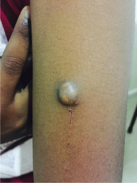
Physical examination revealed a 3cm by 2cm; non-tender, firm mass over the left arm. It was superficial and easily mobile (Figure 1). The neurovascular status of the left hand was noted to be intact; there were no other palpable masses in the extremities and no axillary adenopathy was present. Excision biopsy was performed under regional anesthesia. Grossly the tumor was white in appearance and well circumscribed.

    Figure 1:  Clinical photograph showing well circumscribed pigmented firm
nodule over left arm.
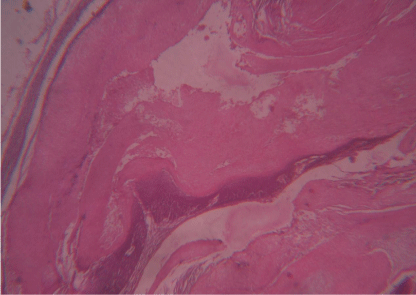
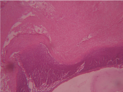
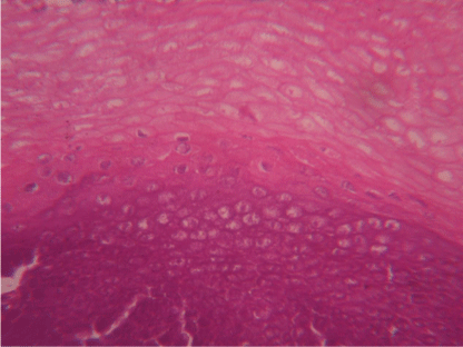
Histopathology revealed a capsulated benign neoplasm over the subcutis, composed of lobules ofirregularly shaped masses of ghost or shadow cells, scattered basophilic cells that undergo abrupt keratinization forming ghost [shadow] cells and areas of calcification. The basophilic cells were oval to polygonal shaped with ill-defined cell borders, a scanty cytoplasm, intensely staining basophilic nuclei with high nuclear-cytoplasm ratio. Ghost cells showed dark granularity and lack of staining in the area previously occupied by their nuclei (Figures 2,3 & 4).

    Figure 2:  Histology of skin [H&E section, magnification 10x 10] showing wellcircumscribed
encapsulated tumor mass in the dermis composed of islands
of basaloid cells and eosinophilic shadow cells.

    Figure 3:  Microscopic view [magnification 25x10] of pilomatricoma showing
shadow cells and basaloid cells.

    Figure 4:  Microscopic view [magnification 40x10] pilomatricoma revealing
ghost cell, transitional cells and basaloid cells.
Discussion
Pilomatricoma, or calcifying epithelioma of Malherbe, is a benign skin neoplasm, which arises from the matricial cells of hair follicle. Malherbe and Chenantais first described this tumor in 1880, referred to as the calcifying epithelioma, though it was thought to arise from sebaceous glands [1]. Later, Forbis and Helwig who further developed the electron microscopic and histochemical features of pilomatricoma discovered that the cell of origin is the outer sheath of the hair follicle root. These lesions are typically present in the head, neck and upper extremities, but also occur over the other areas of the body which is rarely reported. These tumors present most commonly in children and young adults [1,4,5].
Pilomatricoma usually presents as an asymptomatic nodular single lesion. The skin overlying the lesion is usually normal or may have bluish or reddish discoloration [5]. These lesions are characterized by well circumscribed spherical or ovoid shaped and sometimes encapsulated tumor mass [4,5]. There has been association between multiple lesions with Turner’s syndrome, Gardner syndrome, Steinert disease, myotonic dystrophy, Rubinstein-Taybi syndrome, and sarcoidosis [2,4]. Malignant transformation of pilomatricoma is rare [3,7].
The histopathological features of a pilomatricoma shows a welldemarcated tumor, which is surrounded by a connective tissue capsule. Generally, it is located in the dermis or subcutaneous layer. And the tumor is composed of islands of epithelial cells made up of varying amounts of basaloid matrical cells and often shows cystic change [1,3, 4]. As the tumor matures, there is degeneration of these basaloid cells, centrally. This is characterized by formation of anucleated shadow (or ghost) cells due to the central unstained areas of these cells [1,4]. In tumors of recent origin, numerous areas of basophilic cells are usually present, as the lesion ages, the number of basophilic cells decreases because of their transformation into shadow cells, and in long standing tumors, few or no basophilic cells remain [6]. It is important to note, that these ghost cells, are not unique to pilomatricomas. The presence of prominent inflammatory reaction, foreign body giant cells, central calcifications and keratin debris is also characteristic. With the von Kossa stain, calcium deposits are found in approximately 75%of the tumors [6]. Histopathologically it should be differentiated from trichilemmal cysts, which as they keratinize gradually lose their nuclei and often undergo calcification. The peripheral layer of basophilic cells however shows a palisading pattern where as basophilic cells of pilomatricoma do not [1,4,7].
The clinical differential diagnosis of these lesions should include sebaceous cyst, dermoid and epidermoid cysts, metaplastic bone formation, foreign body reaction, hematoma, trichoepithelioma and basal cell epithelioma [3,7]. Surgical excision is the recommended management. Recurrence is uncommon after adequate excision. All patients after surgical excision of tumor need dermatological evaluation with close long-term follow-up [2].
This case [pilomatricoma] is reported because of its rarity and to differentiate it from other soft tissue tumours.
References
- Birman MV, McHugh JB, Hayden RJ, Jebson PJ. Pilomatrixoma of the forearm: a case report. Iowa Orthop J. 2009; 29: 121-123.
- Marrogi AJ, Wick MR, Dehner LP. Pilomatrical neoplasms in children and young adults. Am J Dermatopathol. 1992; 14: 87-94.
- Samina Zaman, Sarosh Majeed, Fakeha Rehman. Pilomatricoma-study on 27 cases and review of literature. D:/Biomedica. 2009; 25: 69–72.
- Kaddu S, Soyer HP, Cerroni L, Salmhofer W, Hödl S. Clinical and histopathologic spectrum of pilomatricomas in adults. Int J Dermatol. 1994; 33: 705-708.
- Schweitzer WJ, Goldin HM, Bronson DM, Brody PE. Solitary hard nodule on the forearm. Pilomatricoma. Arch Dermatol. 1989; 125: 828-829, 832.
- peterson WC, Hult AM. CALCIFYING EPITHELIOMA OF MALHERBE. Arch Dermatol. 1964; 90: 404-410.
- Chuang CC, Lin HC. Pilomatrixoma of the head and neck. J Chin Med Assoc. 2004; 67: 633-636.