
Case Report
Austin J Dermatolog. 2017; 4(1): 1070.
Profuse Atypic Dermatophytosis in a Person Infected By HIV
Ouédraogo MS1,2*, Ouédraogo NA2,3, Andonaba JB4, Tapsoba GP1,2, Korsaga/Somé N1,2, Sakandé J2,5, Barro/Traoré F2, Niamba P1,2 and Traoré A1,2
1Department of Dermatology, Yalgado Ouédraogo University Hospital, Burkina Faso
2University of Ouagadougou I Pr Joseph Ki Zerbo, Ouagadougou, Burkina Faso
3Unit of Dermatology, Raoul Follereau Center, Burkina Faso
4Department of Dermatology, Souro Sanon University Hospital, Burkina Faso
5Department of Laboratory, Yalgado Ouédraogo University Hospital, Burkina Faso
*Corresponding author: Ouédraogo MS, Department of Dermatology, Yalgado Ouédraogo University Hospital, University of Ouagadougou I Pr Joseph Ki Zerbo, 04 BP 8201 Ouagadougou 04, Burkina Faso
Received: April 03, 2017; Accepted: May 12, 2017; Published: May 23, 2017
Abstract
Profuse dermathophytosis can be observed on people infected by HIV. The socio-economic context of Burkina Faso pushes the patients to primarily adopt traditional treatments that modify the clinical aspect of the lesions.
It was about a patient of 47 years old, a farmer, admitted at hospital for squamous pruritic skin lesions of the face and the chest, in a context of general alteration. The lesions were evolving since three years in a chronic and slowly extensive mode. A traditional treatment (black powder of Khaya senegalensis with shea butter) was done without any improvement of the lesions. The patient was seropositive to the HIV type 1, known since two years, and irregularly treated by TDF/ FTC/ EFV since six months with a voluntarily interruption one month ago. The examination showed large closets of various size skin flakes highly pigmented with clear limits, with circinate skin flakes borders on the face and the chest. The mycological examination of the skin flakes isolated a Trichophyton rubrum. The diagnosis of a circinate dermatophytosis profuse chronic modified by the traditional treatments to a person infected with HIV was established. The recovery was obtained after 3 months of antifungal local and general treatment.
The clinical atypia of this dermatophytosis was induced by the traditional treatments. The therapeutic vagrancy was responsible of the chronicity of this table. It is recommended to the people infected by HIV extended antifungal treatment in order to obtain an optimal efficiency. These patients are indeed a slow response to the treatments and a frequent recurrence.
Keywords: Circinate dermatophytosis; HIV; Traditional treatment
Abbreviations
HIV: Human Immunodeficiency Virus; ARV: Antiretroviral; TDF: Tenofovir; FTC: Emticitrabine; EFV: Efavirens; Mg: Milligram; °C: Celsius Degree; Kg/m²: Kilogram/Square Meter; Mm³: Cubic Millimeter; g/dl: Gram/Deciliter
Introduction
The dermatophytosis are mycosis cutaneous infections classified between the five cutaneous affections most frequently observed during the HIV infection [1-4]. Sixteen to 50% of these patients are concerned [2,5]. In Burkina Faso, a sahelian country situated in West Africa, Traoré and al in a hospital survey found that dermatophytosis were occupying the 2nd place of cutaneous infections on patients infected by HIV [4]. Their clinical aspect is variable. The profusion of the lesions is described on patients having less than 200 lymphocytes CD4/mm3 [1,2]. The resistance to the local and general antifungal and/or the relapses after the stopping of the treatment are frequent [1,5]. The traditional phytotherapyis the first solution for the Burkinabe people because it is lower cost than the modern treatments. However it can clinically modify the lesions and even be a source of some complications like eczema, irritation or over infection [6,7]. Khaya senegalensis and Butyrospermum parkii are plants with several properties. They are commonly used for traditional treatment of cutaneous diseases including dermatophytosis. We present a case of an atypical profuse dermatophytosis modified by a traditional treatment on a patient infected by HIV.
Case Presentation
A man of 47 years old was admitted on the 18th August 2014 for skin flakes on the face and the chest with general state altered. The starting was of 3 years by some skin flakes pruritic lesions of centrifugal extension on the chest, and later on the face, the inguinal fold and intergluteal fold. A traditional treatment was prescribed with Khaya senegalensis beverage to be drank, used in bath and in cataplasm (a black powder of barks mixed with shea butter Butyrospermum parkii) to be used on the lesions during one month. The disappearance of the lesions was not effective. An increase of the pruritus was observed. The patient was infected by HIV of type I, diagnosed since two years. He was irregularly treated in the health facility of the area where he was living. His Anti-Retroviral treatment (ARV) started 6 months ago was made of Tenofovir (TDF), Emticitrabine (FTC), Efavirens (EFV). He interrupted the treatment by his own will since a month. The examination showed large placards of variable size skin flakes on their surface with a high pigmentation very clear on the limits whose borders were skin flakes, circinated for most of them, except on the face and the front temporal area of the hair (Figure 1). The lesions were also visible on the neck, the posterior neckline (Figure 2), the antero lateral faces of the chest (Figure 3), the inguinal folds and the pubis (Figure 4), the intergluteal fold, the inferior cadrans of the right buttock, the left trochanterien area and the right knee. We didn’t notice any injury on the nails or palmoplantar zone. The patient was presenting a bad general aspect constituted by an infectious syndrome with a temperature at 38,5°C, a slimming with a body mass index at 14,34 kg/m² (18,5-25kg/m²), by pale conjunctiva and a deep asthenia. The tongue was clean without any injury. There was no edema of the lower limbs or notion of neither dysphagia nor chronic diarrhea. The neuropsychiatric exam was normal. We talked of a circinate profuse and chronic dermatophytosis modified by the traditional treatments on a person infected with HIV, a profuse seborrhea dermatitis and an erythema pellagroide. The blood count showed a normocytic normochromic anemia with an hemoglobin rate at 6,8g/dl (12-17g/dl), a leukopenia at 2300/mm³ (4000-10000/ mm3) with a lymphopenia at 575/mm³ (1500-4000/mm3). The number of lymphocytes CD4+ Tcell was of 22 cells/mm³ and the viral load of 390000 copies/ml. The mycological test of the skin flakes on two different areas (anterior face of the chest, face) showed the presence of mycelium filaments at the direct examination. The exam identified Trichophyton rubrum. The prescribed treatment was made of chlorexidine in foam solution flowing for the toilet, of ketoconazole cream to be applied once a day, an emollient cream made of glycerol, vaseline and paraffin liquid once a day, of ketoconazole tablets 200mg per day, of poly vitamins tablets (Alvityl® 1 tablet a day) and an hyper protidic regime. The evolution after three weeks was marked by a complete disappearance of the skin flakes. However, it was noted the presence of a vesicular erythematosus edge on the lesions of the chest (Figure 5), not elsewhere (Figure 6). After two months treatment, this active edge was persisting. Then, we prescribed sertaconazole cream in two applications per day and terbinafine tablet 250mg per day with a recovery of the lesions one month later.
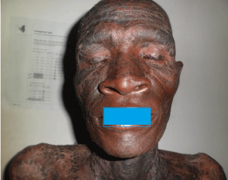
Figure 1: Dermatophytosis on the face.
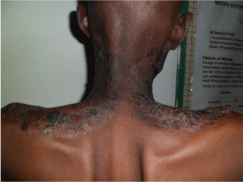
Figure 2: Dermatophytosis on the neck and the posterior neckline.
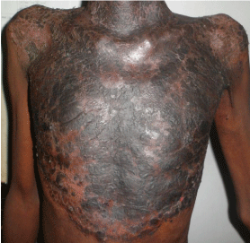
    Figure 3:  Dermatophytosis on the front face of the chest: large placard of skin
flakes very hyperpigmented on the surface.

Figure 4: Dermatophytosis on the inguinal folds and the pubis.
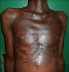
Figure 5: Control after 3 weeks of treatment : active edge below on the right.
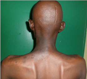
    Figure 6:  Control after 5 weeks of treatment : recovery and temporary
hyperpigmentation.
Discussion
We mention the case of a patient infected by HIV/AIDS for his atypical clinic presentation. It is about the nail injuries and the intertrigos of the small folds which are more revealed on this type of field [5]. The clinical Atypic of this dermatophytosis mainly the high pigmentation and the presence of skin flakes on the whole closets is due to the use of decoctions in baths and the local application of a cataplasm made of dark ash of Khaya senegalensis barks mixed with shea butter. Usually, the lesions present a central recovery [5]. In fact, the 2,6-dimethoxy-p-benzoquinone contained in Khaya senegalensis is known for its allergenicity, irritating effect. The shea butter used as basis or excipient for the cataplasm contains a terpenic derivative (triterpenes) also responsible of dermatitis and eczema [8]. That can explain the exacerbation of the pruritus during its use. The traditional drugs have an effectiveness (anti-inflammatory, antiseptic, antibacterial and pest control for Khaya senegalensis) [9] recognized on various affections. But their dosage and their conditioning probably require to be reviewed (according to the disease to be treated). So that they may have less secondary effects. Their bad use can cause damage to the skin but also modify the clinical aspect of the lesions and delay the diagnosis. Their secondary effects are also reported (eczema of contact, dermatitis of irritation, new appearance of pre-existing dermatitis like the psoriasis). In the most cases, in our African context (80% according to the World Health Organization) people also rely on it because of its low cost [8]. On our patient, the circinate border of some lesions was very significative of the diagnosis of circinate dermatophytosis and the location in the inguinal folds. The extent aspect is also explained by the immunodeprimated patient and also by the inadequation of the treatment and the delay at the specialized consultation. In fact, the profusion of the lesions is described for a certain number of lymphocytes CD4+ T cells inferior to 200 cells per mm3; the reaching of the hair also [1,5]. For our patient, there were about 22 cells/mm³. The Trichophyton rubrum identified on the lesions of our patient is the dermatophytosis mostly the one founded in our country on persons infected by HIV according to Zida and al [10]. It is also one of the pathogenic agent most frequently met on this type of patient [1,5,11]. The profuse seborrheic dermatitis was raised in front of the macular high pigmented aspect of the lesions, the topography on the face (mainly the arcades and the ciliary edge) and the front temporal area of the hair. The erythema sometimes takes an aspect highly pigmented on patients of phototype VI. Only the skin flakes were dry and large. On the persons infected with HIV, the dermatophytosis can have a misleading aspect of seborrheic dermatitis on the face and the chest. The effectiveness of the antimycosis on the lesions without addition of dermocorticoïdeis not in favor of the seborrheic dermatitis, in addition to the isolation of Trichophyton rubrum. The erythema pellagroïde [12], also raised because of the highly pigmented skin flakes of the lesions, the reaching of the photo exposed areas as the face, the hair, the neck, the neckline, the fixed limits and the slimming of the patient. It can be explained by the immune depression but also by a bad nutritional status. In fact, in our country, the basic food is mainly made of corn flour, millet and rice very poor in proteins. Some cases of pellagra have already been reported [13]. There was no notion of ethylism in the medical history of the patient, nor chronic diarrhea or drugs taking disrupting the metabolism of vitamin PP. The patient was not presenting any neurosensory troubles. An association erythema pellagradermatophytosis could be mentioned. But the vitamin tablets given (Alvityl®) contain a few quantity of vitamin PP (8mg), insufficient to treat pellagra. The skin rash lesions of the patient did not completely declined without adjunction of vitamin PP. Concerning the failure of ketoconazole, it can be explained either by a non-observation of the treatment or by a resistance of Trichophyton. The resistance to antifungals is described on person infected by HIV, requiring long durations of treatment [1,5].
This case reminds the problematic of the use of traditional pharmacopeia on the skin and the complications inherent in our society (mainly in the rural area). In fact, the modification of the lesions that it induces can be a source of a diagnostic delay. Then the confidence in it can obstruct the taking care of the chronic diseases as the HIV infection. In our context, the physicians should systematically look for an examination of the use of traditional products. The users of traditional pharmacopeia should be informed of the existence of secondary effects induced by these drugs. It is the opportunity here to draw the attention of the traditional practitioners on the necessity to improve the formulation and conditioning of the products used.
References
- Janier M, Caumes E. Manifestations dermatologiques de l’infection par le virus de l’immunodéficience humaine. EncyclMédChir, Dermatologie. 2002; 17.
- Monsel G, Ly F, Canestri A, Diousse P, Ndiaye B, Caumes E. Prévalence des manifestations dermatologiques chez les malades infectés par le VIH au Sénégal et association avec le degré d’immunodépression. Ann Dermatol Vénéréol. 2008; 135: 187-193.
- Bekkali S, Oumakhir N, El Kesouati J, Ghfir M, Sedrati O. Prévalence des manifestations dermatologiques chez les patients infectés par le VIH au service de dermatologie de l’HMIMV et association avec le degré d’immunodépression. Ann Dermatol Vénéréol. 2013; 140: S35-S39.
- Traoré A, Andonaba JB, Somé A, Niamba P, Barro F, Yaméogo I, et al. Les manifestations dermatologiques au cours de l’infection par le VIH au Centre Hospitalier Universitaire SouroSanou de Bobo Dioulasso : à propos de 203 cas. Burkina Médical. 2005; 8: 51-63.
- M Feuilhade de Chauvin. Mycoses métropolitaines. EncyclMédChir, Dermatologie. 2000; 98-380-A-10- 11p.
- Annuaire statistique du ministère de la Santé Burkina Faso. 2014; 317.
- Korsaga-Somé N, Tapsoba P, Ouédraogo MS, Ilboudo LB, Barro-Traoré F, Niamba P, et al. Nécrolyse épidermique liée à l’application cutanée d’une solution d’hydroxyde de potassium. Pan Afr Med J. 2015; 21: 299-303.
- Niang SO, Tine Y, Diatta BA, Diallo M, Fall M, Seck NB, et al. Negative cutaneous effects of medicinal plants in Senegal. BJD. 2015; 173: 26-29.
- Nikiema A, Pasternak D. Khaya senegalensis (Desr.). Juss A. In: Louppe D, Oteng-Amoako AA, Brink M, (Editors). PROTA (Plant Resources of Tropical Africa / Ressources végétales de l’Afriquetropicale), Wageningen, Netherlands. 2008.
- Zida A, Sawadogo PM, Diallo I, Tapsoba H, Bazié Z, DraboYJ, et al. Aspects épidémiologiques des mycoses cutanéophanériennes chez les patients infectés par le VIH au Centre national de référence du Burkina Faso, Afrique de l’Ouest. J Myc Med. 2016; 26: 133-137.
- Munoz-Pèrez MA, Rodriguez-Pichardo A, Camacho F, Rios JJ. Extensive and deep dermatophytosis caused by Trichophyton mentagrophytes var. Interdigitalis in an HIV-1 positive patient. JEADV. 2000; 14: 61-63.
- Pitche PT. Pellagre et érythèmes pellagroïdes. Cahiers Santé 2005; 15: 205- 208.
- Barro/Traoré F, Diallo B, Tapsoba P, Andonaba JB, Kéré M, Niamba P, et al. La pellagre : aspects épidémiologiques et cliniques dans la région ouest du Burkina Faso. Our Dermatol Online. 2013; 4: 479-483.