Case Report
Austin J Otolaryngol. 2014;1(2): 2.
Blue Tongue as a Presenting Sign of Primary Adrenal Insufficiency
Counts SM and Larsen CG*
Department of Otolaryngology, Head and Neck Surgery, University of Kansas Medical Center, Kansas City, USA
*Corresponding author: Christopher Larsen, M.D, Department of Otolaryngology, Head and Neck Surgery, University of Kansas Medical Center, 3901 Rainbow Blvd. MS 3010, Kansas City, KS 66160
Received: July 10, 2014; Accepted: August 29, 2014; Published: September 02, 2014
Primary adrenal insufficiency, also known as Addison’s disease, is a rare endocrine disorder characterized by inadequate production of the adrenal hormones cortisol and aldosterone. The presentation of this clinical disorder is often insidious, and thus difficult to recognize, leading to potentially fatal outcomes. Patients often present with lethargy, shock, and hyperkaliemia with hyponatremia. We describe herein the first reported case, to our knowledge, of a patient with blue tongue as an early presenting sign of adrenal insufficiency.
A 49 yo woman was referred to the Department of Otolaryngology at the University of Kansas Medical Center for evaluation of bluish discoloration in her tongue. She first noticed this bluish discoloration on her tongue 6 months earlier. There was slow progression of discoloration onto her buccal mucosa. She was completely asymptomatic, denying any pain, dysphagia, bleeding, or fatigue. A previous workup had revealed mild anemia, mildly elevated LFT’s, and some intermittent hyponatremia. Colonoscopy, which was performed to rule out Peutz-Jaghers syndrome, was negative. Her medical history was notable for depression and a distant seizure episode for which she was no longer on antiepileptic medication.
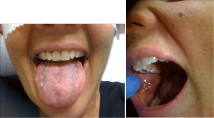
Physical examination revealed patches of bluish-brown discoloration throughout the dorsal tongue and bilateral buccal mucosa (Figure 1). She was also noted to have a slightly tanned appearance to her skin, which she attributed to a recent trip to Hawaii. Some small dark spots were noted in the iris. The remainder of her head and neck examination was within normal limits.
Figure 1 : Blue discoloration in tongue and oral mucosa of a 49 year-old patient.
An office-based biopsy of representative oral mucosal lesion was taken. Serum copper level was mildly elevated but ceruloplasmin was normal (ruling out Wilson’s disease). The biopsy showed “mild chronic inflammation with melanin pigment incontinence and no copper deposition.”
Approximately 2 months later, the patient presented to the Emergency Room complaining of extreme fatigue and malaise. She had significant hypotension, and a basic metabolic panel revealed significant hyponatremia and hyperkaliemia, leading to the diagnosis of primary adrenal insufficiency.
Comment
Oral mucosa discoloration is a relatively uncommon presenting chief complaint, making the evaluation and diagnosis often challenging. The differential diagnosis is widely variable, ranging from lesions as benign as amalgam tattoo and bismuth ingestion to more serious conditions including mucous membrane melanoma, angiokeratoma, and Addison’s disease (Table 1).
    Condition
    Symptoms,    signs
    Diagnosis


    Amalagam    Tatoo
    Isolated    blue, grey, or black macules on the gingivae, the buccal and alveolar mucosa,    the palate, and/or the tongue.
    Clinical    history and physical exam, histopathologic findings of blue or brown pigment


    Argyria    (Silver Ingestion)
    Generalized    bluish-grey discoloration of the skin, nails, and mucous membranes.  Sun exposed areas show greater    discoloration.
    Elevated    serum levels of silver, deposits of silver granules on histology [4]


    Central    Cyanosis
    Bluish    discoloration of the skin, lips, tongue, nails, and mucous membranes.
    Poor arterial    oxygenation


    Blue rubber    bleb nevus syndrome
    Multiple    bluish lesions of the skin and oral mucosa
    Histopathologic    evidence of tortuous, blood filled ecstatic vessels
Table 1: Differential Diagnosis for blue discoloration of the tongue or oral mucosa.
Primary adrenocortical insufficiency was first described by Thomas Addison in 1855 as decreased glucocorticoid and mineralocorticoid function secondary to destruction or dysfunction of the entire adrenal cortex. Morbidity and mortality associated with this condition are often secondary to delay in diagnosis and resultant Addisonian Crisis [1]. Unfortunately, the onset of disease is commonly insidious and nonspecific, typically occurring when 90% or more of the adrenal cortices are destroyed or dysfunctional [2]. A recent survey of adults with Addison’s disease suggested that 60% of these patients sought care from >2 physicians before the diagnosis was made [1].
Hyperpigmentation of the skin and/or mucous membranes is frequently the first presenting symptom. However, to our knowledge blue discoloration of the tongue and mucous membranes has not been previously reported. Mucous membrane hyperpigmentation has usually been reported as brown, tan, or dark spots caused by excess adrenocorticotropic hormone stimulating melanocytes to produce melanin. Other common presenting symptoms include dizziness with orthostasis due to hypotension, progressive weakness, fatigue, poor appetite, and weight loss [3].
References
- Ten S, New M, Maclaren N. Clinical review 130: Addison's disease 2001. J Clin Endocrinol Metab. 2001; 86: 2909-2922.
- Alevritis EM, Sarubbi FA, Jordan RM, Peiris AN. Infectious causes of adrenal insufficiency. South Med J. 2003; 96: 888-890.
- Barnard C, Kanani R, Friedman JN. Her tongue tipped us off. CMAJ. 2004; 171: 451.
- Kim Y, Suh HS, Cha HJ, Kim SH, Jeong KS, Kim DH, et al. A case of generalized argyria after ingestion of colloidal silver solution. Am J Ind Med. 2009; 52: 246-250.
