
Case Report
Austin J Otolaryngol. 2014;1(2): 2.
Temporal Arteritis Presenting as Anterior Tongue Necrosis: A Case Presentation
Patel HH* and Garritano FG
Department of Surgery, Penn State Hershey Medical Center, USA
*Corresponding author: Hetal H Patel, Division of Otolaryngology-Head and Neck Surgery, Department of Surgery, Penn State Hershey Medical Center, USA
Received: August 04, 2014; Accepted: September 17, 2014; Published: September 24, 2014
Abstract
Temporal Arteritis is the most common vasculitis affecting older individuals and presents in multiple different ways, most commonly with symptoms such as fever, headache, scalp tenderness, jaw claudication, and acute change in vision. One known but infrequently reported complication of temporal arteritis is tongue claudication, ischemia and necrosis. This is rarely seen because of the robust and bilateral vascular supply to the tongue musculature. The following case of temporal arteritis presenting as tongue necrosis is reported to highlight the clinical presentation of temporal arteritis with primary tongue involvement. The patient in this case lost a large portion of the anterior tongue as a result of the disease, causing significant dysarthria, dysphagia, and necessitating gastric tube feedings, an outcome which may potentially be preventable in other patients with early diagnosis and appropriate treatment.
Keywords: Vasculitis; Giant cell arteritis; Temporal arteritis; Tongue ischemia; Tongue necrosis
Abbreviations
CT: Computed Tomography; ANA: Anti-Nuclear Antibody; HIV: Human Immunodeficiency Virus; ESR: Erythrocyte Sedimentation Rate; CRP: C-Reactive Protein; MRI: Magnetic Resonance Imaging
Case Presentation
A 68 year old woman presented initially to her primary care physician with bilateral neck edema and pain in the submandibular area. She was diagnosed with sinusitis and treated with amoxicillin. Approximately ten days later she had persistent symptoms and also noted difficulty with tongue mobility, dysarthria, and dysphagia. Because of this the patient presented to an acute care facility for evaluation. Computed Tomography (CT) was performed and found to be concerning for sialadenitis and she was also noted to have an associated leukocytosis on blood chemistry (see Figure 1). She was admitted to the facility and treated with intravenous vancomycin and, cefepime in addition to IV dexamethasone to help reduce tongue edema. A repeat CT scan was performed on treatment day 5 and resolution of the sialadenitis was reported (Figure 1). The patient was transitioned to oral antibiotics and noted to have improvement of her leukocystosis but continued to have tongue edema and pain, dysarthria, dysphagia, and neck pain. She also began to have a gray discoloration of her tongue. Further workup included ANA and HIV titers in addition to evaluation of complement levels, all of which were found to be normal. Biopsy of the tongue was performed to evaluate for the presence of amyloidosis or malignancy but was negative. The patient was noted to have an elevated erythrocyte sedimentation rate (ESR) and C-reactive protein (CRP) level, 55 and 6.58 respectively. Treatment for angioedema and thrush were given also yielding no improvement. Because the patient was not improving clinically and a diagnosis was unable to be established she was transferred to our tertiary care facility for further evaluation and treatment.
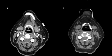
Figure 1 : CT Scans from initial presentation at acute care hospital and b) After 5 day IV antibiotic treatment showing resolution of the left submandibular sialolithiasis.
On presentation at our facility on day13 the patient complained of persistent tongue pain and edema and continued to have significant tongue immobility, dysarthria, and dysphagia. A Magnetic Resonance Imaging (MRI) was performed at this time and although no abnormalities were noted in the neck the tongue was noted to be enlarged with enhancement on T2 imaging. At this point during clinical examination the tongue appeared pale gray, necrotic, and foul smelling, so a tongue biopsy was repeated and resulted in sloughing of the anterior portion of the tongue measuring approximately 6.2 × 6 × 0.8cm (see Figure 2). A biopsy specimen was also taken from the viable tongue. On histopathologic examination the necrotic portion of the tongue was consistent with coagulative necrosis while the viable tongue was only significant for granulation tissue. Rheumatology was consulted for further evaluation and concern was raised for medium-large vessel vasculitis. Ophthalmologic evaluation was obtained to rule out any ocular involvement and temporal artery biopsy was eventually performed. The biopsy revealed a mixed inflammatory infiltrate composed of lymphocytes, histiocytes and multinucleated giant cells with focal destruction of internal elastic lamina and marked intimal thickening significantly decreasing the lumen in bilateral temporal arteries. Histologically, the diagnosis of giant cell temporal arteritis was established and the patient was treated with high dose steroids initially and ultimately discharged home with oral prednisone.
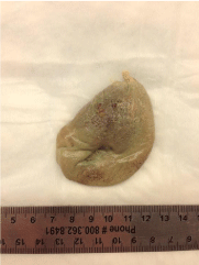
Figure 2 : An image of the pathology specimen submitted after the patient’s tongue was noted to slough during attempted biopsy.
In follow-up approximately one month after appropriate treatment the patient was noted to have improvement in the neck and tongue pain. No further ischemia or necrosis of her tongue was noted. She continued to have significant dysarthria and also required ongoing tube feeds for supplementation because of persistent dysphagia and aspiration following the loss of a significant amount of oral tongue.
Discussion/Conclusion
Temporal arteritis, also known as giant cell arteritis or cranial arteritis is characterized by inflammation of medium to large caliber arteries. Women over the age of 50 are most commonly afflicted and there is often an association with polymyalgia rheumatica. People of Scandinavian descent are afflicted more frequently. The usual presentation is headache and elevated ESR levels. At times patients also complain of tongue and jaw claudication [1]. Vision loss is a feared and commonly recognized complication of temporal arteritis, occurring in approximately 20% of patients [2]. For this reason, once the diagnosis is suspected it is important that therapy be initiated promptly.
Physical examination findings can include prominence of the temporal arteries, with or without the presence of a pulse, weakness in other sites of peripheral pulse palpation, and temporal or scalp tenderness. On laboratory evaluation there is often a rise in acute phase reactants such as CRP and platelet levels, as well as marked elevation of the ESR. Although contrast enhanced CT and MRI are typically negative in this disorder, at least one recent study suggests that 3 Tesla high resolution MRI using contrast has reasonably high sensitivity and specificity in diagnosis [3].
While the diagnosis can be suspected based on the clinical presentation of the patient, it cannot be established until biopsy of the temporal artery is performed. Temporal artery biopsy is generally considered the gold standard of diagnosis, with at least 1 cm of tissue necessary to achieve sufficient sensitivity for the diagnosis [4]. On histology, giant cell arteritis is characterized by an inflammatory infiltrate of mononuclear cells with the development of giant cells. The intimal layer is thickened and the vessel lumen size is decreased [1].
Treatment with high-dose corticosteroids is initiated immediately, at the time of suspected diagnosis. Temporal artery biopsy should be arranged shortly thereafter, as the corticosteroid treatment can alter the microscopic inflammatory histopathology that is characteristic of the disorder and make diagnosis difficult [5]. Regardless, prompt therapy is critical to prevent progression of the disease or the development of additional symptoms, such as visual loss or blindness.
There are reports of patient’s presenting with glossodynia of unknown etiology that eventually progress to have tongue necrosis as a result of an ongoing vasculitis. An initial report of bilateral tongue ischemia resulted in a significant amount of tongue necrosis. Similar to the current report, in this case the patient had a prolonged presentation and treatment was given approximately six to seven weeks after symptom onset [6]. In more recent reports of patients who presented within several days of symptom onset, areas of necrosis tend to be unilateral and relatively small and do not result in significant loss of function [7–9]. Another, more recent report describes a patient with bilateral lingual ischemia who was diagnosed and treated within 10 days of initial symptom onset. In this patient, early treatment with high dose steroids resulted in complete resolution of ischemia and no tongue tissue was lost [10]. Unfortunately, the patient in our case had a delayed presentation to our facility and a delay in initiation of appropriate therapy resulting in significant and persistent morbidity related to her diagnosis.
It is important to have temporal arteritis on the differential diagnosis for patients presenting with glossodynia, tongue claudication, and evidence of tongue ischemia or necrosis. Early suspicion is imperative, not only for the prevention of further tissue loss but also to prevent progression of the disease and potential blindness.
References
- Langford CA, Fauci AS. Chapter 326. The Vasculitis Syndromes. Longo DL, Fauci AS, Kasper DL, Hauser SL, Jameson JL, Loscalzo J, editors. In: Harrison’s Principles of Internal Medicine, 18e [Internet]. New York, NY: The McGraw-Hill Companies; 2012.
- Dasgupta B. Giant Cell Arteritis Guideline Development Group. Concise guidance: diagnosis and management of giant cell arteritis. Clin Med. 2010; 10: 381-386.
- Bley TA, Uhl M, Carew J, Markl M, Schmidt D, Peter HH, et al. Diagnostic value of high-resolution MR imaging in giant cell arteritis. AJNR Am J Neuroradiol. 2007; 28: 1722-1727.
- Ypsilantis E, Courtney ED, Chopra N, Karthikesalingam A, Eltayab M, Katsoulas N, et al. Importance of specimen length during temporal artery biopsy. Br J Surg. 2011; 98: 1556-1560.
- Font RL, Prabhakaran VC. Histological parameters helpful in recognising steroid-treated temporal arteritis: an analysis of 35 cases. Br J Ophthalmol. 2007; 91: 204-209.
- Roseman BB, Granite E. Massive tongue necrosis secondary to temporal arteritis. J Oral Maxillofac Surg. 1984; 42: 682-684.
- Brodmann M, Dorr A, Hafner F, Gary T, Pilger E. Tongue necrosis as first symptom of giant cell arteritis (GCA). Clin Rheumatol. 2009; 28: S47-49.
- Ciantar M, Adlam DM. Glossodynia and necrosis of the tongue caused by giant cell arteritis. Br J Oral Maxillofac Surg. 2008; 46: 231-233.
- Husein-Elahmed H, Callejas-Rubio JL, Rios-Fernández R, Ortego-Centeno N. Tongue infarction as first symptom of temporal arteritis. Rheumatol Int. 2012; 32: 799-800.
- Sainuddin S, Saeed NR. Acute bilateral tongue necrosis--a case report. Br J Oral Maxillofac Surg. 2008; 46: 671-672.