Case Report
Austin J Otolaryngol. 2015; 2(9): 1062.
A Case of Wegener Granulomatosis with Unprecedented Hypopharyngeal and Splenic Involvement
Panwar J*, Panwala HK and Tandur RK
Department of Radiology, Christian Medical College, India
*Corresponding author: Panwar Jyoti, Department of Radiology, Christian Medical College, Vellore, 632004, Tamilnadu, India
Received: October 21, 2015; Accepted: November 16, 2015; Published: November 18, 2015
Abstract
Wegener’s granulomatosis (WG) is a rare form of necrotizing vasculitis which can affect any part of the body. We present a unique case of WG in a 22-year-old male who presented with high grade fever, ear pain, loss of weight and appetite. On subsequent investigation he was found to have multisystem involvement with uncommon ulcerated hypopharyngeal lesion and a large splenic infarct secondary to vasculitis involving the small to medium size vessels. The initial illness and one subsequent exacerbation were treated with oral cyclophosphamide and prednisone. Secondary infections have been managed with use of appropriate antibiotics.
Keywords: Wegener’s granulomatosis; Vasculitis; Hypopharyngeal lesion; Splenic infarct
Introduction
WG is a multisystem autoimmune inflammatory disease can affect virtually any part of the body, but has a predilection for certain organs like upper respiratory tract (sinuses, nose, ears, and trachea), the lungs, and the kidneys with wide spectrum of clinical features, at times being nonspecific [1]. This can cause diagnostic dilemmas with delay in diagnosis and initiation of therapy. Laryngeal (Subglottic) and tracheal involvement is commonly seen however, hypopharyngeal granulomatosis is a rare manifestation of WG [1,2]. Among the viscera, lung and kidneys are commonly involved. Incidence of ante-mortem splenic involvement is very low [3]. We report such a rare case with hypopharyngeal and splenic involvement.
Case Presentation
A 22-year-old male presented to medicine OPD with a 2 month history of intermittent high fever, loss of weight and appetite, ear discharge with decreased hearing and productive cough. Over this time he also developed skin rashes over the lower limbs. He had bilateral myringotomy and grommets insertion elsewhere 1.5 months back for ear pain. He was otherwise fit and well, with no history of diabetes, immunosuppression or smoking and on no regular medications. On physical examination, he was found to have nasal clots, generalized purpuric macules more prominent over the lower legs, coated oral cavity, ulcerated posterior pharyngeal wall and oropharyngeal lesions. On laboratory investigations, Hb: 87 g/L, WCC: 12.7 x 10⁹ g/L, Neutrophil count: 6.5 x 10⁹ g/L, CRP: 167.0 mg/L, ESR: 45 mm, C ANCA & P ANCA (circulating and perinuclear antineutrophil cytoplasmic antibodies): >300/<2 (normal<15 /<15). Urinalysis showed protein (3+) and red blood cells (RBC 3+) in the urine and urine phase contrast microscopy demonstrated dysmorphic RBC with cast formation. Rest of the other blood investigations were within normal limits. He was initially treated with Intravenous antibiotics (meropenem), antipyretic and nasal douches.
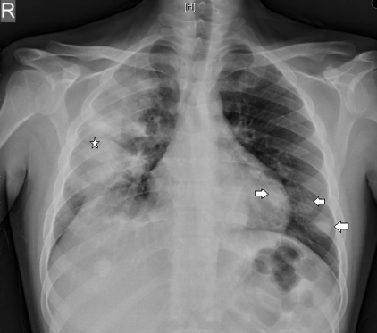
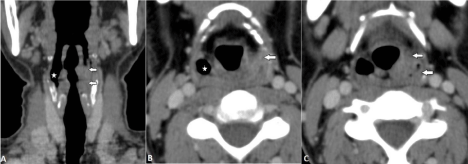
He subsequently underwent anterior and posterior rhinoscopy via rigid nasendoscope revealed blood clots and unhealthy nasal mucosa. Fiberoptic nasopharyngolaryngoscopy showed ulcerated growth of left pyriform sinus and aryepiglottic fold and had subsequent biopsy of this lesion revealed epithelial hyperplasia with extensive ulceration. He also underwent skin biopsy from right leg showed leukocytoclastic vasculitis. In view of significant cough, he had a chest radiograph in the beginning which revealed bilateral lung “infiltrates“ (Figure 1) and subsequently CT (computed tomography) neck, thorax and abdomen was requested to further characterize the supraglottic and lung lesions. CT (Figure 2, 3 & 4) showed left pyriform sinus mass, bilateral lung nodules, splenic lesion and renomegaly. Based on these findings he underwent transbronchial biopsy of right lung showed vasculitis with focal granulomatous inflammation, WG is a possibility. In view of haematuria and proteinuria he had renal biopsy showed mild mesangial proliferation with tubular necrosis. Based on above findings finally a diagnosis of granulomatosis with polyangiitis (WG) with pulmonary, nasal, pharyngeal, renal and dermatological involvement was made and the patient was started on oral cyclophosphamide and prednisone. He was also started on oral fluconazole for oral candidiasis, losartan for proteinuria and other supportive medications for symptomatic relief. He had also received pneumococcal and influenza vaccination and discharged.

    Figure 1:  Posteroanterior chest radiograph shows bilateral nodular opacities
(arrows) more discretely seen on left side in retrocardiac and lower zone
region and a large consolidation on right side (star).

    Figure 2:  A, Coronal, B & C, Axial contrast enhanced CT sections through the supraglottic region show soft tissue in left pyriform sinus (arrows) causing
circumferential wall thickening including the aryepiglottic fold. Note the normal appearing patent right pyriform sinus lumen (star).

    Figure 3:  A, mediastinal window, B & C, lung window axial contrast enhanced CT sections of chest confirms plain radiographic findings. A mass like consolidation
is seen in the right middle and lower lobes (star) and bilateral lower lobes nodules (arrows). Note the pericardial effusion (open arrow) in A.

    Figure 4:  A, arterial phase axial, B venous phase axial and & C, coronal contrast enhanced CT sections of upper abdomen reveal normal proximal (circle) splenic
artery posterior to pancreas. However, distal artery and branches at the hilum are attenuated (arrows); a large non-enhancing portion of spleen in image B (arrow)
with nodular areas of spared normal enhancing splenic tissue. Bilaterally enlarged kidneys (arrows) seen in image C.
Outcome and Follow-up
Initial review seven days after discharge showed good progress. However, within 15 days of initiation of treatment, he presented again with worsening purpuric rash and new onset of arthralgia. He was readmitted for the same and started fluticasone local application for skin lesions. His rashes subsequently improved during hospital stay. He was assessed by rheumatologist for arthralgia and further titration of immunosuppressant and steroid medications was done. Again was discharged in a stable condition and option of biological drug was considered for further future flare up.
Discussion
Since the introduction of the uncommon entity- WG which is also now known as “granulomatous disease with polyangiitis” from 1931, a lot of research has been done regarding the pathophysiology and sites affected. The classical triad of the disease includes, necrotising granulomatous lesions involving 1) the upper or lower respiratory tract, 2) necrotising vasculitis affecting arteries and veins, 3) glomeronephritis in kidney [1-5]. Clinical manifestations are versatile depending on the isolated organ involvement or multisystem involvement. The highest peak age group is 4th to 6th decade [4]. However in patients with upper respiratory tract involvement, the presentation is in younger age groups as seen in our case [4]. Invariably in most of the cases C –ANCA is elevated and confirmation of diagnosis is made by tissue biopsy [1,4].
Primarily pharyngeal involvement is very rare in WG with only few case reports available in literature. In the pharynx, the most common sub-site affected is nasopharynx (60-80 %) [5,6] followed by oropharynx. Only few case reports are available on involvement of hypopharynx as seen in this case. An enhancing soft tissue density lesion in the left pyriform fossa with involvement of the aryepiglottic fold in the current case also showed a rare sub-site involvement of larynx. The most common site affected in the larynx is subglottis (8- 23%) [1,6,7] Supra glottis involvement is rarest of all presentation [2] as seen in our case. To the best of our knowledge only one case report has mentioned supra glottis stenosis in WG [2].
As mentioned above splenic involvement in the WG is rare and usually secondary to small vessel vasculitis affecting the peripheral most branches of splenic artery. Usually, the incidence of splenic lesions in WG is low because of asymptomatic nature in majority of the cases [3,8]. However, the autopsy data suggest high incidence of splenic lesions including infarcts in patients of WG [3]. Different patterns of splenic infarcts are mentioned in the literature in the form of typical peripheral wedge shaped hypodense areas, multiple heterogeneous low attenuation nodules, peripheral low attenuation with central normal enhancement, central low attenuation and peripheral rim enhancement or as seen in our case as a large non enhancing hypodense area except for nodular areas of normal enhancement in periphery [9,10]. This pattern of involvement in WG favors the pan arteritis (involving small to medium size vessels) nature of disease over the splenic granulomas.
Conclusion
Both the radiologists and clinicians should be familiar with wide range of the laryngopharyngeal lesions, bizarre appearance of splenic infarct secondary to vasculitic component and other systemic manifestation of WG. Thus this will alert the physician to facilitate prompt diagnosis and early treatment of this potentially fatal disease.
References
- Rodrigues AJ, Jacomelli M, Baldow RX, Barbas CV, Figueiredo VR. Laryngeal and tracheobronchial involvement in Wegener's granulomatosis. Rev Bras Reumatol. 2012; 52: 231-235.
- Belloso A, Estrach C, Keith AO. Supraglottic stenosis in localized Wegener granulomatosis. Ear Nose Throat J. 2008; 87: E11-14.
- Martusewicz-Boros M, Baranska I, Wiatr E, Bestry I, Roszkowski-Sliz K. Asymptomatic appearance of splenic infarction in Wegener's granulomatosis. Pol J Radiol. 2011; 76: 43-45.
- Srouji IA, Andrews P, Edwards C, Lund VJ. Patterns of presentation and diagnosis of patients with Wegener's granulomatosis: ENT aspects. J Laryngol Otol. 2007; 121: 653-658.
- Morales-Angulo C, García-Zornoza R, Obeso-Agüera S, Calvo-Alén J, González-Gay MA. [Ear, nose and throat manifestations of Wegener's granulomatosis (granulomatosis with polyangiitis)]. Acta Otorrinolaringol Esp. 2012; 63: 206-211.
- Polychronopoulos VS, Prakash UB, Golbin JM, Edell ES, Specks U. Airway involvement in Wegener's granulomatosis. Rheum Dis Clin North Am. 2007; 33: 755-775.
- Langford CA, Sneller MC, Hallahan CW, Hoffman GS, Kammerer WA, Talar-Williams C, et al. Clinical features and therapeutic management of subglottic stenosis in patients with Wegener's granulomatosis. Arthritis Rheum. 1996; 39: 1754-1760.
- Papaioannides D, Nikas SN, Fotinou M, Akritidis NK. Asymptomatic splenic infarction in Wegener's granulomatosis. Ann Rheum Dis. 2002; 61: 185-186.
- Balcar I, Seltzer SE, Davis S, Geller S. CT patterns of splenic infarction: a clinical and experimental study. Radiology. 1984; 151: 723-729.
- Fonner BT, Nemcek AA Jr, Boschman C. CT appearance of splenic infarction in Wegener's granulomatosis. AJR Am J Roentgenol. 1995; 164: 353-354.