
Case Report
Austin J Urol. 2015;2(1): 1020.
Lymphoepithelioma-Like Carcinoma (LELC) of the Kidney Presenting as Non Functioning Kidney - A Case Report
Sriram Krishnamoorthy, Sekar Hariharasudhan and Sunil Shroff
Department of Urology, Sri Ramachandra Medical College, India
*Corresponding author: Sriram Krishnamoorthy, Department of Urology, Sri Ramachandra Medical College & RI, Chennai, India
Received: November 03, 2014; Accepted: February 16, 2015; Published: February 23, 2015
Abstract
Lymphoepithelioma Like Carcinoma (LELC) is a rare poorly differentiated epithelial tumor with a dense inflammatory cell infiltrate. Lymphoepithelioma mainly occurs in the nasopharynx, but can also arise in a variety of other sites including salivary gland, thymus, lung, stomach, skin and urothelium. Primary LELC of the urinary tract is very rare. Cases involving the urinary bladder, ureter and renal pelvis are reported but an isolated LELC of the kidney is published only once before in the literature. We present a 70-year-old man who had right loin pain and increased frequency. He initially had right percutaneous nephrostomy and subsequently underwent right simple nephrectomy for nonfunctioning kidney. The histopathology report was pT1bN0M0 LELC of the Kidney. We present the case because of its rarity and also to highlight the need for recognizing this unusual renal tumor. To our knowledge, this is the second reported case of LELC of the kidney.
Keywords: Lymphoepithelioma like carcinoma; Lymphoepithelioma; Kidney
Introduction
Lymphoepithelioma is a malignant poorly differentiated epithelial tumor with dense inflammatory cell infiltration. More often, it affects nasopharynx but can also rarely affect other organs. Lymphoepithelioma when it occurs in other sites like the salivary gland, thymus, lung, stomach, skin and urothelium is termed as Lymphoepithelioma like carcinoma. LELC of the urinary tract is very rare. Tumors affecting urinary bladder, urethra and renal pelvis are reported, whereas, LELC of kidney is very rare. Differentiation from medullary carcinoma of the kidney and collecting duct carcinoma has its implications on its prognosis and treatment. Only one case of LELC of the kidney has been reported in the literature. To our knowledge, we present the second case of LELC of the kidney.
Case Report
A 70 year-old man presented with a history of right loin pain and increased urinary frequency for a month. He did not have hematuria or other urological complaints. He is a non-smoker, had no co morbid illness. Physical examination was unremarkable. The blood biochemistry and renal function tests were normal. Urine routine showed 1-2 pus cells. Urine culture showed no growth. Ultrasound abdomen showed right moderate hydronephrosis with an ill defined heterogeneous predominantly hypo echoic lesion in the lower pole of the right kidney. A clinical diagnosis of pyonephrosis was made. However, three consecutive urine specimens for AFB were negative. IVU showed a non visualized right kidney (Figure 1). CECT Lower abdomen showed contracted, moderately hydronephrotic right kidney with a small hypodense lesion measuring 3.8 x3.4cm in the inferior pole of the right kidney showing heterogonous enhancement on contrast administration (Figure 2). Surrounding perinephric strandings were observed. Right proximal urethra was mildly dilated with abrupt narrowing and no excretion of contrast in the right urethra. DTPA showed reduced cortical and excretory function (19%) on the right kidney (Figure 3). During the course of evaluation he developed severe cough, fatigability and fever with chills. It was thought to be due to an infected hydroureteronephrosis and a percutaneous nephrostomy was done, suspecting an infective pathology. Around 50ml of turbid urine was drained. Culture specific parenteral antibiotics were administered. He had a daily PCN output of 100ml/day with persistent high grade fever with chills. Hence open right renal exploration and nephrectomy was planned. Intraoperatively, right kidney was densely adherent to the IVC and the retroperitoneum. Nephrectomy was completed with a lot of difficulty. The cut section revealed a slightly yellowish solid tumour involving the entire kidney, mainly the lower pole. On histo-pathological examination, the tumour showed diffuse sheets and cords of undifferentiated cells having large pleomorphic nuclei with coarse chromatin and prominent nucleoli and amphophilic cytoplasm, giving the tumor a syncitial appearance (Fig 4). Individual cells or a group of cells were surrounded by prominent lymphoid infiltrates. The entire tumor demonstrated similar morphology. Immunohistochemistry revealed strong positivity for CD10, Vimentin in the background lymphoid cells and P63, CK7 positive in the epithelial component of the tumor (Figure 4) and negative for CEA and CK20. The histological picture and positivity for P63 were in favor of Lymphoepithelioma like carcinoma. The patient had an uneventful post operative period. Patient was advised adjuvant chemotherapy but refused the same and is on regular follow-up for the past 10 months.
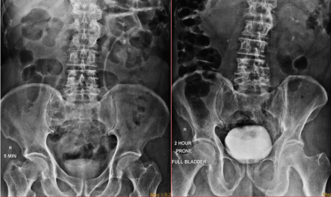
Figure 1: IVU showing non visualized right kidney.
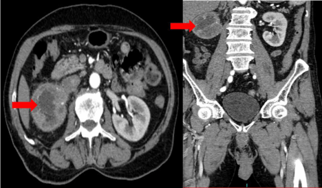
Figure 2: CECT Lower abdomen showing contracted hydronephrotic right kidney with a small hypodense lesion measuring 3.8 x3.4cm in the inferior pole of the right kidney with heterogenous enhancement on contrast administration.
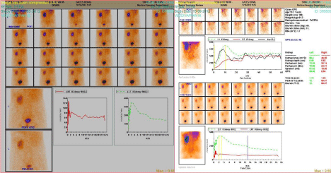
Figure 3: DTPA showed reduced cortical and excretory function (19%) on the right kidney.
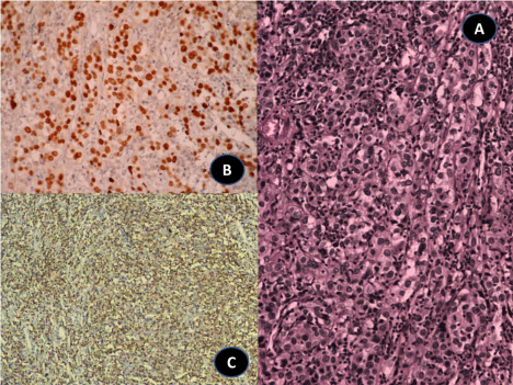
Figure 4: A) Large pink tumor cells admixed with lymphoid cells in the background. H&E x 400. B) The tumors cells show nuclear positivity for p63 and the background lymphoid cells are negative for the same. Immunohistochemistry, p63 x 200. C) The background lymphoid cells are positive for the lymphoid marker CD45 Immunohistochemistry, CD45 x 100.
Discussion
LELC is a rare malignant neoplasm. It can histological mimic lymphomas, poorly differentiated Transitional Cell Carcinomas (TCC) or poorly differentiated Squalors Cell Carcinoma (SCC) with a predominant lymphocytic background.
The term Lymphoepithelioma is used to designate poorly differentiated malignant epithelial tumor of the nasopharynx with histological distinct markedly prominent lymphoid infiltrate [1]. Carcinomas with similar histology arising in other sites are called Lymphoepithelioma-Like Carcinoma (LELC) [2]. LELC occurs in organs such as salivary glands, uterus, cervix, thymus, lung, skin, stomach, bladder, prostate, and the breast [3]. Epstein- Barr Virus (EBV) has been a possible factor in the etiology of Lymphoepithelioma-like tumors, particularly in the thymus gland (thymoma with lymphoid hyperplasia), nasopharynx, but most reports of uroepithelial LELC are negative for EBV [4,5].
LELC of the kidney is rare. The first ever case of LELC of the kidney was reported by Elzevier HW et al. [6]. where a 67 year old female had presented with painless total hematuria. Various authors had published reports of LELC in other parts of urinary tract. LELC of the urinary bladder and urethra also present mostly with hematuria.
Chalik et al. described the first case of LELC of the urethra where a 75 year old man presented with hematuria [7]. LELC of the urinary bladder has been frequently reported in the past. Zuckerberg et al. was the first to report a case of LELC of the urinary bladder [8]. Porcaro et al had reported an overall incidence of 0.4 to 1.3% of all bladder tumors [9]. Dinney et al reported 3 cases of muscle invasive Lymphoepithelioma of the bladder treated mainly with chemotherapy to preserve bladder function [10]. Following this, there had been many such reports of bladder involvement.
Tamas et al. studied a series of 28 cases of LELC of the bladder [3]. To our knowledge, this is the largest series of LELC of the urinary tract published in literature. One of the conclusions of previous smaller studies is that pure or Predominant Lymphoepithelioma-like carcinoma has a better prognosis than focal Lymphoepithelioma-like carcinoma with reported rates of metastasis ranging from only 12 to 15% [11,12].
Amin et al had recommended that the pure forms of LELC are morphologically and clinically different from TCC and merit recognition as a separate clinic-pathological entity [13]. Pure forms are chemo-responsive, which can motivate the treating physician to aim at adopting more organ salvaging approaches.
It is important and imperative for the clinicians and the pathologists to be aware of this rare variant. Misinterpretation of these tumors as TCC or renal cell carcinomas can result in a significant difference in the adjuvant treatment protocol. Our case underwent nephrectomy without chemotherapy and disease free survival of 10 months after surgery.
Conclusion
This case is presented because of its rarity, its atypical form of presentation, to make the clinicians and the pathologists to be aware of this rarer entity and also, in view of its chemo-responsive nature, to stress on the possibility of organ salvaging approaches in the event of LELC affecting the urinary tract.
References
- Carbone A, Micheau C. Pitfalls in the microscopic diagnosis of undifferentiated carcinoma of nasopharyngeal type (Lymphoepithelioma). Cancer. 1982; 50: 1344-1351.
- Han Ki Yun1, Sung Il Yun1,Yoon Hyung Lee1, Kyung Mo Kang2, Eun Kyung Kwak3, Jae Soo Kim1,et al. Lymphoepithelioma-like Carcinoma of the Urinary Bladder. J Korean Med Sci. 2010; 25: 1672-1675.
- Tamas EF, Nielsen ME, Schoenberg MP, Epstein JI. Lymphoepithelioma like carcinoma of the urinary tract: a clinicopathological study of 30 pureand mixed cases. Mod Pathol. 2007; 20: 828-834.
- Terai A, Terada N, Ichioka K, Matsui Y, Yoshimura K, Wani Y. Lymphoepithelioma-like carcinoma of the ureter. Urology. 2005; 66: 1109.e13-1109.e15.
- Gulley ML1, Amin MB, Nicholls JM, Banks PM, Ayala AG, Srigley JR, et al. Epstein-Barr virus is detected in undifferentiated nasopharyngeal carcinoma but not in Lymphoepithelioma-like carcinoma of the urinary bladder. Hum Pathol. 1995; 26: 1207-1214.
- Elzevier HW, Venema PL, Kropman RF, Kazzaz BA. Lymphoepithelioma-likecarcinoma of the kidney. J. Urol. 2002; 167: 2127-2128.
- Chalik YN, Wieczorek R, Grasso M. Lymphoepithelioma-likecarcinoma of the ureter. J. Urol. 1998; 159: 503-504.
- Zukerberg LR, Harris NL, Young RH. Carcinoma of the urinary bladdersimulating malignant lymphoma: A report of five cases. Am J Surg Pathol. 1991; 15: 569-76.
- Porcaro AB, Gilioli E, Migliorini F, Antoniolli SZ, Iannucci A, ComunaleL. Primary lymphoepithelioma-like carcinoma of the urinary bladder: report of one case with review and update of the literature after a pooledanalysis of 43 patients. Int Urol Nephrol. 2003; 35: 99-106.
- Dinney CP, Ro JY, Babaian RJ, Johnson DE. Lymphoepithelioma of the bladder: a clinicopathological study of 3 cases. J Urol. 1993; 149: 840-841
- Cohen RJ, Stanley JC, Dawkins HJ. Lymphoepithelioma-like carcinomaof the renal pelvis. Pathology. 1999; 31: 434-435.
- Antonio Lopez-Beltrán, Rafael LuqueLuis Vicioso, Francisco Anglada, Maria J Requen, Ana Quintero, Rodolfo Montironi. Lymphoepithelioma-like carcinoma of the urinary bladder: a clinicopathologic study of 13 cases. Virchow Arch. 2001; 438: 552-557.
- Amin MB, Ro JY, Lee KM, Ordóñez NG, Dinney CP, Gulley ML, et al. Lymphoepithelioma-like carcinoma of the urinary bladder. Am J Surg Pathol. 1994; 18: 466-473.