
Research Article
Austin J Urol. 2015;2(2): 1025.
Spectrum of Ectopic Pelvic Kidney in Children: A Tenyear Experience
Marte A¹*, Marte G² and Pintozzi L¹
¹Department of Pediatric Surgery, 2nd University of Naples, Italy
²Department of General Surgery, 2nd University of Naples,Italy
*Corresponding author: Antonio Marte, Department of Pediatric Surgery, 2nd University of Naples, Largo Madonna delle Grazie, 1.80138 Naples, Italy
Received: February 09, 2015; Accepted: April 15, 2015; Published: April 21, 2015
Abstract
Introduction: Many patients with ectopic kidneys remain often undiagnosed or asymptomatic throughout life. Ectopic pelvic kidneys present a large spectrum of presentation and symptoms from renal dysplasia to a severe obstruction. One of the most common problem is the UPJ obstruction with stones formation. We present here our experience of pelvic kidney in children, the clinical presentation and the surgical procedures performed
Material and Methods: A total of 17 children, aged from 6 months to 17 years, (14m;3f) were referred to our Institution between January 2004 to June 2014 for pelvic kidney. There were 5 (29.41%) left and 12 (70.5%) right Kidneys.
- 9 patients presented with moderate to severe UPJ obstruction (three cases with 10 to 20mm pelvic stones) with normal/moderately-reduced =25% relative function at MAG3 radionuclide scan of these patients, 5 with clear symptoms of UPJ obstruction underwent a laparoscopic dismembered Anderson –Hynes pyeloplasty. The remaining 4, presented symptomatic intermittent hydronephrosis with colic abdominal pain. These patients underwent pelvis derotation through a minimally invasive transposition of the UPJ.
- 5 patients presented non-functioning kidneys, in 2 cases associated to hypertension, and underwent laparoscopic nephrectomy.
- 2 patients presented asymptomatic pelvic kidney.
- 1 patient presented 3rd G. VUR on the ipsilateral pelvic kidney.
The associate pathology was 1 midshaft hypospadias, 2 criptorchidism , 1 mild mitral insufficiency.
The evaluation of each patient involved their personal and family medical history, ultrasound examination, VCUG, MAG3 renal scan and MRI in selected cases.
Results: After a mean follow-up of 6.9 yrs the majority of the patients are well and none present hypertension. Symptoms resolved in 13 out of 15 surgical patients. 2 patients needed the positioning of a double J stent from 3 to 8 months after pyeloplasty that was removed from 6 to 12 weeks after the procedure
Conclusion: Pelvic kidneys present a large spectrum of symptoms. UPJ obstruction with/without stones, intermittent hydronephrosis, recurrent abdominal pain, UTI is the most frequent symptoms. The majority of our patients needed surgical procedures. In four cases there were associated pathologies as hypospadias, cryptorchidism, and mild mitral insufficiency. Laparoscopic approach seems a useful tool for the treatment of these kidneys. Pelvis derotation can be an easy and effective procedure in moderate, intermittent obstruction.
Keywords: Kidney; Ectopic; Laparoscopy; UPJO; Children
Introduction
Kidneys that fail to migrate to their normal position during the embryo’s life are defined as ectopic. The most common type of this conditions is the pelvic kidney, whereby the organ remains in the pelvic cavity; in this case, the renal pelvis presents an anterior rotation, with a myriad of abnormal vessels originating from both the aorta and the iliac arteries [1,2].
In many patients the condition of renal ectopic remains undiagnosed throughout their life [3]. The diagnosis is often made following the onset of Ureteropelvic Junction Obstruction (UPJO), with or without stone formation. The incidence of ectopic pelvic kidney one normal and one pelvic kidney) is estimated to be 1 out of 2200-3000 newborns, while solitary pelvic kidney is estimated to be 1 in 22000 [4] .
UPJO is one of the most common problems, occurring in 22% to 37% of cases; it may be caused by malrotation and/or high urethral insertion or true UPJ dysplasia [3]. Moreover UPJO in ectopic kidneys presents a wide spectrum of presentations and symptoms varying from renal dysplasia to mild/intermittent or severe obstruction. These latter cases are often complicated by pelvicaliceal stones [5]. The purpose of surgical intervention is to achieve adequate drainage in cases of functioning kidneys or their removal in cases of severe dysplasia or non-functioning kidneys. We present here our experience of pelvic kidney in children, the clinical presentation and the surgical procedures performed
Material and Methods
A total of 17 children, aged from 6 months to 17 years, (14 m;3f) were referred to our Institution between January 2004 to June 2014 for pelvic kidney. There were 5 (29.41%) left and 12 (70.5%) right kidneys.
- 9 patients presented with moderate to severe UPJO (three cases with10 to 20mm pelvic stones) with normal (=40%) or moderately-reduced (=25%) relative function at MAG3 radionuclide scan in anterior view of these patients, 5 with clear symptoms of UPJO underwent a laparoscopic dismembered Anderson–Hynes pyeloplasty. The remaining 4, presented symptomatic intermittent hydronephrosis with recurrent colic abdominal pain. These patients underwent pelvis derotation through a minimally invasive transposition of the UPJ.
- 5 patients presented non-functioning kidneys, in 2 cases associated to hypertension, and underwent laparoscopic nephrectomy.
- 2 patients presented asymptomatic pelvic kidney.
- 1 patient presented 3rd G. VUR on the ipsilateral pelvic kidney.
- Four patients presented concomitants pathologies constituded of 1 midshaft hypospadias , 2 unilateral criptorchidism, 1 mild mitral insufficiency.
The evaluation of each patient involved their personal and family medical history, ultrasound examination, VCUG, MAG3 renal scan and MRI in selected cases.We believe it is noteworthy that in patients with pelvic kidneys, anterior views must be obtained during radionuclide scanning because the pelvis forms a barrier between the radioactively labeled tracer and the gamma camera, thus, reducing the amount of radiation detected and underestimating function [6].
To correct UPJO a standard 3-4 trocar laparoscopy was employed, with the patient in general anesthesia. The first trocar was introduced transumbilically with open access; two lateral 3-mm trocars were introduced under visual guide in the right and left lower quadrants along the midclear line. If necessary a fourth trocar was introduced cephalad, at the level of umbilical line. A transmesocolic approach was employed, without mobilizing the intestine from the anterior surface of the kidney and the renal pelvis. The patients underwent a dismembered pyeloplasty according to Anderson-Hynes, in some cases associated with pyelolithotomy when necessary. The steps for performing the pyeloplasty were the same as those used for the normo-located kidneys. Pyeloplasty was performed in two semi continuous vycril 5/0. Any kidney stone detected was removed under visual guide following pyelotomy, using a 5 mm Johanne under control of image intensifier.
The four patients with intermittent UPJO underwent pelvic derotation and straightening of the ureteropelvic junction. Derotation was achieved by detaching the lower renal pole and by suturing the peritoneum behind the ureteropelvic junction; this caused a forward rotation of the major axis of the kidney, thus straightening the junction.
Five patients with non-functioning or dysplastic kidneys underwent laparoscopic nephrectomy. The removal of hypodysplastic kidneys resulted very easy because the laparoscopic magnification was extremely useful in isolating and removing the hypoplastic residues. The removal of seemingly normal kidneys with impaired renal function was more complex given the number of aberrant vessels and vascular abnormalities. The patient with VUR underwent endoscopic correction with endoscopic dextranomer/hyaluronic acid injection. The two asymptomatic patients undergo regular annual US check. Hypospadias and criptorchidism were corrected at same time of the principal operation or as single procedure in observational cases.
Results
After a mean follow-up of 6.9 yrs all patients are free of symptoms and blood pressure values normalized in those who underwent nephrectomy or pyeloplasty. Symptoms resolved in 13 out of 15 surgical patients. 2 patients needed the positioning of a double J stent from 3 to 8 months after pyeloplasty that was removed from 6 to 12 weeks after the procedure. Mag 3 renal scan remained stable or improved. Mean operating time was 170 minutes (range 40 - 200 minutes), and mean hospital stay was 2.5 days (range 1-7). There were no intraoperative or post-operative complications and none of the patients experienced major complications. In no case was conversion to open surgery necessary. Intraperitoneal drainage was removed after one or two days.
Those who underwent pelvic derotation are free of symptoms, show stable US and diuretic MAG3 renogram (Table 1) & (Figure 1-7).
    N.
    Pt
    Sex
    Age
    Side
    Symptoms
    Pathology
    Procedure
    Split    Function MAG3
    Ass.    Path.
    Split    function results


    1
    D.P.
    M
    6m
    L
    Asymptomatic
    Hypodisplasia
    Lap    nephrectomy
    0%
    L    crypto


    2
    E.P.
    M
    1yr
    R
    Asymptomatic
    L    Pelvic kidney
    Observation
    40%

    stable


    3
    G.C.
    M
    1,6yr
    R
    Asymptmatic
    Non-Functioning.Kidney
    Lap.    Nephrectomy
    0%

    //


    4
    B.C.
    M
    2yr
    L
    Colic    abdominal pain
    Intermittent    hydronephrosis
    Derotation
    37.20%
    Midshaft    Hypospadias
    improved


    5
    N.R.
    M
    3,8yr
    L
    UTI- R.    Abdommial pain
    UPJO
    LAP    DISM. A-H
    38%

    improved


    6
    F.A.
    F
    4,2yr
    R
    Colic    abdominal pain
    Intermittent    hydronephrosis
    Derotation
    37.80%

    improved


    7
    MR.M.
    M
    5yr
    R
    asymptomatic
    R    Pelvic Kidney
    Observation
    40%
    R    cryptor
    stable


    8
    A.V.
    M
    5,5yr
    R
    UTI- R.    Abdominal pain
    OUPJ-Stone
    LAP    DISM. A-H
    30.20%

    Stable-JJ    stent


    9
    G.DG.
    M
    7yr
    L
    Colic    abdominal pain
    Intermittent    hydronephrosis
    Derotation
    36.50%

    stable


    10
    A.Q.
    M
    8,7yr
    R
    Colic    abdominal pain
    Intermittent    hydronephrosis
    Derotation
    35.20%

    stable


    11
    I.T.
    M
    9yr
    R
    Hypertension
    Non-Functioning.Kidney
    Lap.    Nephrectomy
    5%

    //


    12
    C.I.
    F
    9,5yr
    R
    UTI
    UPJO
    LAP    DISM. A-H
    30%
    Mitral    insuff
    Stable_JJ    stent


    13
    M.C.
    M
    10,8yr
    L
    UTI
    VUR    Pelvic kidney
    Deflux    injection
    40%

    stable


    14
    A.F.
    M
    12yr
    R
    UTI
    UPJO
    LAP    DISM. A-H
    39.10%

    stable


    15
    A.P.
    F
    13yr
    R
    Hypertension
    Non-Functioning.Kidney
    Lap.    Nephrectomy
    5%

    //


    16
    L.DR.
    M
    14yr
    R
    Asymptomatic
    Hypodisplasia
    Lap.    Nephrectomy
    0%

    //


    17
    A.DF.
    M
    17yr
    R
    UTI
    UPJO-stone
    LAP    DISM. A-H
    25%

    stable
Table 1: Demographic data.
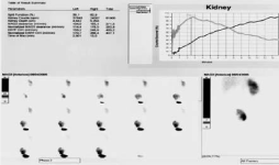
    Figure 1:  Case 14. Anterior view of the MAG-3 nuclear scintigraphy shows
reduction of right renal function (splitright function (%), 39.1).
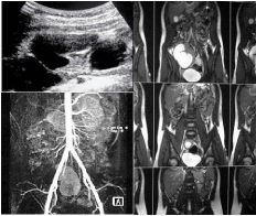
    Figure 2:  Case 14. Us /MRI Imaging confirms the position of the kidney, the
iliac pelvic blood supply, the abnormal pelvicaliceal rotation and the dilation.
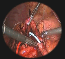
Figure 3: Case 14. Anderson-Hynes laparoscopic pyeloplasty.
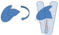
Figure 4: Case 4. Scheme of UPJ derotation.
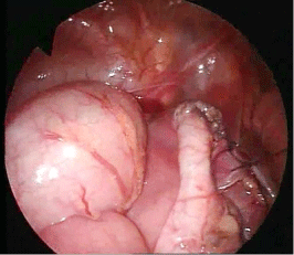
    Figure 5:  Case 4. Final view of laparoscopic UPJ derotation (arrow: suture
line).
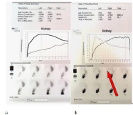
    Figure 6:  Case 4. Preoperative (a) and postoperative (b) anterior MAG3 scan
( Improved splict function).
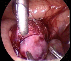
Figure 7: Laparoscopic view of ectopic hypodisplastic kidney.
Discussion
An ectopic kidney may not cause any symptoms and may function normally, even though it is not in its usual position. Many people have an ectopic kidney and do not discover it until they have tests done for other reasons which show a kidney absent from retroperitoneum . In other cases, an ectopic kidney may cause abdominal pain or urinary problems. More severe cases of UPJO may present with persistent abdominal pain that may become more acute during the day, whereas other patients may have intermittent manifestations with totally asymptomatic periods. Severe hypodysplasia of an ectopic kidney may be totally asymptomatic and the diagnosis be made fortuitously in case of abdominal ultrasound examinations for other reasons. These residues should be removed to avoid neoplastic mutations [7,8].
Another important aspect the surgeon must consider is that the ectopic kidney is quite often associated with other malformations such as skeletal (vertebral schisis, skull, rib anomalies), cardiovascular (valvular insufficiency; 30%), pulmonary, and genital system malformations (cryptorchidism, hypospadias). In girls, Rokitansky- Mayer-Kuster-Hauser syndrome is frequent [9].
Our case study shows that ectopic pelvic kidneys in children present a large spectrum of symptoms and confirm the feasibility and safety of the laparoscopic approach for ablative and reconstructive surgery.
With regard to therapeutic options our results show that pelvic kidney is a condition in which there is no unique therapy and each patient requires a specific treatment that needs to be tailored to his condition, providing a series of solutions from no treatment to nephrectomy. UPJO may present as an isolated event or may be associated with renal abnormalities; it can occur in 25-33% of horseshoe kidneys [10] and in 22- 37% of ectopic kidneys [3]. Surgical correction of this complex pathology may require ablation or reconstruction, based on the specific condition of the kidney. Hypodysplastic kidneys can be easily corrected by ablation; in this case, the laparoscopic magnification is extremely useful in isolating and removing the lesion. Instead the removal of seemingly normal kidneys with impaired renal function is much more complex given the number of aberrant vessels and vascular abnormalities that may cause bleedings. In this case, as the ascent of left kidney was been arrested at the level of junction between the common iliacs with the aorta/ inferior vena cava, it is supplied by rather large branches of these vessels [11-13].
Reconstruction may present with several solutions. Given the position of pelvic kidneys, different types of pyeloplasty have been proposed, such as open Anderson Hynes, laparoscopic Anderson- Hynes, the flap technique or robotic assisted Fenger pyeloplasty although, many of the patients described were adults and the results reported inconsistent. [14-17].
In our series we employed two techniques. In the presence of frank obstruction we adopted laparoscopic Anderson– Hynes pyeloplasty, which allowed us to perform also nephrolithotomy. In cases of intermittent hydronephrosis, we used a minimally invasive technique devised by us that consisted in derotating the renal spelvis to place the junction in a straight slope, so as to ensure adequate urine flow. Intermittent hydronephrosis is due to pyeloureteral malpositioning is a distinct condition presenting unspecific symptoms and a relatively preserved renal function [18,19]. An additional problem associated with UPJO surgery of ectopic kidneys is the surgical complexity. Reductive pyeloplasty according to Anderson– Hynes has always been considered the gold standard for the surgical correction of UPJ in otherwise normal kidneys, with a success rate above 90% [1,20]. In patients with horse–shoe kidneys, the success rate reported for open pyeloplasty ranges from 55% to 80% [21,22]. As to laparoscopic pyeloplasty, the current success rates are comparable although no data are available for pyeloplasty according to Anderson Hynes in pelvic kidneys [23,24]. Moreover pyeloplasty in ectopic pelvic kidneys can present persisting varying degrees of hydronephrosis and radiologic obstruction after pyeloplasty that could be attributed to anatomy–related pyelectasis, and so regular follow–up is warranted in this subpopulation.
A final consideration concerns the surgical cases: according to the current literature surgical UPJO in pelvic kidney present a variable incidence of 22–37% of pelvic kidneys and this is in line with our percentage of 29.5%. But if we include also the cases of intermittent hydronephosis then the percentage reaches 52.9%. This higher percentage of surgical cases is explained by the fact we utilized the derotation procedure in patients in which observational therapy could have been a possible alternative [2,25].
Conclusion
Ectopic kidney is an abnormal localization of a kidney due to a developmental anomaly and it occurs as a result of a halt in migration of kidneys to their normal locations during the embryonic period. Treatment of pelvic ectopic kidneys is not always simple and need to be tailored on single cases. Asymptomatic, non–complicated cases can be managed conservatively, but nephrectomy may be necessary if there are otherwise untreatable complications such as chronic hypertension, refractory pyelonephritis and stones. Laparoscopy according to our experience represents the better option when surgical treatment is necessary thanks to its minimally–invasiveness and is extremely useful in the identification of the complex vascularization of these kidneys. The technique we conceived to perform pelvic derotation seems useful and effective in cases of mild, intermittent obstruction, although a longer follow–up is needed to define the long term results.
References
- Bauer SB. Anomalies of the upper urinary tract. Wein AJ, Kavoussi LR, Novick AC, Partin AW, Peters CA, editors. In: Campbell Walsh Urology 9. Philadelphia: Saunders Elsevier, 2007; 3278–3281.
- Ritchey M. Anomalies of the kidney. Belman AB, King LR, Kramer SA, editors. In: Clinical Pediatric Urology. Fourth Edition. Martin Dunitz Ltd. 2002; 537-558
- Gleason PE, Kelalis PP, Husmann DA, Kramer SA. Hydronephrosis in renal ectopia: incidence, etiology and significance. J Urol. 1994; 151: 1660–1661.
- Zafar FS, Lingeman JS. Value of laparoscopy in the managementof calculi complicating renal malformations. J Endourol. 1996; 10: 379–383.
- Pearle MS, Lotan Y. Urinary lithiasis: Etiology, epidemiology, and pathogenesis. Wein AJ, Kavoussi LR, Novick AC, Partin AW, Peters CA edsitors. In: Campbell Walsh Urology, ed 9. Philadelphia: Saunders Elsevier, 2007; 1389–1391.
- Allen D, Bultitude MF, Nunan T, Glass JM. Misinterpretation of radioisotope imaging in pelvic kidneys. Int J ClinPract Suppl. 2005; 147: 111-112.
- Cooke A, Deshpande AV, La Hei ER, Kellie S, Arbuckle S, Cummins G. Ectopic nephrogenic rests in children: the clinicosurgical implications. J Pediatr Surg. 2009; 44: e13-16.
- Coli A, Angrisani B, Chiarello G, Massimi L, Novello M, Lauriola L. Ectopic immature renaltissue: clues for diagnosis and management. Int J ClinExpPathol. 2012; 5: 977-981.
- Chakrabarty S, Mukhopadhyay P, Mukherjee G. Sheares' method of vaginoplasty - our experience. J CutanAesthet Surg. 2011; 4: 118-121.
- Segura JW, Kelalis PP. and Burke EC. Horseshoe kidney in children. J Urol. 1972; 108: 333-336.
- Gray SE, Skandalakis JE. Embryology for surgeons-The embryological basis for the treatment of congenital defects. 1972; 472-474.
- Gülsün M, Balkanci F, Cekirge S, Deger A. Pelvic kidney with an unusual blood supply: angiographic findings. Surg Radiol Anat. 2000; 22: 59-61.
- Sebe P, Chemla E, Varkarakis J, Latrémouille C. Anatomic variations of the vascularization of the pelvic kidney: apropos of a case and review of the literature. Morphologie. 2004; 88: 24-26.
- Basiri A, Mehrabi S, Karami H. Laparoscopic flap pyeloplasty in a child with ectopic pelvic kidney.Urol J. 2010; 7: 125-127.
- Klingler HC, Remzi M, Janetschek G, KratzikC, Marberger MJ. Comparison of open versus laparoscopic pyeloplasty techniques in treatment of uretero-pelvic junction obstruction. EurUrol. 2003; 44: 340-345.
- Bove P, Ong AM, Rha KH, Pinto P, Jarrett TW, Kavoussi LR. Laparoscopic management of ureteropelvic junction obstructionin patients with upper urinary tract anomalies. J Urol. 2004; 171: 77–79.
- Nayyar R, Singh P, Gupta NP. Robot-assisted laparoscopic pyeloplasty with stone removal in an ectopic pelvic kidney. JSLS. 2010; 14: 130-132.
- Arjan DA, Kamla C. Intermittent abdominal pain caused by hydronephrosis. The Indian Journal of Pediatrics. 1965; 32: 14-16.
- Tsai JD, Huang FY, Lin CC, Tsai TC, Lee HC, Sheu JC, et al. Intermittent hydronephrosis secondary to ureteropelvic junction obstruction: clinical and imaging features. Pediatrics. 2006; 117: 139-146.
- Nguyen DH, Aliabadi H, Ercole CJ, Gonzalez R. Nonintubated Anderson-Hynes repair of ureteropelvic junction obstruction in 60 patients. J Urol. 1989; 142: 704-706,
- Persky L, Krause JR. and Boltuch RL. Initial complications and late results in dismembered pyeloplasty. J Urol. 1997; 118: 162-165.
- Das S, Amar AD. Ureteropelvic junction obstruction with associated renal anomalies. J Urol. 1984; 131: 872-874.
- Pitts WR, Muecke EC. Horseshoe kidneys: a 40-year experience. J Urol. 1975; 113: 743-746.
- Helmy TE, Sarhan OM, Sharaf DE, Shalaby I, Harraz AM, Hafez AT, et al. Critical analysis of outcome after open dismembered pyeloplasty in ectopic pelvic kidneys in a pediatric cohort. Urology. 2012; 80: 1357-1360.
- Engelhardt PF, Lusuardi L, Riedl CR, RiccabonaM. The pediatric pelvic kidney--a retrospective analysis. Aktuelle Urol. 2006; 37: 272-276.