Case Report
Austin J Anesthesia and Analgesia. 2016; 4(1): 1043.
Anesthetic Management for Laparoscopic Repair of Morgagni Hernia in a Child with Down Syndrome
Dwivedi D¹*, Arora S¹, Batra YK¹ and Menon P²
¹Department of Anaesthesia & Intensive Care, Post Graduate Institute of Medical Education & Research, India
²Department of Pediatric Surgery, Post Graduate Institute of Medical Education &Research, India
*Corresponding author: Deepak Dwivedi, Assistant Professor, Department of Anaesthesia and Critical Care, Institute of Naval Medicine, INHS ASVINI, Colaba, Mumbai, India
Received: February 19, 2016; Accepted: March 29, 2016; Published: April 01, 2016
Abstract
We report a successful management of a 10 year old, overweight female child of down syndrome with Morgagni hernia. Anesthesia concerns are numerous with association of gastro oesophageal reflux disease, pulmonary hypolplasia, susceptibility to respiratory infections, atlantoaxial instability and sensitivity to anesthetic agents which demand modifications. Desflurane, a less potent inhalational agent, and the minimally invasive surgery technique resulted into favorable outcome with limited pulmonary morbidity and postoperative complications.
Keywords: Morgagni hernia; Down syndrome; Anesthesia; Minimal invasive surgery
Abbrevations
ABG: Arterial Blood Gas; IV: Intravenous Venous; EMLA: Eutectic Mixture of Local Anaesthesia; ECG: Echocardiography; EtCO2: End tidal Carbon Dioxide; SpO2: Pulse Oximetery; FiO2: Fraction of inspired Oxygen; MAC: Monitored Anaesthesia Care; NIBP: Non Invasive Blood Pressure
Case Presentation
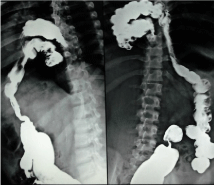
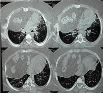
A 10 year old girl with a weight of 67 kg and BMI of 27.8, a known case of Down syndrome with right sided Morgagni hernia was posted for laparoscopic repair. The child, since birth had a history of repeated respiratory tract infections which used to subside in 15 days more than its usual course. One year back she developed respiratory distress following an episode of upper respiratory tract infection and fever which needed hospitalization with mechanical ventilation for 18 days and was diagnosed as a case of Morgagni hernia (right). There was no history of abdominal pain, vomiting. Barium studies (Figure 1) had shown transverse, ascending colon and caecum at high position on right side of abdomen with anterior diaphragmatic hernia. CT scan chest (Figure 2) showed right central and anterior hemi diaphragmatic hernia with mass effect seen on lung parenchyma, Thyroid function test and echocardiography both were normal and pulmonary function test had shown moderate restriction. Chest radiograph revealed widening of the mediastinum with raised right hemi diaphragm. Patient had clinical features of Down syndrome (mental impairment, flat facial features, low set ears, simian crease and mongoloid slant). Mouth opening was adequate without macroglossia and airway was Mallampati class II. Air entry was decreased on right lower zone. Preoperative ABG was normal. Intravenous Venous (IV) access (20G) was secured preoperatively at the dorsum of left hand after the application of EMLA cream followed by administration of 1mg IV midazolam to allay the anxiety. In an operation theatre, all essential monitors were connected (ECG, NIBP, Temperature, ETCO2, SpO2). Inj fentanyl 80 μg, dexamethasone 4mg were administered IV and induced with propofol 2mg.kg-1. Intubation achieved with succinylcholine 100mg IV and airway was secured with 6.0mm cuffed endotracheal tube. Anesthesia was maintained with oxygen (FiO2-0.4), air and desflurane (MAC 1.2%) and positive end expiratory pressure of 4 mmHg was added to adjust normocapnia. Intravenous paracetamol 15mg.kg-1, fentanyl 30μg, ondansetron 4mg and intermittent boluses of vecuronium were administered intraoperatively. Continuous Invasive BP (120/60- 100/68 mmHg) and EtCO2 (35-40 mmHg) were monitored along with intraperitoneal insufflation pressure which were kept between 9-10mmHg. Intraoperative ABG was normal. Surgery included the insertion of a 10 mm umbilical and two 5 mm working ports on both sides of the midline in the upper abdomen. Hernial contents, i.e. transverse colon and a very heavy fat laden omentum were easily reduced. The sac was excised using ultrasonic scalpel without injuring pleura or pericardium. Due to obesity and the thickness of the abdominal wall intracorporeal tissue approximation of defect failed. It was closed by passing conventional 2 inch needle threaded with double 0 silk through the abdominal wall lower lip of defect, and then back to the abdominal wall under endoscopic vision. Multiple such sutures were placed and tied externally under the skin. Surgery lasted for four hours and anaesthesia was reversed with IV neostigmine (50μg.kg-1) and glycopyrrolate (10μg.kg-1). Extubation was successful and recovery was complete and patient was shifted to ICU on 4litres of oxygen, with an uneventful postoperative period.

    Figure 1:  Barium enema lateral view and anterior view, showing transverse
colon, ascending colon and caecum at high position on Right side of abdomen
with anterior diaphragmatic hernia.

    Figure 2:  CT scan showing Right central and anterior hemi diaphragmatic
hernia with mass effect.
Discussion
Morgagni hernia arises congenitally as a result of failure of fusion of septum transversum with costal arches. It arises 90% on the right side and remaining cases are either left sided or bilateral. Morgagni hernia usually contains the transverse colon, omentum, liver and sometimes stomach or small bowel [1]. In our patient the hernia contained transverse, ascending colon and omentum. Presentation varies from severe respiratory distress in infancy to subtle gastrointestinal symptoms or recurrent pneumonia in older children as in the index case, which usually creates a diagnostic dilemma to the clinician [2]. Congenital Morgagni hernia can be associated with other congenital anomalies like, Down syndrome as in our case, congenital heart disease (ASD, VSD), malrotation, chest wall abnormality, genetic syndromes like Turner syndrome, Noonan syndrome [1,3,4].
Recurrent chest infections, pneumonia per se are very common in Down syndrome and the cause remains multifactorial which include immunodeficiency, airway abnormalities, gastro esophageal reflux, and hypotonia [5]. The overall incidence of Down syndrome with congenital Morgagni hernia varies from 20-30% in various studies. A multicenter study by Ahmed et al has reported about 28.3% incidence of Down syndrome with Morgagni hernia [4].
Conventionally, the open surgical approach by transabdominal or transthoracic route was the gold standard for the repair of congenital morgagni hernia. In the recent years, minimal access surgery has evolved as a safe and effective option in terms of lesser postoperative pulmonary morbidity, especially in high risk cases with associated morbidity as in the index case [6-7].
In view of high prevalence of airway abnormalities in a Down syndrome child and increased weight induced intraoperative hemodynamic disturbances with increased incidence of postoperative airway morbidity; desflurane was selected as an inhalational agent of choice. Desflurane having low potency, low blood gas coefficient, low fat-blood solubility coefficient makes it an ideal inhalational agent suitable for subset of high risk cases as our index case to achieve rapid emergence [8].
Conclusion
congenital Morgagni hernia with Down syndrome is a rare clinical entity, diagnosed incidentally in pediatric age group presenting with recurrent pneumonia or tachyapnea. Anesthesia management of such patients for the repair is a challenge in view of associated congenital anomalies and prevalence of airway abnormalities. Recent review of literature shows that outcome is better with lower postoperative morbidity if such patients undergo minimally access surgery or repair.
Acknowledgement
Published with written consent of parent.
References
- Beg MH, Rashidi ME, Jain V. Morgagni Hernia with Down syndrome: A Rare Association. Indian J Chest Dis Allied Sci. 2010; 52: 115-117.
- Jennifer Marin and John Lopoo. An Infant with Trisomy 21 and Tachypnea. Pediatric Emergency Care. 2006; 22: 170-172.
- Mert M, Gunay I. Transsternal Repair of Morgagni Hernia in a Patient with Coexistent Ventricular Septal defect and down syndrome. Acta chir belg. 2006; 106: 739-740.
- Al-Salem AH, Zamakhshary M, Al Mohaidly M, Al-Qahtani A, Abdulla MR, Naga MI. Congenital Morgagni Hernia –A national multiceter study,journal of pediatric surgery. 2014; 49: 503-507.
- Picard E, Ben Nun A, Fisher D, Schwartz S, Goldberg M, Goldberg S. Morgagni hernia mimicking pneumonia in Down syndrome. Journal of Pediatric Surgery. 2007; 42: 1608-1611.
- Mallick MS, Algahtani A. Laparoscopic assisted repair of Morgagni hernia in children. J Pediatr surgery. 2009; 44: 1621-1624.
- De Vogelaere K. Laparoscopic repair of Morgagani diaphragmatic hernia: A new case. Surgical Laparoscopy, endoscopy and percutaneous techniques. 2003: 13: 401-403.
- La Colla L, Albertin A, La Colla G, Mangano A. Faster wash-out and recovery for desflurane vs sevoflurane in morbidly obese patients when no premedication is used. BJ A. 2007; 99: 353–358.