Research Article
Ann Carcinog. 2017; 2(1): 1007.
Effectiveness of Newly Developed Water-Equivalent Mouthpiece during External Beam Radiotherapy for Oral Cancer
Kudoh T¹*, Ikushima H², Kudoh K¹, Furutani S³, Kawanaka T³, Kubo A³, Takamaru N¹, Tamatani T¹ and Miyamoto Y¹
¹Department of Oral Surgery, Tokushima University Graduate School, Japan
²Department of Radiation Therapy Technology, Tokushima University Graduate School, Japan
³Department of Radiology, Tokushima University Graduate School, Japan
*Corresponding author: Takaharu Kudoh, Department of Oral Surgery, Subdivision of Molecular Oral Medicine, Institute of Health Biosciences, Tokushima University Graduate School, 3-18-15 Kuramoto-cho, Tokushima, Japan
Received: March 14, 2017; Accepted: March 30, 2017; Published: April 06, 2017
Abstract
The objective of this study was to research the effectiveness of newly developed water-equivalent mouthpiece during external beam radiotherapy for oral cancer. In external beam radiotherapy for cancer of the tongue, floor of the mouth, and lower gingiva, it is possible to prescribe a low dose to the upper gingiva and hard palate at an open mouth position using a mouthpiece. However, the inhomogeneity correction resulting from the air cavity and the mobility of the tongue produced by an open mouth position should be considered. Therefore, a new mouthpiece was designed to be fixed by the dental arch, and the air cavity of the mouth can be filled with water-equivalent material. In 30 patients with previously treated oral cancer, the simulated homogeneity index of the calculated water-equivalent mouthpiece by a treatment-planning system was significantly better than that of a conventional mouthpiece (p = 0.004). This new mouthpiece facilitates excellent dose distribution while attaining immobilization of the tongue in patients with oral cancer.
Keywords: External beam radiation therapy; Oral cancer; Head and neck cancer; Mouthpiece; Water-equivalent; Immobilization
Abbreviations
EBRT: External Beam Radiotherapy; 3D-CRT: Three- Dimensional Conformal Radiotherapy; CT: Computed Tomography; GTV: Gross Tumor Volume; CTV: Clinical Target Volume; PTV: Planning Target Volume; HI: Homogeneity Index; D max: maximum dose of Planning Target Volume (PTV); D min: minimum dose of PTV; IMRT: Intensity-Modulated Radiotherapy; N: Numbers; WE MP: Water-Equivalent Mouthpiece; C MP: Cotton piece; AL: Antero- Lateral beams; LO: Lateral Opposed beams; WE MP HI: Homogeneity Index of Water-Equivalent Mouthpiece; C MP HI: Homogeneity Index of rolled Cottonpiece; SD: Standard Deviation of mean; SEM: Standard Error of Mean
Introduction
Radiotherapy is widely utilized for oral cancer treatment in an effort to preserve chewing, speech, and swallowing functions and the shape of the oral structures. A mouthpiece is used to depress the tongue away from the hard palate in External Beam Radiotherapy (EBRT) for patients with cancer of the floor of the mouth, lower gingiva, and tongue [1]. It is possible to administer adequate EBRT doses to tumors in patients with cancer of the tongue and floor of the mouth while sparing the upper gingiva and hard palate. Mouthpieces have the benefits of reducing the dose administered to tongue of patients with cancer of the upper gingiva, hard palate, and maxillary sinus and reducing the dose administered to the upper gingiva, hard palate, and maxillary sinus of patients with cancer of the tongue.
According to Willner, et al. an individualized mouthpiece can facilitate accurate and reproducible positioning in patients with head and neck cancer using Three-Dimensional Conformal Radiotherapy (3D-CRT) [2]. They reported that random and systematic deviations in each of the three directions (craniocaudal, anteroposterior, and mediolateral axes) are within the range of ±4 mm and within the range or even less than the deviations described for most thermoplastic masks.
A mouthpiece is used to both depress and fix the tongue during simulation and treatment of EBRT, especially in patients with cancer of the tongue or floor of the mouth [3,4]. However, the position of the mouthpiece may be uncontrollable during treatment [5]. Additionally, interfractional organ motion of the tongue results in uncertainty because of involuntary tongue movement. The silicone mouthpiece, which is fixed by the dental arch, can be steadily immobilized while depressing the tongue. Stereotactic radiotherapy to metastatic brain tumors has a biological advantage over stereotactic radiosurgery [6- 8]. The custom-made mouthpiece worn on the palate increases the setup accuracy in stereotactic radiotherapy to the metastatic brain tumor [9-11]. The silicone mouthpiece also allows the user to easily attain a stable tongue position. However, scattered radiation can be produced because the silicone must be within the treatment field of patients with oral cancer, and severe stomatitis as the reason of an interrupting radiotherapy occurs.
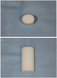
A conventional method using a rolled cotton piece has been used to depress the tongue and immobilize the mandible (Figure 1). This cotton piece is influenced by scattered radiation less than silicone mouthpiece, and is easy to wear, low-cost, and hygienic. However, it is thought to have the disadvantage of positioning accuracy compared with the silicone mouthpiece with respect to fixation of the tongue. Therefore, we developed a new water-equivalent mouthpiece that can improve the inhomogeneity correction while maintaining accurate immobilization. It can be fixed by the dental arch, and the air cavity of the mouth can be filled with water-equivalent material.

    Figure 1: The conventional mouthpiece using a rolled cotton piece.
A: Superior aspect. B: Lateral aspect.
The purpose of this study was to design a mouthpiece that can fills the air cavity of the mouth with a water-equivalent material while immobilizing the tongue without a delay in the commencement of EBRT, and to verify the usefulness of this new mouthpiece by simulation using a treatment-planning workstation.
Materials and Methods
Preparation of water-equivalent mouthpiece to immobilize the tongue
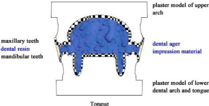
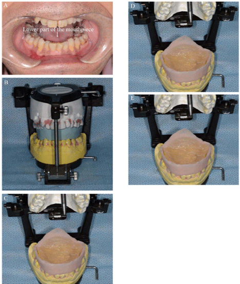
An impression of the upper dental arch was routinely taken. An impression of both the lower dental arch and the tongue together was taken using an upper impression tray to immobilize the tongue. Thermoplastic disks of 1-mm thickness (Erkodur; Erkodent, Pfalzgratenweiler, Germany) were then pressed by a thermoplastic former (Erkopress; Erkodent) to the upper and lower plaster models (Figure 2). The lower part of the mouthpiece was set onto the lower dental arch to immobilize the tongue (Figure 3a). A silicone bite was taken in the open mouth position and cut along the buccal edge of each dental arch for enclosure by the tray resin. The posterior limit was extended as long as possible to fill the air cavity of the mouth.

Figure 2: Schema of a new water-equivalent mouthpiece.
The plaster models and mouthpieces were mounted to an articulator (Figure 3b). The tray resin was enclosed around the silicone bite as thinly as possible from the upper to lower teeth. After the silicone bite was removed, an agar impression material (Dupligel; Shofu, Kyoto, Japan) was poured into the space (Figure 3c). The final mouthpiece was almost equivalent to water, in contrast to silicone material, because thinner resin and water-equivalent agar impression material were used (Figure 3d). Figure 3e shows this mouthpiece in the mouth of a volunteer.

    Figure 3: A: Lower part of the mouthpiece. B: Plaster models and
mouthpieces mounted to an articulator. C: Agar impression material poured
into the space. D: Water-equivalent mouthpiece mounted to an articulator. E:
Water-equivalent mouthpiece in the mouth of a volunteer.
Analyses of the effect of the new water-equivalent mouthpiece on dose distribution
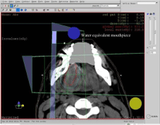
In total, 111 patients with oral cancer were treated by EBRT from September 2002 to August 2011 in Tokushima University Hospital. Thirty dentulous patients who underwent irradiation of cancer of the tongue, lower gingiva, and floor of the mouth by the conventional method using a rolled cotton piece in the open mouth position were included in this institutional review board–approved clinical investigation. The numbers of primary cancer sites involving the tongue, lower gingiva, and floor of the mouth were 13, 9, and 8, respectively (Table 1). Planning Computed Tomography (CT) images were reconstructed to a 3-mm thickness with a 1-mm slice thickness using an Asteion (Toshiba Medical Systems, Tochigi, Japan). The CT data were exported to a treatment planning workstation (XiO; Elekta, Inc., MO, USA). The calculated water-equivalent electron density mouthpiece was delineated on the CT images using commercial treatment planning software possessing the ability to delineate an outline with any electron density. Figure 4 shows the delineation of the calculated water-equivalent mouthpiece. According to our policy, the Gross Tumor Volume (GTV) – the Clinical Target Volume (CTV) margin, Planning Target Volume (PTV) – CTV margin, and leaf margin were set to 5, 5, and 3 mm, respectively. A 4-MV photonpenetrating machine was used (MEVATRON KD2/7450; Siemens, Tokyo, Japan). The field arrangements were as follows: a 15° opposed lateral wedge pair technique if the GTV was across the midline, and a 30° anterolateral wedge pair technique if the GTV was not across the midline. A bolus was used to improve the dose distribution if the PTV was close to the skin surface. Dose homogeneity was assessed with the Homogeneity Index (HI), which is defined as the maximum PTV dose (Dmax) / minimum PTV dose (Dmin) to evaluate the usefulness of the water-equivalent mouthpiece and conventional method using a rolled cotton piece.

Figure 4: Delineation of water-equivalent mouthpiece.
    Primary site
    Tongue
    Lower gingiva
    Floor of the mouth


    N
    13
    9
    8


    N: Number
Table 1: The numbers of primary cancer sites.
Statistical analysis
All statistical analyses were performed using PASW Statistics 18 (SPSS Japan Inc., Tokyo, Japan). The HI calculated from XiO of both the conventional method using a rolled cotton piece and the new mouthpiece was tested using Student’s t-test. The difference in the HI between with and without the bolus and in each beam direction was tested using Levene’s test. Intergroup differences were considered statistically significant when the p value was <0.05.
Results
According to the degree of invasion of the primary lesion, 21 patients were planned to undergo the 15° opposed lateral wedge pair technique and 9 patients were planned to undergo the 30° anterolateral wedge pair technique. Because the Dmin was <65% of the prescribed doses without a bolus, there were 13 patients with a bolus (data not shown).
Inhomogeneity correction: analysis of calculated waterequivalent mouthpiece method
The Dmax, Dmin, and HI of the water-equivalent mouthpiece method and conventional method using a rolled cotton piece in different beam directions and with and without a bolus were calculated (Table 2). Table 3 shows the HI of each method.
    WE MP
    C MP
    Beam directions
    Bolus


    Dmax
    Dmin
    HI
    Dmax
    Dmin
    HI


    212
    167
    1.27
    212
    167
    1.27
    AL
    With


    223
    160
    1.39
    224
    160
    1.40
    AL
    With


    227
    161
    1.41
    228
    159
    1.43
    AL
    With


    210
    154
    1.36
    208
    151
    1.38
    AL
    With


    213
    160
    1.33
    212
    159
    1.33
    AL
    Without


    218
    156
    1.40
    218
    156
    1.40
    AL
    Without


    218
    168
    1.30
    218
    168
    1.30
    AL
    Without


    215
    156
    1.38
    217
    156
    1.39
    AL
    Without


    220
    156
    1.41
    224
    154
    1.45
    AL
    Without


    219
    153
    1.43
    219
    153
    1.43
    LO
    With


    231
    154
    1.50
    231
    155
    1.49
    LO
    With


    233
    158
    1.47
    235
    161
    1.46
    LO
    With


    228
    153
    1.49
    228
    153
    1.49
    LO
    With


    213
    155
    1.37
    214
    145
    1.48
    LO
    With


    216
    154
    1.40
    216
    156
    1.38
    LO
    With


    219
    153
    1.43
    219
    153
    1.43
    LO
    With


    219
    154
    1.42
    219
    150
    1.46
    LO
    With


    211
    145
    1.46
    218
    136
    1.60
    LO
    With


    206
    165
    1.25
    207
    165
    1.25
    LO
    Without


    207
    166
    1.25
    207
    147
    1.41
    LO
    Without


    214
    167
    1.28
    214
    152
    1.41
    LO
    Without


    210
    162
    1.30
    224
    165
    1.36
    LO
    Without


    214
    159
    1.35
    215
    154
    1.40
    LO
    Without


    214
    156
    1.37
    214
    157
    1.36
    LO
    Without


    213
    153
    1.39
    213
    152
    1.40
    LO
    Without


    230
    157
    1.46
    230
    157
    1.46
    LO
    Without


    224
    158
    1.42
    228
    158
    1.44
    LO
    Without


    222
    148
    1.50
    223
    150
    1.49
    LO
    Without


    225
    157
    1.43
    226
    148
    1.53
    LO
    Without


    223
    154
    1.45
    223
    154
    1.45
    LO
    Without


    WE MP:    Water-Equivalent Mouthpiece; C MP: Cotton Piece; Dmax (Gy): maximum dose of Planning    Target Volume (PTV); Dmin (Gy): minimum dose of PTV; HI: Homogeneity Index;    AL: Antero-Lateral beams; LO: Lateral Opposed beams.
Table 2:  Dmax, Dmin and HI of the water-equivalent and cotton piece by two
different beam directions and with/without of bolus.
    Mean
    SD
    SEM


    WE MP HI
    1.389
    0.072
    0.013


    C MP HI
    1.418
    0.074
    0.013


    WE MP HI:    Homogeneity Index of Water-Equivalent Mouthpiece; C MP HI: Homogeneity Index    of rolled Cotton piece; SD: Standard Deviation of mean; SEM: Standard Error    of Mean.
Table 3: HI of the ideal water-equivalent mouthpiece and rolled cotton piece.
The mean HI using the calculated water-equivalent mouthpiece was 1.39, and that of the conventional method using a rolled cotton piece was 1.42. The HI of the calculated water-equivalent mouthpiece was significantly better than that of the conventional method using a rolled cotton piece (Student’s t-test, p: 0.004). The HIs of the calculated water-equivalent mouthpiece in two different beam directions and with and without the bolus are shown in (Tables 4 & 5). There were no significant differences with the two different beam directions (Levene’s test, p: 0.819) or with or without the bolus (Levene’s test, p: 0.079).
    N
    Mean
    SD
    SEM


    AL
    9
    1.361
    0.051
    0.017


    LO
    21
    1.401
    0.077
    0.017


    AL:    Antero-Lateral beams; LO: Lateral Opposed beams; N:    Number; SD: Standard Deviation of mean; SEM: Standard Error of Mean.
Table 4: HI of ideal water-equivalent mouthpiece by the different beam directions.
    N
    Mean
    SD
    SEM


    Without bolus
    17
    1.369
    0.075
    0.018


    With bolus
    13
    1.415
    0.061
    0.017


    N: Numbers; SD:    Standard Deviation of mean; SEM: Standard Error of Mean.
Table 5: HI of ideal water-equivalent mouthpiece with/without bolus.
Discussion
Positioning accuracy is required to minimize the CTV-PTV margin during EBRT. Because this new mouthpiece immobilizes the tongue, the inaccuracy caused by involuntary movement and flexibility of the tongue is none. The method used to prepare the mouthpiece can be standardized although it is custom-made. Additionally, the delay in the commencement of radiotherapy is shortened because the mouthpiece can be fabricated using a conventional upper impression tray and conventional dental materials. Finally, this new mouthpiece fills the air cavity of the mouth with water-equivalent material to improve the inhomogeneity correction. Therefore, the dose homogeneity using this new mouthpiece is expected to be better than that using the conventional method with a rolled cotton piece.
A two-dimensional treatment plan was used until 2002 in Tokushima University Hospital. Thereafter, 30 eligible dentulous patients were selected to undergo the conventional method using a rolled cotton piece to simulate the dose volume histogram using this new mouthpiece. Only two treatment field arrangements were used. The PTV of the primary lesion (not involving the neck area) was evaluated because the influence of the inhomogeneity correction mainly occurred around the mouthpiece. The mean HI of the calculated water-equivalent mouthpiece was better than that of the conventional method using rolled cotton piece although this study was small sample size. And there were no significant differences with the two different beam directions. On the other hand, there was a trend towards the significance with or without the bolus. For all 13 patients with a bolus, the thickness of the bolus was set to be minimum so that Dmin was to be more than 65% of the prescribed dose. Therefore, it was thought that the patients with a bolus had a trend toward high HI in spite of compensation by bolus. A previously described mouthpiece was used to depress the tongue and immobilize the mandible in patients with head and neck cancer [2,4,5]. The mouthpiece used during EBRT in patients with head and neck cancer is fabricated from high-density material more than soft tissues (i.e., silicone, or similar materials). The delineation of the tissue near the mouthpiece is influenced by the scattered radiation, even if the image is fused with magnetic resonance imaging to delineate the contour.
There are two precautions associated with this new waterequivalent mouthpiece. First, this new water-equivalent mouthpiece cannot be stabilized during each treatment session in edentulous patients because it can be fixed with the teeth. Therefore, it is not applicable to edentulous patients. Second, the posterior border should be limited to the anterior border of the soft palate to reduce the gag reflex.
EBRT shift from 3D-CRT to Intensity-Modulated Radiotherapy (IMRT). Patients undergoing IMRT exhibit comparable acute and significantly less severe late toxicity than do those undergoing 3D-CRT [12]. Therefore, IMRT for oral cancer has usually been performed to spare the parotid gland [13-15]. Wagner, et al. refrained from the use of a mouthpiece during treatments such as IMRT to reduce the dose and increase the accuracy of the treatment in patients with head and neck cancer [5]. At Tokushima University Hospital, IMRT for head and neck cancer is currently performed with a closed mouth in patients with an unstable mandible. However, there are patients in whom the tongue contacts the palate on treatment-planning CT images in the closed mouth position. The dose for normal tissues within the PTV margins is high in these patients. The normal adjacent tissues near the tumor can be positioned for separation from the opposite side of the jaw using this new mouthpiece. Thus, the inhomogeneity correction may be improved because of the use of the water-equivalent material. The positioning accuracy of this new mouthpiece is considered to be equivalent to that of a silicone mouthpiece by fixation using the dental arch.
Postoperative radiation alone or combined with concurrent chemotherapy is an established adjuvant treatment in patients at high risk for disease recurrence. Such risk factors include positive margins, extracapsular nodal extension, lymphovascular invasion, and perineural invasion [16]. The optimal management of oral cancer typically involves surgical resection followed by adjuvant radiotherapy or chemoradiotherapy in the setting of such risk factors, and the outcomes of patients who were treated with a radiation-based approach were consistently inferior to those treated with surgery followed by adjuvant radiotherapy or chemoradiotherapy [17]. Definitive IMRT using this new mouthpiece would allow for better locoregional control in patients with oral cancer because a better dose distribution can be attained. Furthermore, this new mouthpiece is thought to be effective against oral cancer and other head and neck cancers, such as cancer of the maxillary sinus, soft palate, and base of the tongue.
In conclusion, the calculated HI for 30 previously treated patients with oral cancer significantly improved with the use of the herein-described water-equivalent mouthpiece. The mouthpiece is custom-made for immobilization of the tongue, but the preparation method can be standardized. In addition, the mouthpiece can be rapidly fabricated by a general practitioner without a delay in the commencement of EBRT. This new mouthpiece is expected to be used in not only 3D-CRT but also IMRT for head and neck cancer.
Acknowledgement
We are grateful to Dr. Omar Rodis for providing constructive comments and useful feedback and to Dr. Makoto Fukui for assisting with the statistical analysis.
References
- Cox J, Ang K. Radiation Oncology: Rationale, Technique, Results 8th edition (St. Louis, MO: Mosby). 2003; 241-244.
- Willner J, Hädinger U, Neumann M, Schwab FJ, Bratengeier K, Flentje M. Three dimensional variability in patient positioning using bite block immobilization in 3D-conformal radiation treatment for ENT-tumors. Radiother Oncol. 1997; 43: 315-321.
- Brady P. Principles and Practice of Radiation Oncology 5th edition (Philadelphia, PA: Lippincott Williams &Wilkins). 2008; 902-903.
- Juliana RV, Fabio AA, Jose DP, Karina WB, Milena P, Maria Leticia GS, et al. Impact of intraoral stent on the side effects of radiotherapy for oral cancer. Head and Neck. 2013; 35: 213-217.
- Wagner D, Anton M, Vorwerk H. Dose uncertainty in radiotherapy of patients with head and neck cancer measured by in vivo ESR/alanine dosimetry using a mouthpiece. Phys Med Biol. 2011; 56: 1373-1383.
- Ikushima H, Tokuuye K, Sumi M, Kagami Y, Murayama S, Ikeda H, et al. Fractionated stereotactic radiotherapy of brain metastases from renal cell carcinoma. Int J Radiat Oncol Biol Phys. 2000; 48: 1389-1393.
- Manning MA, Cardinale RM, Benedict SH, Kavanagh BD, Zwicker RD, Amir C, et al. Hypofractionated stereotactic radiotherapy as an alternative to radiosurgery for the treatment of patients with brain metastases. Int J Radiat Oncol Biol Phys. 2000; 47: 603-608.
- Jyothirmayi R, Saran FH, Jalali R, Perks J, Warrington AP, Traish D, et al. Stereotactic radiotherapy for solitary brain metastases. Clin Oncol (R Coll Radiol). 2001; 13: 228-234.
- Masi L, Casamassima F, Polli C, Menichelli C, Bonucci I, Cavedon C. Cone beam CT image guidance for intracranial stereotactic treatments: Comparison with a frame guided setup. Int J Radiat Oncol Biol Phys. 2008; 71: 926-933.
- Bednarz G, Machtay M, Werner-Wasik M, Downes B, Bogner J, Hyslop T, et al. Report on a randomized trial comparing two forms of immobilization of the head for fractionated stereotactic radiotherapy. Med Phys. 2009; 36; 12-17.
- Baumert BG, Egli P, Studer S, Dehing C, Davis JB. Repositioning accuracy of fractionated stereotactic irradiation: Assessment of isocentre alignment for different dental fixations by using sequential CT scanning. Radiother Oncol. 2005; 74; 61-66.
- Chen WC, Hwang TZ, Wang WH, Lu CH, Chen CC, Chen CM, et al. Comparison between conventional and intensity-modulated post-operative radiotherapy for stage III and IV oral cavity cancer in terms of treatment results and toxicity. Oral Oncol. 2009; 45: 505-510.
- Claus F, Duthoy W, Boterberg T, De Gersem W, Huys J, Vermeersch H, et al. Intensity modulated radiation therapy for oropharyngeal and oral cavity tumors: clinical use and experience. Oral Oncol. 2002; 38: 597-604.
- Ahmed M, Hansen VN, Harrington KJ, Nutting CM. Reducing the risk of xerostomia and mandibular osteoradionecrosis: the potential benefits of intensity modulated radiotherapy in advanced oral cavity carcinoma. Med Dosim. 2009; 34: 217-224.
- Daly ME, Le QT, Kozak MM, Maxim PG, Murphy JD, Hsu A, et al. Intensity-modulated radiotherapy for oral cavity squamous cell carcinoma: patterns of failure and predictors of local control. Int J Radiat Oncol Biol Phys. 2011; 80: 1412-1422.
- Yao M, Chang K, Funk GF, Lu H, Tan H, Wacha J, et al. The failure patterns of oral cavity squamous cell carcinoma after intensity-modulated radiotherapy-the University of Iowa experience. Int J Radiat Oncol Biol Phys. 2007; 67: 1332-1341.
- Sher DJ, Thotakura V, Balboni TA, Norris CM, Haddad RI, Posner MR, et al. Treatment of Oral Cavity Squamous Cell Carcinoma With Adjuvant or Definitive Intensity-Modulated Radiation Therapy. Int J Radiat Oncol Biol Phys. 2011; 81; 215-222.