Case Report
Ann Carcinog. 2018; 3(1): 1013.
Large Diameter Esophageal Leiomyomas
Simoglou C¹* and Gymnopoulos D²
¹Department of Cardiothoracic Surgery, General University Hospital of Evros, Democritus University of Thrace, Greece
²Department of Cardiothoracic Surgery, Private Hospital St. Luke’s, Greece
*Corresponding author: Christos Simoglou, Department of Cardiothoracic Surgery, General University Hospital of Evros, Democritus University of Thrace, Greece
Received: February 16, 2018; Accepted: May 22, 2018; Published: May 29, 2018
Abstract
Leiomyomas are the most common benign tumor of the esophagus. Featured mainly in middle age and is twice as frequency in men. Leiomyomas are the most common benign tumours of the esophagus. The most frequent symptoms are dysphagia and epigastric pain, although 50% of patients remain asymptomatic and the tumour is sometimes discovered incidentally. We report the case of a 51 years old male patient with esophageal leiomyoma 7 cm diameter and emphasis on the operative management by open surgery. In this study we present our experience using open surgery.
Keywords: Dysphagia; Leiomyoma; Esophagoscopy; Esophageal tumor
Introduction
Benign tumors of the esophagus are rare lesions that constitute less than 1% of esophageal neoplasms. Leiomyomas represent a hyper proliferation of interlacing bundles of smooth muscle cells that are well-demarcated by adjacent tissue or by a smooth connective tissue capsule. They usually arise as intramural growths, most commonly along the distal two thirds of the esophagus. They are multiple in approximately 5% of patients [1]. The majority of leiomyomas have been discovered incidentally during evaluation for dysphagia or during autopsy. Bleeding rarely occurs in cases of benign disease but typically is observed with leiomyosarcoma, the malignant counterpart of this tumor. The potential for malignant degeneration of leiomyomas is extremely small. In the distal esophagus, leiomyomas may reach large proportions and may encroach on the cardia of the stomach [2].
Benign tumors of the esophagus are rare and leiomyoma is the most common benign tumor of the esophagus. It is reported that most leiomyomas originate the inner circular muscle layer of the distal and midthoracic esophagus, particularly at the esophagogastric junction. Middle-aged men are most frequently affected. The main symptoms usually are dysphagia and epigastric pain, but they are not specific for the disease. The size of the esophageal leiomyoma may change; a size of 1 to 29 cm has been defined in the literature. But most of them were smaller than 5 cm in diameter. Tumor that size larger than 7cm is rare [3]. It was easily misdiagnosis as mediastinal mass, esophageal cancer and esophageal stromal tumor. Its clinical feature and management are different with other smaller esophageal leiomyoma [4]. Here, a patient with large calcified esophageal leiomyoma who was treated in our institute is presented, initial discuss its diagnosis and management against the background of previously published cases and series.
Case Report
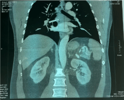
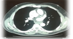
A 51-year-old man was admitted to our clinic without any symptoms. There was no nausea, vomiting, or weight loss. Results of a physical examination and standard laboratory tests were normal. A chest radiograph showed a mass in the right low mediastinum, and a filling defect was apparent on esophagography. Computerized tomography (CT) scanning of the chest revealed an irregularly shaped mass, 7cm mass, at the right lower posterior mediastinum narrowing the esophagus lumen. (Figures 1 & 2). Growth over the organ borders or infiltration in neighboring structures was not detected. There were two enlarged lymph nodes diameter 1, 6 cm and 2, 5 cm respectively, and there was no evidence for distant metastases. A right thoracotomy was performed. Opening the mediastinal pleura and manufacture of performs with careful detachment of the muscle layer and the mucosa of the esophagus. The muscular layer of the esophagus was repaired with Vicryl 3/0 sutures. Check for bleeding and air leak from the lung parenchyma. During surgical exploration, an unregular calcified mass in the distal esophagus. The mass was enucleated completely. Histopathologic examination revealed a tumor of 70x40x40 cm and calcified spindle cell fascicles without mitosis or atypical was observed. The patient was hospitalized seven days in our clinic and relief of symptoms, with no perioperative morbidity or mortality.

    Figure 1:  Computerized Tomography (CT): Medistinal mass of the post
mediastinum (esophageal leiomyoma).

    Figure 2:  Computerized Tomography (CT): large esophageal leiomyoma
7cm.

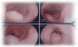
    Figure 3:  Esophagoscopy: In the middle of the esophagus, smooth swelling
without altering mucosal.
Discussion
Leiomyomas are benign tumors descending from smooth muscle cells of the esophagus. They are the most common benign tumors of the esophagus and they may occur in all parts of the esophagus, but 60% occur in the distal third, 30% in the middle, and 10% in the proximal esophagus. Leiomyomas are the commonest benign mesenchymal tumors of the esophagus contributing about two-thirds of all benign lesions of the esophagus [5]. Leiomyomas usually arise as intramural growths, most commonly along the distal two thirds of the esophagus. They are multiple in approximately 5% of patients [6]. If a leiomyoma is suspected at esophagoscopy, a biopsy should not be performed as it would cause scarring at the biopsy site, which would hamper definitive extramucosal resection at surgery (Figure 3).
Esophageal leiomyomas rarely cause symptoms when they are smaller than 5 cm in diameter. Large tumors can cause dysphagia, vague retrosternal discomfort, chest pain, esophageal obstruction, and regurgitation. Rarely, they can cause gastrointestinal bleeding, with erosion through the mucosa. Other than the nonspecific symptoms associated with esophageal leiomyomas, very few physical findings are usually noted [7]. In extremely rare cases where severe esophageal obstruction is caused by a leiomyoma, weight loss and muscle wasting may be observed.
Histologically, leiomyomas comprise of bundles of interlacing smooth muscle cells, well-demarcated by adjacent tissue or by a definitive connective tissue capsule. They are composed of fascicles of spindle cells that tend to intersect with each other at varying angles. The tumor cells have blunt-ended elongated nuclei and show minimal atypical with mitotic (Figure 3) [8].
Conclusion
Esophageal leimyoma is begin, rare tumor of esophagus. The tumor is often asymptomatic and the surgical removal is the treatment of choice. Attention must be paid to avoid trauma of laceration of mucosa due to high morbidity of this complication. Diagnosis of esophageal leiomyomas requires both endoscopic and radiologic examinations [9]. Our patient healed satisfactorily and the long-time quality of life was good during follow-up and it was coincident with the literature.
References
- Vermorken JB, Mesia R, Rivera F, Remenar E, Kawecki A, Rottey S, et al. Platinum-based chemotherapy plus cetuximab in head and neck cancer. N Engl J Med. 2008; 359: 1116-1127.
- Vermorken JB, Stöhlmacher-Williams J, Davidenko I, Licitra L, Winquist E, Villanueva C, et al. SPECTRUM investigators. Cisplatin and fluorouracil with or without panitumumab in patients with recurrent or metastatic squamous-cell carcinoma of the head and neck (SPECTRUM): an open-label phase 3 randomized trial. Lancet Oncol. 2013; 14: 697-710.
- Bossi P, Bergamini C, Siano M, Cossu Rocca M, Sponghini AP, Favales F, et al. Functional genomics uncover the biology behind the responsiveness of head and neck squamous cell cancer patients to cetuximab. Clin Cancer Res. 2016; 22: 3961-3970.
- Siano M, Espeli V, Mach N. Gene expression prediction of panitumumab efficacy in head and neck squamous cancer.
- Rischin D, Spigel DR, Adkins D, Wein R, Arnold S, Singhal N, et al. PRISM: Phase 2 trial with panitumumab monotherapy as second-line treatment in patients with recurrent or metastatic squamous cell carcinoma of the head and neck. Head Neck. 2015; 38: E1756-E1761.
- Custodio A, Feliu J. Prognostic and predictive biomarkers for epidermal growth factor receptor-targeted therapy in colorectal cancer: beyond KRAS mutations. Crit Rev Oncol Hematol. 2013; 85: 45-81.