Case Report
Ann Carcinog. 2025; 5(1): 1021.
Mucinous Tubular and Spindle Cell Carcinoma of Kidney - A Case Report
Tbouda M¹*, Jendouzi O³, Aggouri H¹, Oqbani K², Miry A² and Abbaoui S²
¹Department of Pathology, Oued Eddahab Military Hospital, Agadir, Morocco
²Department of Urology, University Hospital Center, Agadir, Morocco
³Departement of Radiology, Oued Eddahab Military Hospital, Agadir, Morocco
*Corresponding author: TBOUDA Mohammed, Department of Pathology, Oued Eddahab Military Hospital, Agadir, Morocco Email: dc.med.tbouda@gmail.com
Received: July 05, 2025 Accepted: July 23, 2025 Published: July 25, 2025
Abstract
Mucinous tubular and spindle cell carcinoma is a rare renal tumor that usually progresses favorably. It is often discovered by chance during radiological investigation for a particular cause. Diagnosis is often based on anatomopathology. Treatment is surgical. We report here the case of a patient with tubulomucinous and fusiform renal carcinoma treated by total nephrectomy.
Keywords: Carcinoma; Myxoid; Histology; Immunohistochemistry; Kidney; Surgery
Introduction
Mucinous tubular and spindle cell carcinoma (MTSCC) is a rare renal tumor. Since MTSCC was recognized as a distinct entity by the World Health Organization (WHO) in 2004, fewer than 100 cases have been reported in the literature, making it an extremely rare disease [1]. This subtype of renal cell carcinoma (RCC) primarily affects adults and is generally considered an indolent variant with a more favorable prognosis compared to other forms of RCC [2–3].
Diagnosis is typically based on histopathological examination, often supplemented by immunohistochemical analysis. The standard treatment is surgical, usually involving radical nephrectomy. The overall prognosis is favorable.
We report the case of a 58-year-old female patient diagnosed with MTSCC.
Observation
The patient, aged 58, with no previous pathological history, presented to the urology department with chronic abdominal and lumbar pain dating back more than 3 months, with occasional hematuria, all evolving in a context of preserved general condition. Clinical examination revealed lumbar tenderness with no palpable mass. The rest of the clinical examination was unremarkable.
The patient underwent a C-/C+ uroscan, which revealed a welllobar medio-lobar renal mass measuring
The mass exhibited minimal enhancement at the arterial phase with more enhancement in the venous phase (equivocal enhancement) and no renal vascular invasion.
Biological tests were unremarkable. The patient underwent total nephrectomy under general anesthesia.
The specimen was sent to the pathological anatomy laboratory.
Macroscopic examination revealed a well-limited medio-lobar tumour of yellowish-white color, dotted with a few haemorrhagic and cystic changes, with no visible tumour necrosis. The renal hilum and peri-renal fat were not invaded.
Histological examination reveals a carcinomatous tumour proliferation consisting of cells of variable size, organized in tubes and cellular cords, with a fusocellular component. The tumor stroma is abundantly myxoid.
On immunohistochemical study, tumor cells strongly express CK7, EMA, PAX8 and AMACR antibodies.
CD10 and CD117 antibodies are negative.
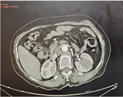
Figure 1: chromatogram of sample solution (80 μg/mL).
Discussion
Kidney tumours are uncommon tumours, ranking third after prostate cancer and bladder tumours. Most renal tumours are malignant. The most common histological type is clear-cell carcinoma.
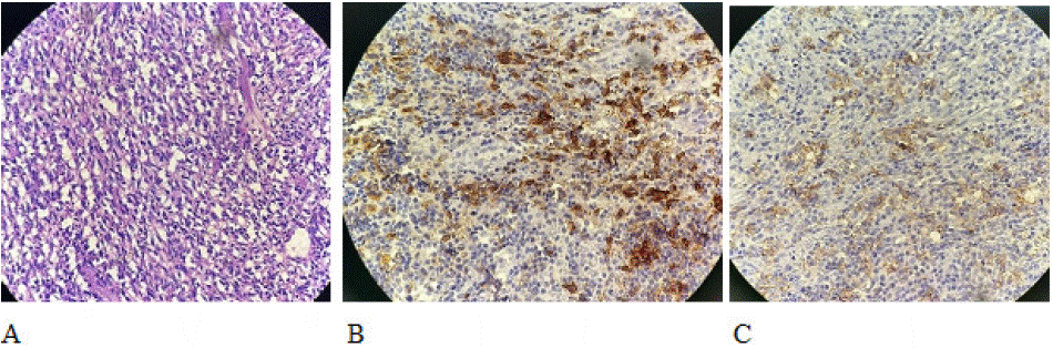
Figure 2: Tubular and spindle cell tumor proliferation (HE x100) (A). Expression of tumor proliferation antibodies EMA (B) and CK7 (C).
MTSCC, is a rare subtype of RCC, comprising 1-4% of all renal cell carcinoma [4-5]. This rare subtype was initially described by Lopez-Beltran et al [4]. This type of tumor most often affects adult women. Pediatric cases have been reported in the literature. The youngest reported case was in a 13-year-old boy with rapid disease progression and eventual fatality due to metastasis [6].
MTSCC, is often asymptomatic and discovered by chance following a radiological examination for another cause, but can sometimes be responsible for lumbar or abdominal pain, as in our patient's case.
The radiological work-up allows us to characterize the tumour, carry out an extension and operability assessment. In numerous instances, the identification of MTSCC can be suggested by analyzing a combination of CT/MRI features. Notably, the presence of slow enhancement with a plateau in dynamic contrast-enhanced CT/MRI, coupled with intermediate to high T2 signal intensity that contrasts with low apparent diffusion coefficient values on MRI, is indicative of this specific diagnosis [7].
However, the diagnosis of certainty is based on the anatomopathological study of the surgical specimen. Macroscopically, the tumour is often well-limited and yellowish-white in color, sometimes dotted with microcysts and sometimes with haemorrhagic changes.
Microscopic examination reveals a tumoral proliferation made up of tubular structures and pseudopapillaries mistaken for papillary carcinoma, with territories of spindle cells. The stroma is characteristically myxoid and abundant. Atypia are minimal. Diagnostic confirmation is usually based on an immunohistochemical study showing intense positivity to CK7, EMA and AMACR antibodies.
Treatment is essentially surgical, involving total or partial nephrectomy if the tumour is small. Progression is usually favorable.
Conclusion
MTSCC is a rare renal tumor. It is usually asymptomatic. Diagnosis is essentially anatomopathological. It is based on a morphological study supplemented by an immunohistochemical study.
Treatment is surgical. Evolution is favorable.
References
- Kobari Y, Yoshida K, Minoda R, Fukuda H, Hata K, Unagami K, et al. Longtime survival of a renal transplant recipient with metastatic mucinous tubular and spindle cell carcinoma: A case report. In Vivo. 2023; 37: 1394–1398.
- Sharma K, Dhua A, Agarwala S and Kaushal S. Mucinous tubular and spindle cell renal cell carcinoma (MTSC-RCC) with an unusual presentation in a child. J Kidney Cancer VHL. 2022; 9: 6-9.
- Mahmood ZH, Mohemed FM, Fatih BN, Qadir AA and Abdalla SH. Cancer publications in one year (2022); a cross-sectional study’: BMJ; 2023; 1.
- Lopez Beltran A, Scarpelli M, Montironi R and Kirkali Z. 2004 WHO classification of the renal tumors of the adults. Eur Urol. 2006; 49: 798-805.
- Shen SS, Ro JY, Tamboli P, Truong LD, Zhai Q, Jung SJ, et al. Mucinous tubular and spindle cell carcinoma of kidney is probably a variant of papil lary renal cell carcinoma with spindle cell features. Ann Diagn Pathol. 2007; 13: 13-21.
- Sharma K, Dhua A, Agarwala S and Kaushal S. Mucinous tubular and spindle cell renal cell carcinoma (MTSC RCC) with an unusual presentation in a child. J Kidney Cancer VHL. 2022; 9: 6.
- Cornelis F, Ambrosetti D, Rocher L, Derchi LE, Renard B, Puech P, et al. CT and MR imaging features of mucinous tubular and spindle cell carcinoma of the kidneys. A multi institutional review. Eur radiol. 2017; 27: 1087-1095.