1Department of Pediatric and Preventive Dentistry, K.G.M.U, India
2Department of Pediatric and Preventive Dentistry, Chandra Dental College, India
3Department of Community Dentistry, K.G.M.U, India
*Corresponding author: Ansari AA, Department of Pediatric and Preventive Dentistry, King George’s Medical University, Lucknow-226003, U.P, India
Received: October 30, 2014; Accepted: December 03, 2014; Published: December 04, 2014
Citation: Ansari AA, Pandey P, GuptaVK and Pandey RK. Bilateral Fusion of the Mandibular Primary Incisors with Hypodontia: A Case Report. Austin J Clin Case Rep. 2014;1(12): 1057. ISSN : 2381-912X
Fusion is a condition in which the crowns of two separate teeth have been joined together during the crown development. Although, its etiology is not known, it is believed that some physical forces or pressures cause the contact of developing teeth. Bilateral mandibular fusion of the primary incisors is a rare event, occurring with a prevalence of 0.01 to 0.04 percent. Fusion may cause esthetic and space problems, occlusal disturbances, and delayed eruption of permanent successors. The aim of this article is to highlight the rarity of occurrence of bilaterally fused mandibular primary central and lateral incisors; to identify congenital absence of successor teeth; and to discuss different treatment modalities in cases of fusion. An unusual case of bilateral fusion of primary mandibular incisors in a six-year-old boy is reported in this article. Clinical observation along with radiographic evaluation was used to arrive at diagnosis. Bilateral fusion of primary mandibular central and lateral incisors of incomplete type with congenital absence of left permanent mandibular central incisor is diagnosed. The rarity of absence of a single permanent central incisor makes the case further interesting.
Keywords: Fusion; Bilaterally fused teeth; Hypodontia; Partial anodontia; Primary fused teeth
Fusion is defined as the union of two independently developing primary or permanent teeth [1]. This union of two separate tooth germs may be either complete or incomplete [1]. Fused teeth have separate or shared pulp chambers and canals [2]. Clinically fused anterior teeth frequently have a groove or notch on the incisal edge that goes in buccolingual direction and radiographically with separate pulp chambers [3]. Although its etiology is unknown, it is believed that some physical forces or pressures from the adjacent tooth follicles, as well as hereditary conditions or racial differences; may be the cause of fusion [4]. Bilateral dental fusion in the primary dentition is a rare dental anomaly with its prevalence ranging from 0.01 to 0.04% [5]. Eighty three percent of the cases of bilateral fusion in the primary dentition are found in the mandible, out of which, 70% show the involvement of the lateral incisors and canines [6] with rare incidence of bilateral fused central and lateral incisors. The data for prevalence of bilateral fusion in Indian population are not available; the previous survey for unilateral fusion of primary teeth revealed 0.14%, as confirmed by Reddy and Munshi [7]. The most common problem related to fused teeth is hypodontia of the permanent dentition which has been observed in 50% of affected subjects [8]. The purpose of this article is to highlight the rarity of the condition; to identify congenital absence of succedaneous teeth and to discuss different treatment options in cases of the fused teeth. The case presents a six-year-old boy with bilateral fusion in his mandibular primary central and lateral incisor teeth with absence of only left permanent mandibular central incisor.
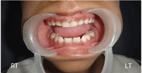
A six-year-old boy reported to the Department of Pediatric and Preventive Dentistry with the complaint of crowding of the teeth within his lower jaw. His medical history was irrelevant with his condition. There was no family history of dental anomalies and no consanguinity was reported in the parents. Intraoral examination revealed bilateral presence of unusually large teeth in the lower incisor region. Both sides were strongly suggestive of conjoined primary central and lateral incisors. Deep labio-lingual grooves were associated with both the enlarged teeth (Figure A).
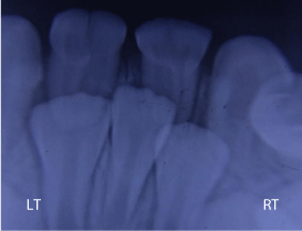
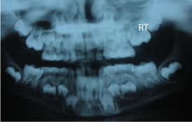
An Intra Oral Periapical Radiograph (IOPA) was advised. Radiographic evaluation of the IOPA revealed fused 71-72; and 81-82 with two crowns and two roots fused together and two separate root canals, one in each of the fused teeth indicating that the fusion was of incomplete type. Absence of a single incisor was also observed on the radiograph (Figure B). To confirm the findings and to detect any other abnormality an Orthopantomograph (OPG) was advised. The OPG confirmed the same findings (Figure C). It was very difficult to confirm whether the central incisor present in the middle was left or right at this stage as the three incisors were closely situated and were still within the bone. A thorough examination of the IOPA revealed that the middle incisor was right and the left incisor was missing. It was confirmed from the following ways 1. Morphology of the mamelons and crown found in the middle was much more like that of right mandibular permanent central incisor 2. This central incisor was present on the right side of the midline (Figure B) and 3. The path of eruption of the tooth present resembled more closely to that of right mandibular permanent central incisor. Hence, the patient was diagnosed to be a case of bilaterally fused mandibular primary central and lateral incisors of incomplete type with congenital absence of left permanent mandibular central incisor.
Incidence of bilateral fused central and lateral incisors, which was observed in our case, is even rarer than incidence of bilateral fusion of primary lateral and canine teeth in primary dentition, which itself is a very rare condition. Only three cases of bilateral fusion of mandibular primary central and lateral incisors in an Indian population have been reported yet [9-11]. Hypodontia of the permanent dentition, which was seen in our case is the most common problem related to the fused teeth. Gellin [12] reported that when the double tooth involved central and lateral incisors, only 37.5% of the cases presented with missing permanent successors. According to some studies no effect on the permanent successors was observed when the double teeth involved the primary mandibular central and lateral incisors [13-15]. Tiwari N and Pandey RK [10] reported bilateral fusion of mandibular primary incisors (71-72 and 81-82) with absence of both permanent central incisors while absence of both lateral incisors was reported with bilateral fusion of mandibular primary incisors (71-72 and 81- 82) by Chachra S and Sharma AK [9]. Kumar P et al. [11] reported bilateral fusion of primary mandibular central and lateral incisors with no missing permanent tooth. Contrary to these findings, only one succedaneous central incisor was found absent in our case despite being presence of bilateral fusion of primary anterior teeth. To the best of our knowledge, bilateral fusion of primary mandibular central and lateral incisors with missing single permanent mandibular central incisor has not been reported in Indian population yet. The etiology of the absence of a single permanent central incisor could not be traced.
Clinical problems that are caused due to fusion are esthetics, malocclusion, diastema formation, susceptibility to caries due to the presence of deep fissures and delayed resorption of the root due to greater root mass and increased area of root surface of the fused teeth [16]. These problems require both cosmetic and orthodontic considerations. If the fused tooth is free from caries, its treatment may not be required [17]. Only universal preventive advice should be given to the parent and the child, while in case of presence of caries, a restoration should be placed for the retention of the function and esthetics [17]. The presence of pulpal involvement requires endodontic treatment in the same way as for a multi-rooted tooth [18] but the approach of access opening may be different depending upon crown morphologies. Primary fused teeth may be retained as they are. If extraction is required, it is important to determine whether the corresponding succedaneous tooth/teeth is/are present or not [16]. In the present case since the patient had no caries and had already attained the age of six years, which is generally the age of exfoliation of the primary mandibular incisors, no treatment was planned and the child and parent were only advised to maintain good oral hygiene. In this case, one permanent central incisor was missing. The parent was informed about the orthodontic problems e.g. midline diastema, midline shifting, spacing etc. in this case that may follow the eruption of permanent incisors. Hence, instructions were given regarding the importance of long-term follow-up of the patient.
Tooth fusion is one of the rare anomalies of the shape of the tooth. To make a diagnosis clinical observation along with an orthopantomograph and periapical radiographs are necessary. A long-term follow-up is very important in most of the situations with preventive procedures and minimal intervention.
Intra oral photograph of the patient showing fused 71, 72 and 81, 82.

IOPA radiograph showing fused 71, 72 and 81, 82 and absence of left permanent central incisor.

OPG of the patient.
