
Special Article - Cataract Clinical Cases and Images
Austin J Clin Ophthalmol. 2015;2(2): 1046.
A Case with Lens Coloboma Treated by Cataract Surgery
Jia-Kang Wang1,2,3,4* and Sheng-Hsiang Ma5
1Department of Medicine, National Yang Ming University,Taiwan
2Department of Ophthalmology, Far Eastern Memorial Hospital, Taiwan
3Department of Health Care Administration and Department of Nursing, Oriental Institute of Technology, Taiwan
4Department of Medicine, National Taiwan University,Taiwan
5Department of Medicine, China Medical University, Taiwan
*Corresponding author: Jia-Kang Wang, Department of Medicine, National Yang Ming University, Taipei, Taiwan, 21, Sec 2, Nan-Ya South Road, Pan-Chiao District, New Taipei City, 220, Taiwan
Received: February 16, 2015; Accepted: April 24, 2015; Published: April 27, 2015
Abstract
Purpose: To present a case of with lens coloboma treated by cataract operation successfully.
Methods: A case report
Results: A five-year-old boy was referred to our clinic due to abnormal visual acuity test at school. He was a healthy, well-developed boy with unremarkable systemic disease. His Best Corrected Visual Acuity (BCVA) was 20/20 in the right eye. Because measurement of the refraction failed, the bare visual acuity and corrected with pinhole were both counting finger only in the left. Intraocular pressure, anterior segment, and fundus examination were normal in both eyes. No strabismus was found. Nasal side deficiency of lens substituted by a membrane was found in the left eye. Normal crystalline lens was noted in the contralateral eye. Lens coloboma with congenital cataract was diagnosed. After making 3-mm limbal wound, congenital cataract was removed by anterior CCC and lens aspiration. Posterior CCC and anterior vitrectomy on the lens side was performed to prevent posterior capsular or anterior hyaloid opacity. As the defect in his lens was enormous, intracapsular placement of Intraocular Lens (IOL) was not feasible. A 3-piece acrylic soft IOL (MA60MA, Alcon lab., Texas, USA) was placed in the sulcus. Mild temporal decentration of IOL was noted after the operation, without changes of IOL position during follow-up. Because amblyopia was diagnosed by poor BCVA as 0.025 fully corrected by myopia -3D and astigmatism -2.5D one month after the operation, occlusion therapy with correcting eyeglass started as 6 hours a day on the contralateral eye. His BCVA improved to 0.16 seven months after the operation
Conclusions: Lens coloboma is a rare congenital disease with anomaly in lens shape. Surgical treatment can achieve visual improvement.
Keywords: Lens coloboma; Cataract surgery; Intraocular lens
Introduction
Lens coloboma is defined as the anomaly of lens shape, and it is mainly a result of defects in zonules and ciliary body during ocular development [1]. Lens coloboma is a congenital problem, and may come along with other ocular defects or systemic disorders. Surgical treatment is required for visual improvement. Here, we presented a case of with lens coloboma and congenital cataract treated by cataract operation successfully.
Case Report
A five-year-old boy was referred to our clinic due to abnormal visual acuity test at school. He was a healthy, well-developed boy with unremarkable systemic disease. His Best Corrected Visual Acuity (BCVA) was 20/20 in the right eye. Because measurement of the refraction failed, the bare visual acuity and corrected with pinhole were both counting finger only in the left. Intraocular pressure, anterior segment, and fundus examination were normal in both eyes. No strabismus was found. Nasal side deficiency of lens substituted by a membrane was found in the left eye (Figure 1). Normal crystalline lens was noted in the contralateral eye. Lens coloboma with congenital cataract was diagnosed. After making 3-mm limbal wound, congenital cataract was removed by anterior CCC and lens aspiration. Posterior CCC and anterior vitrectomy on the lens side was performed to prevent posterior capsular or anterior hyaloid opacity. As the defect in his lens was enormous, intracapsular placement of Intraocular Lens (IOL) was not feasible. A 3-piece acrylic soft IOL (MA60MA, Alcon lab., Texas, USA) was placed in the sulcus. Mild temporal decentration of IOL was noted after the operation, without changes of IOL position during follow-up (Figure 2). Because amblyopia was diagnosed by poor BCVA as 0.025 fully corrected by myopia -3D and astigmatism -2.5D one month after the operation, occlusion therapy with correcting eyeglass started as 6 hours a day on the contralateral eye. His BCVA improved to 0.16 seven months after the operation.
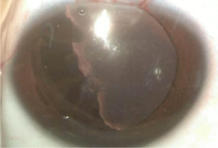
Figure 1: Lens coloboma presented as nasal side deficiency of lens.
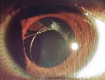
    Figure 2:  Mild temporal decentration of a sulcus-placed three-piece acrylic
soft intraocular lens.
Discussion
Lens coloboma results from the failure of fetal fissure closure during ocular development, usually unilaterally. Once the zonule or ciliary body fails to develop, the tension will be released and the lens contracts segmentally with a notch. So, it is actually the coloboma of zonule rather than defect in lens itself [1].
Lens coloboma occasionally combines with other ocular defect, including iris, choroid or optic disc coloboma [2,3]. Besides, there is also a higher incidence of cataract and retinal detachment in these patients. The retinal detachment commonly involves nasal quadrant, and it is the result of ciliary body defect, which might induced abnormal adherence between lens and peripheral retina. Moreover, fluid might gain access to the subretinal area through the break in ciliary body [1]. Apart from ocular defects, lens coloboma might be one of the manifestations in systemic diseases, including Marfan syndrome and Marshall syndrome [4-6]. Several case reports had shown the association between Marfan syndrome and lens coloboma, and it is postulated that increased Transforming Growth Factor β (TGFβ) might be the link behind [5]. In our case, the patient had lens coloboma without accompanying congenital cataract, and no other systemic diseases were noted.
In order to enhance surgical planning, a detailed imaging of the lens, zonulae and ciliary body is needed. High-resolution ultrasound biomicroscopy has been suited before for in vivo study of the human zonular apparatus and lens. The use of scheimpflug images can be obtained with the pentacam to depict a more accurate view of the lens coloboma morphology and to anticipate the potential complications during surgery [7]. Because the five-year old patient can not cooperate, we did not perform pentacam examination.
Treatment of lens coloboma is mainly surgical but limited literatures are available. Nordlund and colleagues reported seven cases of lens coloboma treated by phacoemulsification and IOL placement. Six of the seven patients showed improvement of visual acuity with only one patient developed retinal detachment after surgery [8]. The surgical management of a lens coloboma is frequently defying since there is a high risk of capsular fornix aspiration, zonular dialysis extension, vitreous herniation into the anterior chamber, IOL decentration and closure of the capsular opening. Hence, the implantation of a capsular tension ring is preferred before the emulsification of the nucleus to prevent capsular bag deformity, enable nucleus rotation and avoid capsular collapse; therefore, reducing surgical complications. In another study by Gurler and colleagues, they presented 13 cases of lens coloboma under treatment of phacoemulsification, along with Capsular Tensor Ring (CTR) and IOL placement. Significant improvement in visual acuity was noted after surgery without major complication. Thus, the paper concluded that phacoemulsification with CTR and IOL implantation is a safe and effective option [9].
In our case, the patient cannot afford CTR owing to poor economy of the family. Owing to enormous defects in lens, and intracapsular IOL placement was not feasible. Thus, we chose a sulcus 3-piece IOL as alternative. Posterior capsulotomy and anterior vitrectomy were also performed to avoid posterior capsular opacity, which is a common complication in children undergoing cataract surgery [10]. In our patient, visual acuity only mildly improved due to prolonged deprivation amblyopia.
In summary, lens coloboma is a rare congenital disease with anomaly in lens shape. Surgical treatment can achieve visual improvement.
References
- Onwochei BC, Simon JW, Bateman JB, Couture KC, Mir E. Ocular colobomata. Surv Ophthalmol. 2000; 45: 175-194.
- Bavbek T, Ogut MS, Kazokoglu H. Congenital lens coloboma and associated pathologies. Doc Ophthalmol. 1993; 83: 313-322.
- Li J, Ma X, Hu Z. Lens coloboma and associated ocular malformations. Eye Sci. 2011; 26: 108-110.
- Mehrotra AS, Solanki N, Sabharwal KK. Bilateral coloboma of lens in Marfan's syndrome. Indian J Ophthalmol. 1985; 33: 201-202.
- LeBlanc SK, Taranath D, Morris S, Barnett CP. Multisegment coloboma in a case of Marfan syndrome: another possible effect of increased TGFbeta signaling. J AAPOS. 2014; 18: 90-92.
- Schlote T, Volker M, Knorr M, Thiel HJ . Lens coloboma and lens dislocation in Stickler (Marshall) syndrome. Klin Monbl Augenheilkd. 1997; 210: 227-228.
- Camarena JC, Ayup-Arguijo E, Chavez-Mondragon E, Ramirez-Miranda A. Surgical management and scheimpflug analysis of an atypical lens coloboma. Case Rep Ophthalmol. 2012; 3: 317-320.
- Nordlund ML, Sugar A, Moroi SE. Phacoemulsification and intraocular lens placement in eyes with cataract and congenital coloboma: visual acuity and complications. J Cataract Refract Surg. 2000; 26: 1035-1040.
- Gurler B, Coskun E, Okumus S1, Pinero DP, Ozcan E1, Erbagci I. Surgical outcomes of isolated lens coloboma with or without cataract among young adults. Can J Ophthalmol. 2014; 49: 145-151.
- Ram J, Brar GS, Kaushik S, Gupta A. Role of posterior capsulotomy with vitrectomy and intraocular lens design and material in reducing posterior capsule opacification after pediatric cataract surgery. J Cataract Refract Surg. 2003; 29: 1579-1584.