
Abstract
We report the case of a 78-year-old man who presented with fever and unilateral leg pain. Because of rapid progression toward septic shock, the patient was transferred to the ICU. The patient died within 48 hours after ICU admission. Post mortem blood cultures appeared to be positive for Escherichia Coli.
Keywords: Necrotizing fasciitis; Soft skin tissue infection; Escherichia coli infection; Feptic shock; Fatal sepsis
Abbreviations
ICU: Intensive Care Unit; NF: Necrotizing Fasciitis; NSTI: Necrotizing Soft Tissue Infection; COPD: Chronic Obstructive Pulmonary Disease; CT: Computed Tomography; INR: International Normalized Ratio; CNF: Cytotoxic Necrotizing Factor; PCR: Polymerase Chain Reaction; MRI: Magnetic Resonance Imaging; EXPEC: Extra-Intestinal Pathogenic E. Coli; CRP: C-Reactive Protein
Introduction
Necrotizing Fasciitis (NF) is an uncommon life-threatening Necrotizing Soft Tissue Infection (NSTI) which is caused by virulent toxin-producing bacteria. NSTI’s are defined as infections of any of the layers within the soft tissue compartment. Their prevalence is rare (around 1000 cases worldwide annually) but up to 20% of these patients die [1]. The latter results from delayed diagnosis due to a difficult differential diagnosis with other soft tissue infections and due to the rapid progression and evolution into septic shock. Patients with NF can be divided into 2 groups according to the causative pathogen: Type 1 is caused by polymicrobial infections and type 2 is caused by monomicrobial infections [2]. Escherichia coli have been isolated from polymicrobial or Fournier’s gangrene, but have rarely been reported in monomicrobial necrotizing fasciitis.
Case Presentation
A 78-year-old man consulted his general practitioner because of fever and unilateral leg pain since one day, for which he was treated with paracetamol. His medical history revealed COPD Gold III, peripheral vascular disease (stenting of the aortic bifurcation) and primary myelofibrosis (since 5 years). As he had persisting fever (up to 38.8°C) he was referred to the emergency department.
On arrival, his blood pressure was 108/56 mmHg and heart rate was 119 beats per minute. His right thigh and abdomen were examined by echography. As this could not show relevant abnormalities, CT scan of the abdomen was performed, showing no focus of infection (no psoas abscess, no signs of fasciitis). Deep venous thrombosis and phlebitis were also excluded. Cultures were taken and broad spectrum antibiotics (piperacillin/tazobactam) were started, given the observed leucopenia (2810 cells/mm3), thrombocytopenia (18000 cells/μL) and a spontaneous rise in INR (1.6) in the initial lab results.
The next day, the patient was transferred to the intensive care unit because of rapid progression towards septic shock. On arrival, the patient was awake and complained of pain on the medial side of the right thigh. Although still hemodynamically stable, peripheral cyanosis was present with metabolic lactic acidosis (lactate 8.23 mmol/l) developing overnight and finally leading to a circulatory arrest (based on pulseless electrical activity). After successful resuscitation, including intubation and administration of 1 mg of epinephrine (twice), spontaneous circulation could be restored after 7 min of advanced life support. High doses of vasopressors were needed in order to maintain adequate blood pressures. The skin lesions on the thigh enlarged rapidly, forming enormous fluid containing bullae (Figure 1,2). The high doses of catecholamines triggered episodes of atrial fibrillation, which were treated with amiodarone. Despite early initiation of renal replacement therapy, the addition of hydrocortisone and amikacin, septic shock did not decline. Within 24 hours the patient was in multiple organ failure with ischemic hepatitis, hyperbilirubinemia, and persisting lactic acidosis (under dialysis) being most prominent. The patient died within 48 hours after ICU admission. Post mortem blood cultures appeared to be positive for Escherichia Coli. The Cytotoxic Necrotizing Factor (CNF) 1 toxin gene could not be identified by Polymerase Chain Reaction (PCR) [3].
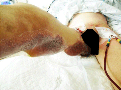
    Figure 1:  E.coli necrotizing fasciitis case. The skin lesions on the thigh
enlarged rapidly, forming enormous fluid containing bullae.
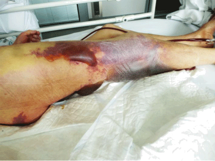
    Figure 2:  E. coli necrotizing fasciitis case: The skin lesions on the thigh
enlarged rapidly, forming enormous fluid containing bullae.
Literature Review
We searched Embase/PubMed and Google scholar for case reports and series on necrotizing fasciitis caused by E. Coli. Filters were set to “human”, “published from 2006-2016”. Gallois et al described a fatal case of a 29-year-old woman with chronic ulcerative pancolitis and liver cirrhosis (Child-Pugh C), who was under immunosuppressive therapy. She experienced fatal NSTI caused by a virulent strain of E. coli carrying numerous extraintestinal virulence factors, notably Cytotoxic Necrotizing Factor (CNF) 1. E. coli is a versatile pathogen and may cause diverse extraintestinal diseases. This particular feature is associated with the acquisition of virulence attributes not present in commensal strains which could contribute to the fatal outcome [4].
Grimaldi et al believe that a combined host-pathogen genetic analysis can explain the phenotype. They present a life-threatening case of NF in a 83-year-old male patient [5]. The strain belonged to the phylogenetic B2 group, which is the main group of extraintestinal pathogenic E. coli [6]. The close relationship between genetic profile of the patients and the virulence of the micro-organism underscores the importance of a personalized approach in the treatment of this disease. NF was also reported in immunocompetent patients. Taif et al. describe the case of a 26- year- old woman who presented with severe thigh pain, swelling and irritability since a few hours and right abdominal pain since 2 days. Urgent MRI and CT scan showed features of necrotizing fasciitis in the thigh spreading from an inflamed appendix. During emergent surgical exploration a perforated appendix was found with disseminated infection in the intraperitoneal and retroperitoneal spaces as well as the right thigh. The patient rapidly deteriorated. Despite surgery and maximal supportive measures, she died in the immediate postoperative period. Blood cultures revealed Staphylococcus aureus and Streptococci, while tissue culture showed growth of Escherichia coli [7]. Shaked et al. describe the clinical characteristics and outcomes of seven cases diagnosed at a single institution during an 18-month period. All patients had some form of immunosuppression and the lower limb was most commonly involved. In all cases, E. coli was isolated as a monomicrobial pathogen from blood, fascia, or both of them. All patients died during hospitalization, three within the first 48h. The RAPD (Random Amplification of Polymorphic DNA) assay showed a high degree of genetic diversity among the “flesh- eating” strains and controls. The cnf1 toxin gene was identified in two out of three cases, but not in the controls. Strains harboring this virulence factor have been designated necrotoxic E. coli [8].
Three cases of fatal E. Coli soft tissue infections after liver transplantation were presented by Janny et al. The 3 patients were immunosuppressed as a result of pre-transplant cirrhosis and the postoperative administration of immunosuppressive therapy. Skin and soft tissue infections developed within the first week after liver transplantation. The 3 patients presented with fever and skin lesions with or without bullae. Despite prompt appropriate antibiotic therapy and surgical debridement, the outcome was rapidly fatal (24h on average). E. coli was isolated from subcutaneous tissues in 2 cases and from several blood cultures in the third one. The 3 isolates belonged to distinct phylogenetic groups, and did not harbor most of the virulence factors usually reported in EXPEC isolates [9].
Endo et al. report a remarkable case of sequential necrotizing fasciitis caused by the monomicrobial pathogens streptococcus equisimilis and secondly, by extended-spectrum beta-lactamaseproducing Escherichia coli. A 85-year-old man was admitted because of necrotizing fasciitis of his right thigh. Streptococcus equisimilis was detected as a mono-microbial pathogen, and the infection was cured by amputation of the patient’s right leg and the administration of antibiotics. However, 5days after discontinuing antibiotic therapy, he developed necrotizing fasciitis on his right upper limb and died. ESBL-producing E. coli was the only bacterial species isolated from blood and skin cultures. This case demonstrates that ESBL-producing E. coli can cause monomicrobial necrotizing fasciitis, particularly during hospitalization and that a different bacterial species can cause disease shortly after a previous episode [10].
Discussion
Early diagnosis of NF is crucial but often very difficult as it is a rare disease with a non- specific clinical presentation. It is key to be suspicious and intercept cases early to ensure an early surgical intervention and hence improve the prognosis. The early manifestations are mostly local symptoms and signs of inflammation in the affected region such as swelling, pain, redness and tenderness occasionally accompanied by fever. All these signs are indicative for a local infection such as cellulitis or erysipelas, but the differentiation between non- necrotizing and necrotizing soft tissue infections remains difficult. The most constant clinical feature seems to be ‘pain out of proportion’. Sudden deterioration or progression despite antibiotic treatment is another important clue. In our case, the patient had both early and late manifestations. The patient progressed to sepsis and shock with tachycardia, hypotension and acute renal failure. Skin changes such as discoloration, crepitus, blistering, bullae and fluid discharge were present and are also late features.
Antimicrobial therapy and recurrent surgical debridement are the cornerstones of management. Hemodynamic support, nutrition, and wound care are also important. If the infection begins to spread despite treatment, amputation may be life-saving. Some authors propose a scoring system to facilitate early diagnosis and differentiation between non-necrotizing and necrotizing fasciitis, the Laboratory Risk Indicator for Necrotizing Fasciitis Score (LRINEC) [11]. It is based on CRP, white blood cell count, sodium, creatinine, glucose and hemoglobin levels. Patients with a LRINEC score of 6 or more should be carefully evaluated for the presence of necrotizing fasciitis.
Conclusion
Gram-negative necrotizing fasciitis is a rare but serious infection, most often caused by E Coli. Clinical presentation frequently combines bullous skin lesions with septic shock, which may quickly lead to death. Both host factors and virulence factors of the causative organism appear to be major determinants promoting negative outcome. The close relationship between genetic profile of the patients and the virulence of the micro-organism underscores the importance of a personalized approach in the treatment of this disease. Empiric antibiotic therapy against both gram-positive and gram-negative organisms is indicated as soon as the diagnosis is considered, with early surgical debridement if necessary.
References
- Anaya DA, Dellinger EP. Necrotizing soft-tissue infection: diagnosis and management. Clin Infect Dis. 2007; 44: 705-710.
- Audard V, Pardon A, Claude O, Jablonski M, Remy P, Desvaux D, et al. Necrotizing fasciitis during de novo minimal change nephrotic syndrome in a kidney transplant recipient. Transpl Infect Dis. 2005; 7: 89-92.
- Clermont O, Bonacorsi S, Bingen E. Rapid and simple determination of the Escherichia coli phylogenetic group. Appl. Environ. Micro- biol. 2000: 66: 4555-4558.
- Gallois C, Hauw-Berlemont C, Richaud C, Bonacorsi S, Diehl J-L, Mainardi J-L, et al. Fatal necrotizing fasciitis due to necrotic toxin-producing Escherichia coli strain. New Microbes and New Infections. 2015; 8: 109-112.
- Grimaldi D, Bonacorsi S, Roussel H, Zuber B, Poupet H, Pitlik S, et al. Unusual “flesh- eating” strain of Escherichia coli. J Clin Microbiol. 2010; 48: 3794-3796.
- Russo TA, Johnson JR. Proposal for a new inclusive designation for extraintestinal pathogenic isolates of Escherichia coli: ExPEC. J. Infect. Dis. 2000; 181: 1753-1754.
- Taif S, Alrawi A. Missed acute appendicitis presenting as necrotizing fasciitis of the thigh. BMJ Case Reports. 2014.
- Shaked H, Samra Z, Paul M, Madar-Shapiro L, Cohen J, Pitlik S, Bishara J, et al. Unusual “flesh-eating” strains of Escherichia coli. J Clin Microbiol. 2012; 50: 4008-4011.
- Janny S, Bert F, Dondero F, Nicolas Chanoine MH, Belghiti J, Mantz J, et al. Fatal Escherichia coli skin and soft tissue infections in liver transplant recipients: report of three cases. Transpl Infect Dis. 2013: 15: 49-53.
- Endo A, Matsuoka R, Mizuno Y, Doi A, Nishioka H. Sequential necrotizing fasciitis caused by the monomicrobial pathogens Streptococcus equisimilis and extended-spectrum beta-lactamase-producing Escherichia coli. J Infect Chemother. 2016.
- Wong CH, Khin LW, Heng KS, Tan KC, Low CO. The LRINEC (Laboratory Risk Indicator for Necrotizing Fasciitis) score: a tool for distinguishing necrotizing fasciitis from other soft tissue infections. Crit Care Med. 2004; 32: 1535-1541.