Editorial
RM is a 26-year-old male patient with a known history of seizure disorder and substance abuse who presented to the local emergency room via EMS after being found with a decreased level of consciousness. He was noted to have two Fentanyl patches on his body and Narcan was given by EMS prior to arrival to the emergency room.
In the ER the patient had a seizure and an episode of emesis. The decision was made to intubate the patient. Two additional Fentanyl patches were noted in the oropharynx and were removed. During intubation there was no noted obstruction of the vocal cords or evidence of aspiration. He was then admitted to the intensive care unit.
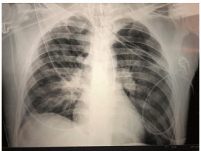
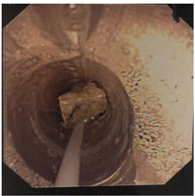
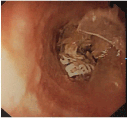
During his time in the ICU he was noted to have intermittent episodes of high peak pressure. Ventilator setting changes were made which only partially helped. Respiratory therapy also noted difficulty passing the suction catheter, but this too was intermittent and seemed to coincide with the peak airway issues. His initial CXR Figure 1 showed some pulmonary edema and a right basilar infiltrate. This right basilar infiltrate did worsen some in the ICU. The findings on his CXR did not account for his ventilator issues. Decision was made to do a fiberoptic bronchoscopy. This revealed a Fentanyl patch at the distal end of the endotracheal tube just above the level of the carina (Figure 2,3). The patch was removed by holding onto the patch with biopsy forceps while extubating the patient. The patient was immediately reintubated. Bronchoscopy after patch removal did not demonstrate any evidence of additional foreign bodies. The airway exam demonstrates diffuse airway edema. After bronchoscopy was completed it was also noted that a piece of a patch was pulled out by NGT. This prompted an EGD. This demonstrated esophagitis thought to be secondary to the Fentanyl patches, but no actual patches were found. A duodenal ulcer was also noted. The patient was successfully extubated after being treated for overdose and aspiration pneumonia. He was discharged from the ICU and hospital.

    Figure 1:  CXR image showing good placement of an endotracheal tube and
a right peripheral infiltrate.

Figure 2: Fentanyl patch within the endotracheal tube.

    Figure 3:  Fentanyl patch located in the distal trachea. The lettering is visible
on the patch itself.
The ER staff did not see any evidence of obstruction of the vocal cords or patches at the level of the vocal cords. Suspect the patch was dragged into the trachea by the endotracheal tube. This patient had been sucking the gel out of the patches in addition to lining his mucosal membranes. Inspection of the patch after removal shows that it has been peeled apart. This allowed for it to be dragged along into the trachea. It is not surprising that the patch caused the ventilator issues. Without direct visualization this patch would not have been found as these patches are not radiopaque and would not show up on standard CXR.
Review of the literature shows that it is not new for patients to abuse Fentanyl, but this is a unique situation. In addition to just placing patches on the skin patients can use mucosal surfaces to more rapidly absorb the medication or to ingest the gelatinous reservoir of these patches which contains large amounts of Fentanyl, far more than one would get from transdermal absorption and it’s quicker. Case series have been reported for oral exposure [1,2]. There has been a case report involving a fatality from chewing and aspirating a Fentanyl patch [3].
Patients continue to be creative in ways to abuse drugs. Transdermal patches are no exception. Patients are known to line mucosal surfaces, ingest patches whole or to ingest the gelatinous center portion of the patch which will deliver a large amount of drug quickly. If patients present with overdose or decreased level of consciousness with suspected Fentanyl patch use or abuse, you need to ensure that an inspection of the oral cavity occurs. This could potentially help to decrease the risk of aspiration either by the patient or with a little help from an endotracheal tube.
References
- Mrvos R, Feuchter AC, Katz KD, Duback-Morris LF, Brooks DE, Krenzelok EP. Whole Fentanyl patch ingestion: A multi-center case series. J Emerg Med. 2012; 42: 549-552.
- D’Orazio JL, Fischel JA. Recurrent respiratory depression associated with Fentanyl transdermal patch gel reservoir ingestion. J Emerg Med. 2012; 42: 543-548.
- Kim JW. Aspiration of Fentanyl Patch. J Addict Med. 2010; 4: 244-245.
