Clinical Image
Austin J Emergency & Crit Care Med. 2014;1(1): 1.
Rigler's Sign
Wei-Ting Lin1,2 and Chien-Ming Chao3,4*
1Department of Trauma, Chi Mei Medical Center, Tainan, Taiwan
2Department of Physical Therapy, Shu Zen College of Medicine and Management
3Department of Intensive Care Medicine, Chi Mei Medical Center, Liouying, Tainan, Taiwan
4Department of Nursing, Min-Hwei College of Health Care Management, Tainan, Taiwan
*Corresponding author: Dr. Chien-Ming Chao, Department of Intensive Care Medicine, Chi Mei Medical Center, Liouying, Tainan
Received: June 13, 2014; Accepted: July 23, 2014; Published: July 29, 2014
What is your diagnosis?
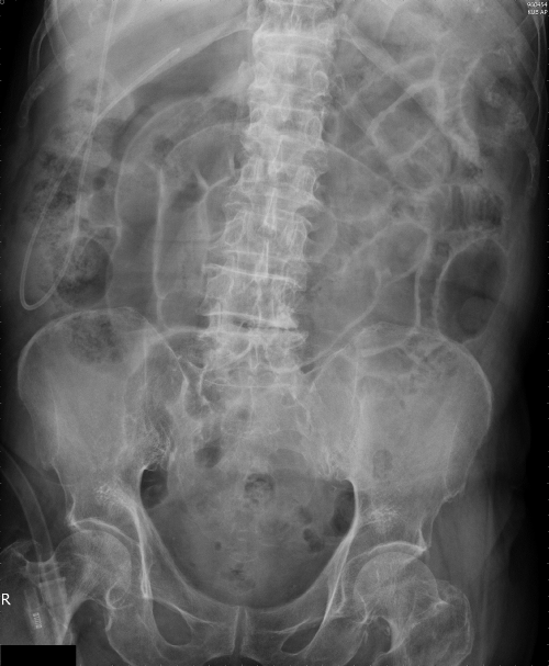
A 91-year-old man presented with progressive abdominal pain for one day. Radiography of the abdomen was done (Figure 1).
Figure 1: Radiography of the abdomen revealed Rigler's sign, which indicated the visibility of bowel wall outlined by intraluminal and extraluminal air (white arrows)
Diagnosis: Hollow organ perforation caused by perforated gastric ulcer.
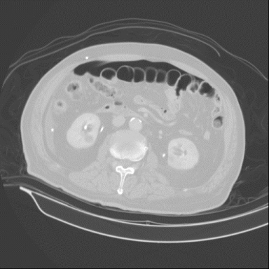
Radiography of the abdomen showed Rigler's sign which is defined as the visibility of bowel wall outlined by intraluminal and extra luminal air and indicates pneumoperitoneum. Computed tomography of abdomen confirmed the diagnosis of pneumoperitoneum (Figure 2). Emergent explorative laparotomy was performed and disclosed a 1-cm ulcer perforation at the anterior wall of pyloric ring. The post-operation course was smooth, and he was discharged uneventfully one month later. Acute abdomen caused by hollow organ perforation is usually difficult to diagnose by plain films that are always the initial diagnostic modality in this clinical condition. However, several characteristic sign of pneumoperitoneum in plain film, such as Rigler's sign, may sometimes help clinicians make the prompt diagnosis.

