Clinical Image
Austin J Emergency & Crit Care Med. 2015;2(3): 1019.
Contained Rupture of a Large Hydatid Cyst
Blasco-Navalpotro MA, Flordelís-Lasierra JL* and Chamorro-Borraz N
Intensive Care Unit, Severo Ochoa University Hospital,Spain
*Corresponding author: Flordelís-Lasierra JL, Intensive Care Unit, Severo Ochoa University Hospital, Av. Orellana s/n. Leganés 28911, Madrid, Spain
Received: March 10, 2015; Accepted: March 17, 2015; Published: March 19, 2015
Clinical Image
What is your diagnosis?
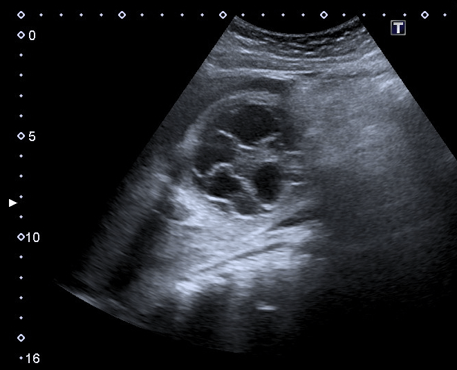
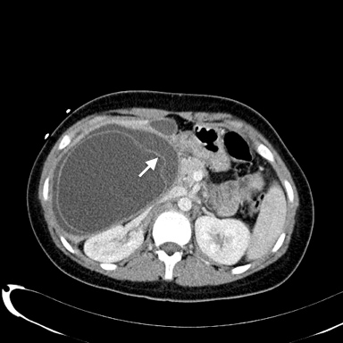
A 15 -year-old woman with a history of pollen allergy and consumption of vegetables from a rural area was admitted to our hospital emergency presenting generalized itching and abdominal pain. The blood tests only revealed 17,710 leucocytes/mcl with 13% eosinophils and immunoglobulin E 5000 KUI/L. An abdominal ultrasound and computed tomography were performed. The findings were compatible with a contained rupture of a large liver hydatic cyst (Figures 1,2) [1,2]. She was admitted to intensive care and urgent partial pericystectomy with previous cyst sterilization with hypertonic saline solution was performed [3]. Evolution was satisfactory undergoing treatment with albendazole. The hydatid serology test was positive.

    Figure 1:  The abdominal ultrasound showed an anechoic (cystic) lesion
with membranes floating on the cyst fluid. Within the clinical context referred
previously, such image is compatible with a cyst type 3 of International
classification of ultrasound images in cystic echinococcosis [1].
Our case reveals that, besides the direct or communicating breakage, the contained rupture of a hydatid cyst should be diagnosed and treated also with priority, in order to avoid open rupture and development of anaphylactic shock [4].

    Figure 2:  The abdominal computed tomography with intravenous contrast
scan revealed a cystic lesion in liver segments V and VI (148X 100 mm) with
a curvilinear linear image inside it (arrow) that corresponds with the endocyst
detachment.
References
- WHO Informal Working Group. International clasification of ultrasound images in cystic echinococcosis for application in clinical and field epidemiological settings. Acta Trop. 2003; 85: 253-261.
- Lewall DB, McCorkell SJ. Rupture of echinococcal Cyst: diagnosis, classification, and clinical implications. Am J Roentgenol. 1986; 146: 391-394.
- Rinaldi F, Brunetti E, Neumayr A, Maestri M, Goblirsch S, Tamarozzi F. Cystic echinococcosis of the liver: A primer for hepatologist. World J Hepatol. 2014; 6: 293-305.
- Murali MR, Uyeda JW, Tingpej B. Case 2-2015: A 25-year-old man with abdominal pain, syncope, and hypotension. N Engl J Med. 2015; 372: 265-273.