
Case Presentation
Austin J Emergency & Crit Care Med. 2015;2(3): 1023.
How an Ultrasound can Save a Life
Martino IF¹*, Marra S², Pagani L², Ceresa IF¹ and Bressan MA¹
¹Departement of Emergency, IRCCS Poliniclinico San Matteo, Italy
²School of Emergency Medicine, University of Pavia, Italy
*Corresponding author: Martino IF, Department of Emergency, IRCCS Poliniclinico San Matteo, piazzale Golgi 19, Pavia, Italy
Received: May 05, 2015; Accepted: June 11, 2015; Published: June 13, 2015
Abstract
Emergency Ultrasound, performed during the visit, led us towards a correct diagnosis and gave us the ability to save the patient’s life.
Keywords: Ultrasound; Dyspnea; Pericardial effusion
Case Presentation
A 51 year old woman’s access to our emergency department (DEA) with increasing dyspnea and tachypnea.
Over the previous days, the patient was already examined twice in other First Aid structures.
The access in the first emergency department (DEA) was caused by dyspnea and coughing; the arterial blood gas examination (EGA), without oxygen therapy, revealed a mild hypoxia and moderate hypocapnia. The chest X-ray showed pneumonia at the base of the right lung with minimal pleural effusion, enlarged heart and enlarged pulmonary hilum.
The patient was discharged with a diagnosis of pneumonia and antibiotic therapy was prescribed. Three days later, after a syncope, she showed up at the emergency unit a second time; the EGA showed hypoxia with hypocapnia and the patient was discharged and told to continue the antibiotic therapy.
When she arrived in our DEA, her vital signs were normal, except for a respiratory rate of 33breaths/minute, showing the symptoms of tachypnea. The patient’s medical history included a right breast cancer with rib metastasis and axillary lymph node involvement and a previous pericarditis.
During the examination, it was noted that the chest breath sounds had disappeared at the base of the right lung, the heart tones were muffled and the abdomen was normal. The EGA revealed: hypoxia, hypocapnia and increased lactates. The ECG showed: sinus rhythm, low voltage, complete right bundle branch block. Considering the severe suspect of pulmonary embolism, it was decided to perform a chest angio-CT, but the patient reported an iodinated contrast medium allergy, therefore an examination with bedside integrated ultrasound was commenced.
Subcostal projection highlighted that the inferior vena cava was markedly enlarged, non collapsible, with a pericardial effusion of approximately 4 cm and crucial collapse of the right ventricle (Image 1). Bilateral chest effusion right >left was present. The patient’s general condition underwent a rapid deterioration, with hemodynamic instability, hypotension and high frequency atrial fibrillation (165 beats per minute). Pericardiocentesis was urgently performed, with 1100 cc of blood spillage.
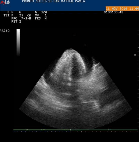
image 1: Pericardial blood effusion.
Conclusion
Many studies [1-7] have shown that clinical ultrasound bedside evaluation of critically ill patients leads to a more rapid diagnosis and to a more appropriate and prompt treatment.
This case report shows that the non-execution of ultrasound in previous examinations has prevented a correct diagnosis (in both cases, dyspnea was imputed to pneumonia). On the other hand, the ultrasound performed during our visit in the clinical suspect of pulmonary embolism, while looking for right ventricular dilatation, showed massive pericardial effusion and oriented us towards the correct diagnosis and prompt treatment of the patient.
The paper shows the beneficial effects of echocardiography used in emergency, not only in ischemic heart diseases. Moreover echocardiography can help to ensure a correct therapy.
Emergency ultrasound is not as descriptive and detailed as that of the sonographer who works in an outpatient clinic; the ultrasound performed by the emergency physician aims at problem solving. It gives fast and clear answers to specific questions that arise while the physician is treating the patient. Therefore, we believe that the competence to perform emergency ultrasound should be considered a prerequisite to operate in the emergency department and cannot be delegated to any other medical specialist other than the doctor who is treating the critical patient.
References
- Lichtenstein DA. Lung ultrasound in the critically ill. Ann Intensive Care. 2014; 4: 1.
- Lichtenstein DA, Mezière GA. Relevance of lung ultrasound in the diagnosis of acute respiratory failure: the BLUE protocol. Chest. 2008; 134: 117-125.
- Lichtenstein D, van Hooland S, Elbers P, Malbrain ML. Ten good reasons to practice ultrasound in critical care. Anaesthesiol Intensive Ther. 2014; 46: 323-335.
- Lichtenstein DA. How can the use of lung ultrasound in cardiac arrest make ultrasound a holistic discipline. The example of the SESAME-protocol. Med Ultrason. 2014; 16: 252-255.
- Seif D, Perera P, Mailhot T, Riley D, Mandavia D. Bedside ultrasound in resuscitation and the rapid ultrasound in shock protocol. Crit Care Res Pract. 2012; 2012: 503254.
- Wright J, Jarman R, Connolly J, Dissmann P. Echocardiography in the emergency department. Emerg Med J. 2009; 26: 82-86.
- Citilcioglu S, Sebe A, Ay MO, Icme F, Avci A, Gulen M, et al. The relationship between inferior vena cava diameter measured by bedside ultrasonography and central venous pressure value. Pak J Med Sci. 2014; 30: 310-315.