Research Article
Austin J Emergency & Crit Care Med. 2015;2(4): 1025.
Emergency Ultrasound in Critical Patients: The “Bedside Ultrasonography” as the “Third Hand” of Emergency Surgeon
Giuseppe Frazzetta*¹, Faraci Cristofaro¹, Mancuso Salvatore Igor¹, Fragati Giuseppe¹, Di Giovanni Silvia², Costanzo Luigi¹
¹U.O.C. General Surgery and Emergency Hospital "M. Chiello" of Piazza Armerina, Italy
²U.O. Medicine Acceptance and Emergency Hospital "Umberto I" Enna, Italy
*Corresponding author: Giuseppe Frazzetta, U.O.C. General Surgery and Emergency Hospital "M.Chiello" of Piazza Armerina, Italy
Received: May 05, 2015; Accepted: June 22, 2015; Published: June 24, 2015
Abstract
Introduction: Ultrasound (US) is a safe and useful diagnostic tool for the detection and the study of several organs. Absence of radiations and collateral effects make it be a repeatable test with low costs, feasible and quickly. Ultrasound scan can be performed to patient's bedside avoiding losing precious time for diagnostic and therapeutic decision making in transferring him in radiologic room or department.
Aim: To illustrate and support the precious aid of emergency ultrasound in emergency surgeon's hands.
Materials and Methods: we selected 45 patients admitted to our department of general and emergency surgery for abdominal and thoracic acute problems. A retrospective cohort study was made on a period of six month.
Conclusion: Bedside ultrasonography in critical patients is safe and feasible; it could be the “third hand” of emergency surgeon integrating the bounds of clinical examination.
We stand that all emergency surgeon or physician should be trained in bedside emergency ultrasound.
Keywords: Ultrasound; Critical; Ultrasonography; Surgeon; Bedside
Introduction
Ultrasound is a safe and useful diagnostic tool used for the detection and the study of several organs and their diseases. Because of absence of radiations and collateral effects it is a repeatable test with low costs, feasible and quickly. One of the great advantages of the ultrasound scan is the possibility to perform the exam to patient's bedside avoiding transferring him in radiologic room or department and losing precious time for diagnostic and therapeutic decision making [1]. In recent time not only radiologist appreciate the potentiality of ultrasound, but overall emergency physicians and surgeons start to perform ultrasonography scans both as Focused Assessment Sonography for Trauma (FAST) and monitoring hemodynamic conditions specially in critically ill patients. Increasingly, the ultrasound scans integrates the clinical examination giving precious dates on areas closed to physical exploration [2]. Ultrasound power makes the “bedside ultrasonography” often the first diagnostic instrumental approach to several both abdominal and lung pathologies and not only but also in the neurological evaluation because of the advances in technology that allow to penetrate the skull [3-4]. Often the bedside ultrasound is the “third hand” of the emergency physician or surgeon. In this cases series we report our daily experience in a small centre illustrating the precious aid of this fantastic diagnostic tool [5].
Materials and Methods
For this retrospective cohort study, we collect the data, from October 2014 to April 2015 from the emergency department of general and emergency surgery of “M. Chiello” hospital in Piazza Armerina. In a period of six month we selected 45 patients, as heterogeneous population, admitted to our department for abdominal and thoracic acute problems. They were divided in four groups: Abdominal acute problems (Group 1), Blunt trauma (Group 2), Post-operative complication (Group 3), and Thoracic acute problems (Group 4). The main age was 63 years old with a range between 8 and 92 years old. 7 patients were admitted for thoracic pain and evolving dyspnea: 3 had pleural effusion. 2 patients developed pneumothorax, one had spontaneous pathogenesis and one iatrogenic due to central vein catheterization; 2 patients developed pulmonary edema and lung disease. 11 patients, suffered from upper abdominal pain, vomiting and fever, had biliary colic pain; 17 patients were admitted for lower right or left abdominal-pelvic pain, sub-continuous fever and vomiting, 2 of these had missed evacuation of gasses or feces, dehydrations and other occlusive clinical signs. 2 patients were admitted for left colic pain with associated resistance to abdominal palpatory examination. 6 patients were recovered for jaundice; 2 had blunt abdominal trauma and 2 were reported for postoperative surgical complications. All these patients underwent first of all to ultrasonography evaluation (Table 1). In our experience all these patients were primary valued in emergency rooms and often underwent to a summary radiologic evaluation in emergency setting; often the primary examination was superficial and too fast so all the patients admitted to our department were be retested to ultrasound evaluations. Only expert surgeon with advanced echographyc technique skills carried out all the exams. Where use always the same echographyc instrument. All the exams were carried out using Convex probe 3-5 MHz and linear probe 7-9 MHz for both abdomen and thorax. Patient's position was chosen in agreement with general clinical conditions, patient's degrees of collaborations, previous surgical procedures if present and however to patient's bedside. Few times patients were prepared the day before with fasting diet and medicine to reduce abdominal meteorism; according to the age and the collaboration of the patients, we use standard scansions to detect abdomen and thorax; in some cases, patients were studied in sitting or prone position. In all cases, we do not use contrast enhancement. Median time elapsed between admission and echographyc evaluation was about 1.3 ± 8.5 hours. Mean operative time was 10 ± 30 min. Mean ASA (American Society of Anaesthesiologists) score was III.
    Indications to admission
    N° pz
    Ecographyc findings
    Diagnosis
    Decision making


    Group 1
      Abdominal acute problems
      Upper pain, fever, vomiting, biliary colic    pain
    11
    -Gallbladder distension and wall thickening    > 6 mm
      -pericholecystic fluid and peri-intrahepatic free gasses
      -stagnant or purulent gallbladder contents
    Acute lithyasic cholecystitis
      - associated pericholecystic abscesses
      -gangrenous cholecystic perforation
    -Cholecystectomy
      -Perihepatic drainage


    Lower right or left abdominal-pelvic pain,    fever, vomiting, missed evacuation dehydrations
    17
    -Free intrabdominal fluid
      -pelvic abscesses
      -thickening appendicular wall
      -intestinal dilatations and bowel immobility
      -Colonic wall thickening
      -per colic free fluid
      -“Pseudo kidney” images
    -Acute appendicitis
      -Ovarian rotation and pelvic diseases
      -Intestinal occlusion
      -Acute diverticulitis
      -Per colicabscesses
    -Appendectomy pelvic lavage and drainage
      -Gynaecologic counselling and transfer
      -Intestinal resection
      -Bowel viability monitoring and discharging
      -Colic resection


    Jaundice
    6
    -Biliary tree dilatations
      -Obstructive iperechogenic images
      -intrahepatic masses
    -Choledocholithyasis
      -Liver tumours
    ERCP


    Group 2
      Blunt trauma
    2
    -Non homogenous spleen aspect
      -per splenic free fluid
      -Psoas disomogeneus aspect
    -Evolving spleen rupture
      -Psoas hematoma
    -Splenectomy
      - Monitoring


    Group 3
      Post-operative complications
    2
    -Disomogeneus wall collection
    -Hematoma of rectum muscle
      -Seroma mesh related
    Non Operative Management


    Group 4
      Thoracic acute problems
      Chest pain,
      dyspnea, blunt chest trauma
    7
    -Lung point
      -A Lines
      -B Lines
      -Pleural free fluid
    -Pneumothorax (2)
      -Pleural effusion (3)
      -Lung diseases (2)
    -Thoracentesis (3)
      -Chest drainage (2)
      -Medical management (2)


    Total
    45
Table 1: Summary of ecographyc findings driving decision making.
Results
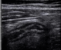
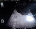
In 11 patient of Group 1 were found gallbladder enlargement, with wall thickening and free fluid or perihepatic collection and in all cases was made diagnosis of acute cholecystitis, associated in two cases to hepatic abscesses and gangrenous cholecystic perforation in other two patients (Figure 1-2). In all these patients were performed cholecystectomy and perihepatic lavage and drainage [6]. In 17 patients, admitted for lower abdominal pain, the echographyc findings were: intra abdominal fluid, pelvic collection, thickening small bowel or colonic wall or distention and was made diagnosis of acute appendicitis in 11 cases whom underwent to appendectomy (Figure 3); ovarian torsion and pelvic pathologies were found in two cases whom underwent to gynaecologic evaluation avoiding useless surgical procedures (Figure 2). In two patients were demonstrated echographyc signs of mechanical occlusion and in one cases was necessary the surgery but in one due to adhesion we preferred Non Operative Management and echographyc monitoring of bowel viability [7-8] (Figure 3). One of two patients with colonic wall thickening developed the “pseudo-kidney” signs after few days as expression of paracolic abscesses and was performed a colonic resection after TAC evaluation. 6 patients had jaundice and were studied to detect the obstructive cause, that in 5 cases was a biliary stones, but in one case was an intrahepatic mass: all these underwent to Endoscopic-Retrograde-Cholangiopancreatography [9-11] (Figure 4).

Figure 1: Acute cholecystitis.

Figure 2: Hepatic abscesses.

Figure 3: Acute appendicitis.

Figure 4: Pelvic disease.
In Group 2 one patients with finding of free increasing perisplenic fluid, anaemia and haemodynamic instability, after blunt abdominal trauma underwent to splenectomy; the other one to Non Operative management and monitoring of psoas muscle hematoma [12]. In Group 3 two patients developed post-operative complication and echographyc detection showed dishomogeneous wall collection due to hematoma of rectum muscle and seroma mesh related, no surgical procedure were carried out (Figure 5). In Group 4 were collected patients with chest pain, dyspnea or chest blunt trauma; in these cases we performed ultrasounds evaluation finding pleural effusion, treated by thoracentesis, “lung point” as expression of pneumothorax treated by chest drainage [13-15].

Figure 5: Bowel obstruction.
The only echographyc evaluation has been sufficient to establish that in 30 cases (66.7%) was necessary immediate surgery, in 9 cases (20%) was sufficient monitoring and non-operative management or transfer the patient to other specialists' care and in 6 cases (13.3%) endoscopic management was required (Table 2).
    Decision Making
    Procedures
    Cases
    %


    Surgical Management
    Cholecystectomy and peritoneal drainage
      Appendectomy
      Splenectomy
      Bowel resections
      Thoracentesis
      Chest drainage
    11
      11
      1
      2
      3
      2
    66.7


    Endoscopic Management (ERCP)
    Biliary stent
      Common bile duct “toilette”
    1
      5
    13.3


    Non Operative Management (NOM)
    Monitoring (hematoma-seroma-bowel motility)
      Medical menagement
      Transfer to other specialists
    5
      2
      2
    20


    Total

    45
    100
Table 2: Management and procedure performed.

Figure 6: Choledocholythiasis.

Figure 7: Abdominal wall hematoma.
Discussion
Ultrasound evaluation was performed as soon as possible to assure the best medical or surgical management, avoiding delay in diagnosis. In some cases ultrasound data were integrated with a second level diagnostic step, but we report the echographyc findings that ride and drive the decision making process. In all cases, the contribution of ultrasonography was precious both too led to a certain diagnosis and to drive toward the correct decision. The only echographyc evaluation has been sufficient to establish if it was necessary immediate surgery, monitoring and non-operative management or transfer the patient to other specialists' care. Only in two cases was necessary a CT scan to integrate ultrasound evaluation. In these way with a noninvasive diagnostic tool is possible in few minutes integrate with a great number of data the information of clinical evaluation with the advantage to expand the areas where the human sense are less sensitive. One of the most important aspects consists in the possibility to perform the echographyc evaluation when you want, how much time is necessary and overall where you want without moving the patient just to his bedside [16].
Conclusion
Bedside ultrasound evaluation in critical care patients with blunt chest/abdominal trauma, biliary, splenic, bowel problems is a useful tool for the emergency physician and surgeon. Often critical patient may be not able to collaborate giving the essential clinical history and in this case the ability to survey specific organs is lifesaving; however in greater cases US can guide the decision making process and even if it is not infallible could avoid diagnostic mistakes and delay [17-18]. As a “handily” technique requires a good instrument but overall a serious operator's trained skill and eye. However bedside ultrasonography evaluation of critical patients is safe and feasible, representing a powerful weapon in emergency department: it could be the “third hand” of emergency surgeon, so we stand that all emergency surgeon or physician should be trained in bedside ultrasound evaluation [19-20].
References
- Kirkpatrick AW, Šustic A, Blaivas M. Introduction to the use of ultrasound in critical care medicine Critical Care Medicine. 2007; 35: S123-S125.
- Melanson SW, Heller M. The emerging role of bedside ultrasonography in trauma care. Emerg Med Clin North Am. 1998; 16: 165-189.
- Soult MC, Weireter LJ, Britt RC, Collins JN, Novosel TJ, Reed SF, et al. Can routine trauma bay chest x-ray be bypassed with an extended focused assessment with sonography for trauma examination? Am Surg. 2015; 81: 336-340.
- Kirkpatrick AW, Sirois M, Laupland KB, Liu D, Rowan K, Ball CG, et al. Hand-held thoracic sonography for detecting post-traumatic pneumothoraces: the Extended Focused Assessment with Sonography for Trauma (EFAST). J Trauma. 2004; 57: 288-295.
- Agrusa A, Romano G, Frazzetta G, Amato G, Chianetta G, Di Giovanni S, et al. The aid of “bedside ultrasonography” for the emergency surgeon: the experience of a single centre. Critical Ultrasound Journal. 2014; 6: A3.
- Romano G, Agrusa A, Frazzetta G, De Vita G, Chianetta D, Di Buono G, et al. Laparoscopic drainage of liver abscess: case report and literature review. G Chir. 2013; 34: 180-182.
- Guttman J, Stone MB, Kimberly HH, Rempell JS. Point-of-care ultrasonography for the diagnosis of small bowel obstruction in the emergency department. CJEM. 2015; 17: 206-209.
- Jang TB, Schindler D, Kaji AH. Bedside ultrasonography for the detection of small bowel obstruction in the emergency department. Emerg Med J. 2011; 28: 676-678.
- Rickes S, Treiber G, Mönkemüller K, Peitz U, Csepregi A, Kahl S, et al. Impact of the operator's experience on value of high-resolution transabdominal ultrasound in the diagnosis of choledocholithiasis: a prospective comparison using endoscopic retrograde cholangiography as the gold standard. Scand J Gastroenterol. 2006; 41: 838-843.
- Costi R, Gnocchi A, Di Mario F, Sarli L. Diagnosis and management of choledocholithiasis in the golden age of imaging, endoscopy and laparoscopy. World J Gastroenterol. 2014; 20: 13382-13401.
- Stott MA, Farrands PA, Guyer PB, Dewbury KC, Browning JJ, Sutton R. Ultrasound of the common bile duct in patients undergoing cholecystectomy. J Clin Ultrasound. 1991; 19: 73-76.
- Kirkpatrick AW. Clinician-performed focused sonography for the resuscitation of trauma. Crit Care Med. 2007; 35: S162-172.
- Lichtenstein DA. Ultrasound in the management of thoracic disease. Crit Care Med. 2007; 35: S250-261.
- Theerawit P, Touman N, Sutherasan Y, Kiatboonsri S. Transthoracic ultrasound assessment of B-lines for identifying the increment of extravascular lung water in shock patients requiring fluid resuscitation. Indian J Crit Care Med. 2014; 18: 195-199.
- Wilkerson RG, Stone MB. Sensitivity of bedside ultrasound and supine anteroposterior chest radiographs for the identification of pneumothorax after blunt trauma. Acad Emerg Med. 2010; 17: 11-17.
- Wang HP, Chen SC. Upper abdominal ultrasound in the critically ill. Crit Care Med. 2007; 35: S208-215.
- Fagley RE, Haney MF, Beraud AS, Comfere T, Kohl BA, Merkel MJ, et al. Critical care basic ultrasound learning goals for american anesthesiology critical care trainees: recommendations from an expert group. Anesth Analg. 2015; 120: 1041-1053.
- Bobbia X, Hansel N, Muller L, Claret PG, Moreau A, Genre Grandpierre R, et al. Availability and practice of bedside ultrasonography in emergency rooms and prehospital setting: a French survey. Ann Fr Anesth Reanim. 2014; 33: e29-33.
- Soucy ZP, Mills LD. American Academy of Emergency Medicine Position Statement: Ultrasound Should Be Integrated into Undergraduate Medical Education Curriculum. J Emerg Med. 2015. pii: S0736-4679(15)00243-7. 2014.12.092. [Epub ahead of print].
- Rozycki GS, Newman PG. Surgeon-performed ultrasound for the assessment of abdominal injuries. Adv Surg. 1999; 33: 243-259.