Case Report
Austin J Emergency & Crit Care Med. 2015; 2(6): 1034.
Occult Pneumothorax in Minor Thoracic Trauma: Role of Ultrasonography – A Case Series
Marco B*, Sossio S and Giuseppe R
Department of Emergency and Trauma Center, Ospedale Maurizio Bufalini of Cesena, Italy
*Corresponding author: Marco Barozzi, Department of Emergency and Trauma Center, Ospedale Maurizio Bufalini of Cesena, UOC Medicina d’urgenza – Pronto Soccorso, Viale Ghirotti, 286 – 47522 – Cesena (FC), Italy
Received: August 04, 2015; Accepted: October 01, 2015; Published: October 05, 2015
Abstract
Bed–side ultrasound is a feasible, rapid and not expensive technique in order to detect lesions and injuries in traumatic patients.
We describe a series of minor blunt chest traumas in which echography was useful to diagnose pneumothorax not detected by conventional x–ray. This finding was subsequently confirmed by CT scan with important impact on treatment and prognosis of these patients.
Keywords: Pneumothorax; Chest ultrasonography; Thoracic trauma; Chest–XR
Introduction
Thoracic trauma is the third most common type of trauma [1]. Contrast–enhanced MDCT study is considered to be the diagnostic method of choice for severe thoracic trauma: in fact the most relevant traumatic diagnoses (pneumothorax, hemothorax, pulmonary laceration, tracheobronchial injury and aortic lesions) can be made in a short time with high accuracy, providing high sensitivity and specificity.
Minor blunt thoracic traumas are usually studied with chest XR with or without ribs series to detect the most common injuries as rib fractures, pneumothorax and pleural effusion. But the limitations of this exams are well known: about 50% of post traumatic pneumothorax are misdiagnosed because of anterior location (that is difficult to detect in a single supine projection); the same for minimal pleural effusion.
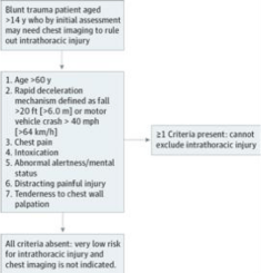
Moreover rib fractures, which are the most common injuries in blunt thoracic trauma, are often underestimated, but it’s well known the risk of thoracic and extra thoracic associated lesions even in presence of a single isolated rib fracture [2], especially in patients with Nexus Chest Di = 1 [3] (Figure 1).

Figure 1: The NEXUS score DI.
Bed-side ultrasound of the chest is a well known technique able to detect also small anterior pneumothorax: the absence of gliding sign, associated with the presence of lung point and step sign are all indicative of presence of pneumothorax with high sensitivity and specificity. This technique is simple, not expensive, and repeatable without exposure to radiations [4].
We describe a series of minor blunt chest traumas in which thoracic echography has been useful to diagnose pneumothorax not detected by conventional x-ray.
Case Presentation
From January 2014 to June 2015 we found 6 cases of occult X-Ray pneumothorax seen at the Maurizio Bufalini Hospital Emergency Department in Cesena (Italy), a first level Trauma Center in which about 65000 patients and nearly 300 cases of major trauma are managed every year. All patients had mild chest trauma without associated injury, a low risk mechanism of trauma as codified in the ATLS manual [5] (Table 1), a low NEXUS score [3], without clinical sign.
    Falls
      >20 feet (6 meters) in    adults and
      >10 feet (3 meters)
      or 2-3 times height in    children

      High-risk auto crash
      Intrusion >12 inches    occupant site or 18 inches any site
      Ejection (partial or    complete) from vehicle
      Death in same passenger    compartment
      Vehicle telemetry data consistent with high    risk of injury
Table 1: ATLS - High risk mechanism of injury or evidence of high-energy impact.
All patients underwent a chest X-ray and ribs series performed by radiologist; a FAST (Focused Assessment with Sonography in Trauma) echography extended to the chest was performed by the emergency physician.
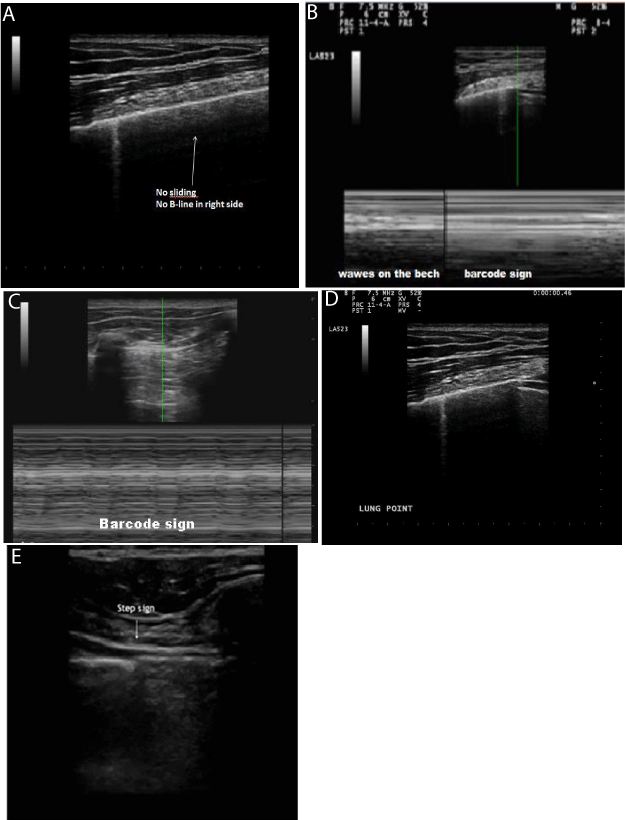
We used a phased-array transducer (3.5–5 MHz) to allow FAST echography, pleural effusion and adequate thoracoabdominal intercostal scanning, and a linear array transducer (7.5–10 MHz) with a preset for superficial tissues for the evaluation of pneumothorax. Patient is studied bedside in supine position. The pleural line is studied moving the probe by vertical lines parallel to the sternum (perpendicular to the ribs), starting from the parasternal line at the second to third intercostal space and moving between intercostal spaces towards the midclavicular line and reaching the axilla. PNX is generally identified in the “deep sulcus area” that is the iuxtacardiac space also involving the costophrenic angle. Pleural line appears approximately one-half centimeter deep to the ribs as an echogenic horizontal line. With patients breathes pleural line slides back and forth producing a glistening or shimmering appearance on ultrasound called as “lung sliding”. The lung sliding is not detectable in case of pneumothorax (Figure 2a). The lung sliding can be also graphically depicted by using M-mode-Doppler. A normal image will depict ‘‘waves on the beach”; in case of pneumothorax M-mode Doppler shows only repeating horizontal linear lines, demonstrating a lack of lung sliding or absence of the ‘‘beach’’ called as “stratosphere sign” or “barcode sign” (Figure 2b,2c). The presence of lung sliding and B lines (comet tails) rules out a pneumothorax. It may be possible to detect the lung lead point or area where an incomplete pneumothorax touches the chest wall [6]. This point of transition between the area of lung sliding and the absence of sliding may be detected by looking at the pleural line in several intercostal spaces. The step sign, a downward bending of pleural line in correspondence of lung point, may also indicate a pneumothorax (Figure 2d,2e).

    Figure 2:  (a-e): Our series of chest Ultrasonography for Pneumothorax
evolution.
Absent lung sliding and no B-lines in a case of pneumothorax (Figure 2a).
The “stratosphere sign” or “barcode sign” at M-Mode-Doppler in a case of
pneumothorax (Figure 2b, 2c).
The lung lead point in a case of pneumothorax. In this point lung sliding
suddenly appears and then disappears at the inter-space being examined
(Figure 2d).
The step sign, a downward bending of pleural line in correspondence of lung
point, may also indicate a pneumothorax (Figure 2e).
If a pneumothorax was suspected at ultrasonography, a thoracic CT was performed to confirm the diagnosis. See Table 2 for patient’s characteristics.
    Age
    Sex
    Etiology
    Nexus Score
    Rib Fractures
    Confirmed PNX at TC scan
    Chest Drainage
    Figure


    61
    M
    Motor Vehicle
    3
    Yes
    Yes
    Yes
    Figure 2a


    42
    M
    Minor fall
    2
    Yes
    Yes
    No
    Figure 2b


    15
    M
    Minor fall
    2
    No
    Yes
    Yes
    Figure 2c


    56
    F
    Minor fall
    3
    Yes
    Yes
    No


    37
    M
    Bike accident
    2
    Yes
    Yes
    No
    Figure 2d, 2e


    68
    F
    Minor fall
    3
    Yes
    Yes
    No
Table 2: Patients characteristics.
In our series cases a chest tube replacement was required in two patient; the other patients were treated only with observation and repeated x-ray and ultrasonography until the resolution of PNX.
All patients were then discharged without complications.
Discussion
Post-Traumatic pneumothorax is a potentially life-threatening condition because of the risk of progression to tension or massive pneumothorax and respiratory distress.
CT scan with a high priority for detection of chest lesions is the gold standard for diagnosis in thoracic traumas, but accessibility, costs, and radiation exposure often limit its use.
CT scan is widely used in patients with high suspicion of traumatic injury (high NEXUS score) and in patients with high risk mechanism of injury and evidence of high-energy impact.
Minor thoracic traumas are routinely managed with: CXR, rib radiograph series, and FAST examination (Focused Assessment with Sonography for Trauma) for detention of abdominal injury.
Supine AP chest radiograph is the most practical initial study in the initially evaluation of trauma victims especially when cervical spine immobilization is mandatory. Supine AP chest X-ray is insensitive for detecting pneumothorax. The reported proportion of pneumothoraces that are occult compared with those actually present on supine AP chest radiograph is variable and ranges from 29% to 72% [7-9]. Several Studies indicate that ultrasonography is more accurate than chest radiography for detection of pneumothorax and pleural effusion with a sensitivity ranging from 92% to 100% among patients with blunt injuries [10-11].
Additional data from reviews and meta-analysis reveal that performance of ultrasonography for the detection of pneumothorax is excellent and is superior to supine chest radiography [12] and support the adoption of chest ultrasonography for routine use in patients with clinically suspected pneumothoraces.
For this reason some trauma centers have begun performing an extended FAST exam (EFAST), evaluating for pneumo- and hemothorax in addition to intraperitoneal injuries [13].
In our institution approximately 70% of emergency physicians have a good expertise in EFAST technique. They all attended a 1– to 3– day training course with mentoring and practice on live models, followed by a six months period in which examinations were managed independently, recorded and later reviewed by the mentors. In our experience, such a kind of training has shown to be adequate to achieve a good level of performance.
Despite scientific evidence and its easy learning curve, thoracic ultrasound is generally underused in the emergency department. We believe that the underutilization of chest ultrasonography in emergency department depends on several causes. First of all, the belief that the lung is not suitable for ultrasound is still alive; so many doctors, in particular the seniors, still look at it with “great suspicion” and poor will to overcame this lack of confidence. We believe that we should fight against this misunderstanding and make strong efforts to spread the culture of ultrasound within emergency physicians. A second cause could be the limited availability of ultrasound equipment in the emergency department (economic reasons). Eventually, because of overcrowding in E.D., emergency doctors often may think to have no time to perform chest ultrasonography, even if this exam takes only few minutes.
Our case series supports the importance of chest ultrasonography and Extended FAST especially in patients with minor chest trauma (low NEXUS score) or with a low risk mechanism of injury which usually are discharged without a CT scan.
Conclusion
We recommend that any thoracic trauma victim presenting to the emergency department should be screened with thoracic ultrasonography to avoid missing a pneumothorax, because a negative AP chest radiograph can dangerously delay pneumothorax recognition. We think that this recommendation should be stronger for patients with minor chest trauma studied with a single AP chest X–ray and discharged without a CT scan.
References
- Freixinet Gilart J, Ramírez Gil ME, Gallardo Valera G, Moreno Casado P. [Chest trauma]. Arch Bronconeumol. 2011; 47 Suppl 3: 9-14.
- Karadayi S, Nadir A, Sahin E, Celik B, Arslan S, Kaptanoglu M. An analysis of 214 cases of rib fractures. Clinics (Sao Paulo). 2011; 66: 449-451.
- Rodriguez RM, Anglin D, Langdorf MI, Baumann BM, Hendey GW, Bradley RN, et al. NEXUS chest: validation of a decision instrument for selective chest imaging in blunt trauma. JAMA Surg. 2013; 148: 940-946.
- Volpicelli G. Sonographic diagnosis of pneumothorax. Intensive Care Med. 2011; 37: 224-232.
- Advanced Trauma Life Support - Student Course Manual -American College of Surgeons 8th Edition. Chicago. 2008.
- Perera P, Mailhot T, Riley D, Mandavia D. The RUSH exam: Rapid Ultrasound in SHock in the evaluation of the critically lll. Emerg Med Clin North Am. 2010; 28: 29-56, vii.
- Hill SL, Edmisten T, Holtzman G, Wright A. The occult pneumothorax: an increasing diagnostic entity in trauma. Am Surg. 1999; 65: 254-258.
- Neff MA, Monk JS Jr, Peters K, Nikhilesh A. Detection of occult pneumothoraces on abdominal computed tomographic scans in trauma patients. J Trauma. 2000; 49: 281-285.
- Xirouchaki N, Magkanas E, Vaporidi K, Kondili E, Plataki M, Patrianakos A, et al. Lung ultrasound in critically ill patients: comparison with bedside chest radiography. Intensive Care Med. 2011; 37: 1488-1493.
- Soldati G, Testa A, Sher S, Pignataro G, La Sala M, Silveri NG. Occult traumatic pneumothorax: diagnostic accuracy of lung ultrasonography in the emergency department. Chest. 2008; 133: 204-211.
- Lichtenstein DA, Mezière G, Lascols N, Biderman P, Courret JP, Gepner A, et al. Ultrasound diagnosis of occult pneumothorax. Crit Care Med. 2005; 33: 1231-1238.
- Alrajhi K, Wo MY, Vaillancourt C. Test characteristics of Ultrasonography for the Detection of Pneumothorax: a systematic review and meta-analysis. Chest. 2012; 141: 703–708.
- Kirkpatrick AW, Sirois M, Laupland KB, Liu D, Rowan K, Ball CG, et al. Hand-held thoracic sonography for detecting post-traumatic pneumothoraces: the Extended Focused Assessment with Sonography for Trauma (EFAST). J Trauma. 2004; 57: 288-295.