
Clinical Image
Austin J Emergency & Crit Care Med. 2015; 2(6): 1035.
The Electrocardiographic Phenomenon of Osborn Wave
Petrov DB*
Department of Cardiology, “Pirogov” Emergency Hospital, Bulgaria
*Corresponding author: Daniel Bogdanov Petrov, Department of Cardiolgy, “Pirogov” Emergency Hospital, 21 Totleben Ave., Sofia 1606, Bulgaria
Received: September 29, 2015; Accepted: October 08, 2015; Published: October 10, 2015
Clinical Image
A 61-year-old man presented with severe hypothermia (rectal temperature of 26°C), after being found by emergency medical services on his basement floor. An electrocardiogram revealed atrial fibrillation with a ventricular response rate of 77 beats/minute. The QRS complexes are narrow and are deformed at their terminal portions by a slurred wave occurring prior to the inscription of the ST-T waves; this is the Osborn wave (arrows). The patient was taken to the intensive care, but he died, despite all resuscitation efforts and rewarming methods.
Osborn waves, also known as J waves, camel-hump, and hypothermic waves, are best seen in the inferior and lateral precordial leads. The J waves are inscribed at the terminal portion of the QRS complexes, before the ST segment and T wave, and they appear secondary to an exaggerated outward potassium current leading to repolarization abnormality. Osborn waves should not be mistaken for portions of QRS complex or ST segment elevation.
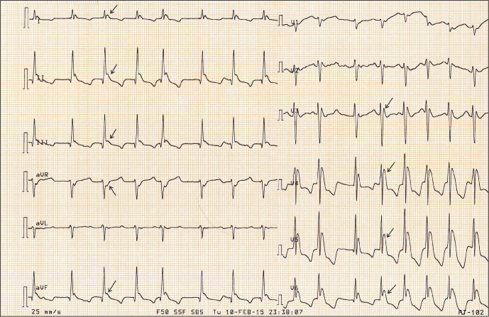
Figure 1: Image