Case Report
Austin J Emergency & Crit Care Med. 2015; 2(6): 1038.
Initial Assessment and Stabilization of the Airway for a Rare Case: Tracheobronchial Injury Occurred with Neck Scarf
Behcet AI*, Nogay S, Zengin S, Yavuz E and Yildirim C
Department of Emergency Medicine, Gaziantep University, Turkey
*Corresponding author: Behcet AI, Department of Emergency Medicine, Gaziantep University, Turkey
Received: September 22, 2015; Accepted: November 17, 2015; Published: November 19, 2015
Abstract
Tracheobronchial injuries are rare but potentially life-threatening. Tracheobronchial injury should be suspected in traumatic patient who has an opened wound with air entry and exit, dyspnea, hemoptysis, subcutaneous emphysema, upper rib fractures, chest tube with leakage and providing no expansion. A blunt cervical trachea injury due to scarf that covered the neck and seen rare was discussed in present case.
Keywords: Tracheobronchial injury; Neck; Scarf; Emergency department
Introduction
The incidences of the tracheobronchial injuries vary according to series. While, the most of tracheal injury related to penetrating trauma occurres at the cervical trachea, mediastinal trachea is injured more in blunt trauma [1,2]. Signs and symptoms are closely related to severity and the location of injury. Patients apply to emergency department (ED) with Stridor, subcutaneous emphysema, hemoptysis, pneumothorax, severe respiratory distress, hoarseness and pneumomediastinum [3]. The diagnosis is made by history, symptoms, signs and evaluation of the radiological data. The first step in treatment is to keep the airway open [4]. The purpose of this case report was to illustrate the importance of recognizing subtle signs and symptoms in the evaluation of a young with a potentially lifethreatening injury to the tracheobronchial injury secondary to blunt neck trauma. The options for initial assessment and stabilization of the airway in the emergency department (ED) are reviewed.
Case Presentation
A 27-year-old healthy male applied to our ED with circular abrasions on the neck, pain on the throat and voice hoarseness. The patient arrived by emergency medical service transport 60 minutes after accidentally snatched the end of his weft to the rubber band (2 cm in width and 1.5 cm in thickness) of the water dynamo while he was pulling water from well that was work in with electricity. The other end of the scarf that was surrounded the neck stretched the patient’s neck when dynamo pulled the free end for 2 or 3 second until it stopped automatically.
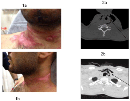
Immediately after accident, the patient complained of difficulty breathing pain on the neck and voice hoarseness. The patient had no other injuries or current illnesses. Medical history included no important special feature. On physical examination, the patient was comfortably sitting but moderate compelled talking and difficulty breathing. Vital signs were as follows: heart rate, 110 beats per minute; respiratory rate, 18 breaths per minute; blood pressure, 130/85 mm Hg; temperature, 37.0C; and oxygen saturation in room air, 94%. There was no sign of trauma on the other part of body. All surrounding of the neck was tender, swelling and included abrasions (Figure 1a,1b). Crepitus was determined on the anterior-superior of left shoulder. The thyroid and cricoid cartilage landmarks appeared normal on inspection. Cervical spine examination involved no tender. The lungs were clear to auscultation, the heart rhythm was regular; heart sounds were normal; and there was no murmur. The chest wall was non-tender without crepitus. No abnormalities were obtained in neurological examination. The laryngeal framework was stable on palpation of the larynx. Pneumomediastinum and subcutaneous gas especially throughout the left neck was obtained in Computed tomography (CT) scan of neck and thorax (without contrast) (Figure 2a,2b). The cervical spine was normal. Thorax and neck surgery were consulted. The patient was hospitalized to the care unit with cricothyrotomy procedure equipment at the bedside.

Figure 1: Text Here.
The patient was treated with intravenous dexamethasone and piperacillin/tazobactam. On the 10th hours after the injury, microlaryngoscopy, bronchoscopy, and esophagoscopy were performed. The patient underwent careful mask induction without positive-pressure ventilation. The subglottis, trachea, mainstream bronchi, vocal cords mobility, and mid portions of the esophagus were determined normal. The patient was continued on intravenous piperacillin/tazobactam for the next seven days. No complications developed. Due to a good recovery control flexible laryngoscopy at the bedside was not obtained. . The patient was discharged home after 8th days of admission with 875 mg amoxicillin & clavulanic acid (2x1 for 7 days) and 30 mg lansoprazole (1x1 for one month) medications. At follow-up 2 weeks later, the patient had no complaints of noisy or difficulty breathing or voice change. No invasive interventions such as microlaryngoscopy and bronchoscopy were planned.
Discussion
Since the scarf caused injury in our case, we evaluated it as blunt trauma. In the literature, there is no evidence of scarf-derived tracheobronchial injury. As the airway is totally seen open in first bronchoscopy, it gave us an idea about intact peribronchial adipose tissue, while since only air densities are seen between mediastinal soft tissue planes on tomography, it gave us an idea about intact mediastinal pleura. Although we could not localise the exact place of the injury on imaging, since there are subcutaneous emphysema, respiratory distress, hoarseness symptoms in our patient, we thought about cervical trachea as the place of injury.
Because there is no increase in the complaints of the patient, bronchoscopy was not tried again. No tissue loss, no need for positivepressure ventilation, limited subcutaneous emphysema, no additional injuries on esophagus and main vascular system has speeded up the healing and resulted in no need for invasive approach. In follow-ups, there were also no complications as atelectasia, pneumonia, abscess and empyema.
Grillo HC stated that of 71% of the penetran injuries, cervical trachea was injured, on the other hand, of 63% of the blunt injuries; mediastinal trachea was injured [1]. Bertelsen and Howitz had found that in 1178 post blunt thorax trauma case serials, 2.8% of them was tracheobronchial injury and %18 of these patients also had major vascular injury, more than 80% of tracheobronchial rupture was 2.5 cm close to carina, almost 90% of bronchi injuries were on main bronchi [3]. Lee RB et al. had stated that after penetran thorax trauma, the incidence of tracheobronchial injury was 0.5%-2% and 25% of these had affected the intrathoracic airways [5].
It was stated that in blunt trauma, injury mechanism on tracheobronchial tree usually occurs because of sudden increase of air pressure in main bronchi and trachea, expansion of thorax diameter towards laterals, insufficient support structure on right main bronchi and in “çamasir ipi tarzindaki yaralanma” compression of cervical trachea to vertebral objects [6,7].
Stridor, subcutaneous emphysema, severe respiratory distress, hemoptysis, hoarseness are the most frequent symptoms in cervical tracheal injury. Symptoms and findings in mediastinal tracheobronchial injuries are dependent on exposure of mediastinal pleura and peribronchial adipose tissue. In mediastinal pleura ruptures, tension pneumothorax being the first, severe clinical situations can be seen. If mediastinal pleura is kept intact, pneumomediastinum stands out and respiratory distress is rarely seen [6,8]. Injuries in which peribronchial adipose tissue is kept intact, since the airway is partly open, ventilation continues and these patients cannot be diagnosed. In 1-4 weeks, there is a risk of atelectasia, pneumonia, and abscess and empyema development in those patients. Fallen lung finding, pneumothorax continuation in spite of chest tube and air leakage are the findings of tracheobronchial injury. Endotracheal entubation would mask the tracheal injury [9]. In penetrance cervical injury, additional injuries on esophagus (43%) and main vascular system (14%); in blunt injuries, rib fracture possibilities should be investigated [4]. Almost 50% of the cases can be diagnosed within 48 hours. Important part of cases with left main bronchi rupture (app. 25-75%) can be discharge from hospital in early period without diagnosis [10,11]. Especially in acute period, in 10% of mediastinal tracheobronchial injuries, there can be no radiological findings and existing findings in 30% of patients can be missed out [4,8]. First diagnostic tests for stabile patients are lung and cervical radiography. Exact diagnosis can be done with bronchoscopy. Pneumomediastinum, refractive pneumothorax, air leakage to chest tube, apparent subcutaneous emphysema, atelectasia are the indications of bronchoscopy [4]. If injury cannot be detected in the first bronchoscopy but suspicion continues, bronchoscopy should be repeated. Developments in computer tomography do not put away the need for bronchoscopy. More than half of the injuries, entubation is needed. If entubation is insufficient, tracheotomy is done [7]. Before tracheobronchial repairmen, additional injuries (such as cervical and abdominal injuries, main cardiovascular injuries, esophageal and pharyngeal injuries) should be certainly investigated [4,11]. In patients who have linear laceration, airway diameter less than 1/3 of the airway (hava yolu çapinin 1/3’den daha küçük olan), who does not need positive-pressure ventilation, have full re-expansion of the lung with tube thoracostomy, limited or retrograding subcutaneous emphysema, loss of air leakage right after chest entubation, conservative treatment can be applied [1]. Patients who cannot take conservative treatment should get surgical treatment immediately. Mortality rates in tracheobronchial injuries are generally below 30% (3.5-67%) [12].
References
- Grillo HC. Tracheal and bronchial trauma. In: Grillo HC, editor. Surgery of the trachea and bronchi. London B.C. Hamilton Inc. 2004; 271–90.
- Hahn B. Tracheobronchial rupture. J Emerg Med. 2007; 33: 193-194.
- Bertelsen S, Howitz P. Injuries of the trachea and bronchi. Thorax. 1972; 27: 188-194.
- Balci AE. Trakeobronsiyal yaralanmalar. Turkiye Klinikleri. J Surg Med Sci. 2007; 3: 52-62.
- Lee RB. Traumatic injury of the cervicothoracic trachea and major bronchi. Chest Surg Clin N Am. 1997; 7: 285-304.
- Özdülger A. Trakeobronsiyal Yaralanmalar. TTD Toraks Cerrahisi Bülteni. 2010; 1: 45-54.
- Karmy-Jones R, Wood DE. Traumatic injury to the trachea and bronchus. Thorac Surg Clin. 2007; 17: 35-46.
- Ishibashi H, Ohta S, Hirose M, Akimoto T. Blunt tracheal transection and long tear in posterior membranous trachea. Eur J Cardiothorac Surg. 2006; 30: 945-947.
- Stark P. Imaging of tracheobronchial injuries. J Thorac Imaging. 1995; 10: 206-219.
- Hahn B. Tracheobronchial rupture. J Emerg Med. 2007; 33: 193-194.
- Bingol-Kologlu M, Fedakar M, Yagmurlu A, Fitoz S, Dindar H, Gokcora IH. Tracheobronchial rupture due to blunt chest trauma: report of a case. Surg Today. 2006; 36: 823-826.
- Hancock BJ, Wiseman NE. Tracheobronchial injuries in children. J Pediatr Surg. 1991; 26: 1316-1319.