Case Report
Austin J Emergency & Crit Care Med. 2015; 2(7): 1039.
Snowflakes in the Liver
Ceruti S¹* and De Vivo S²
¹Department of Intensive Care Unit, Geneva University Hospitals, Switzerland
²Department of Anesthesiology and Intensive Care, University Hospital “Federico II”, Italy
*Corresponding author: Samuele Ceruti, Department of Intensive Care Unit, Geneva University Hospitals, Geneva, Switzerland
Received: November 10, 2015; Accepted: December 20, 2015; Published: December 24, 2015
Case Presentation
A 43-year-old male patient was admitted to the Emergency Department ward for general malaise with abdominal pain in the right upper quadrant, breath employee, already present two months before and actually increase. There was no sign of fever, pathologic lymphadenopathy, jaundice or other symptoms. At physical examination the patient appeared lucid, well spatial/temporaloriented, afebrile, hemodynamically stable (AP 100/70 mmHg, HR 68 bpm), weight 73 Kg, height 180 cm; all examinations were in the normal range, except for mild deep pain in right upper quadrant and the right shoulder.
Past history
He was born in Portugal, lived in Switzerland from 1983 to 2000, then in France from 2000 to 2007 and again in Switzerland. In 1983 the patient had suffered from Brucellosis, treated with antibiotic therapy for 9 months; an abdominal CT-scan in 1992 revealed multiple hepatic calcifications. In 2001 he had a single episode of nephrolithiasis treated pharmacologically.
Laboratories and radiological findings
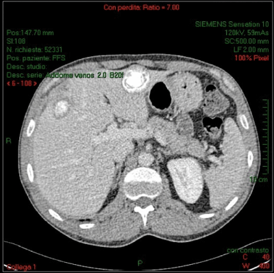
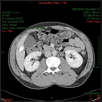
The laboratory exams performed show an elevation of GGT (68 U/l), CRP (31,9 mg/l) and ferritin (771 ng/ml), while other routine exams were in the normal range. An abdominal CT-scan showed calcification with a “snowflake appearance” in the liver (Figures 1,2); for our radiologic team these are suggestive for signs of chronic hepatic Brucellosis. The patient was seen twice at our department to undergo liver fine-needle biopsy of the major hepatic lesion, negative for helminths (Trichinellaspp, Toxocariasis, Echinococcusgranulosus, Fasciolaspp, Anguillosisspp, Cisticercosisspp), Protozoa, Parasites and Bacteria.

Figure 1:

Figure 2:
Medical treatment
Suspecting bacterial super infection of the calcified liver lesions, the patient was treated first with Ceftriaxone 2 g qd IV and Metronidazol 500 mg tid PO without improvement of the patient’s clinical condition. After 2 weeks, his serology became positive for Brucellaspp; he was then treated with Doxicycline 100 mg bid PO and Gentamicin 120 mg bid IV, again without any significant clinical improvement. After 8 weeks of therapy, the treatment was switched on Rifampicin 300 mg tid PO and Ciprofloxacin 750 mg bid PO.
The lesion and clinical signs persist for 2 weeks; then a sub- capsular draining of one of these lesions with general sedation was made, with 8 ml of pus removed, leaving a local drainage in order to wash the lesion twice a day. All the material was sent to laboratory, showing amplification of genetic material derived from Brucella after amplification with specific primers, while cultural exams remained negative for microorganism. Five days after, the drainage tube was removed. Then an abdominal echography was performed which showed no signs of intra-parenchymal or sub-capsular fluid collection. The patient was discharged.
The next week the patient felt the same pain exacerbated in the same region, worsening with minor movement; a new abdominal echography showed new fluid collection (3,6 * 3,6 * 2,3 cm) in the IV-hepatic segment. An abdominal-CT-scan with contrast showed a small increase of the sub-diaphragmatic inflammatory collection (axial diameters of 3 * 6 cm), without any other pathological finding.
Surgical treatment
After unsuccessful drainage and antibiotic therapy for 14 weeks, we decided to choose a surgical approach. The patient was operated for removal with enucleation all pathological lesions; they were sent to the pathology in order to identify the etiological and pathological origin of every single lesion. Subsequent analysis performed show us areas of diffuse hepatic necrosis with focal calcifications and peripheral macrophages, without clear margins and a granulome-like organization, picture compatible with multiple Brucellomas [1].
The patient was discharged after one week, with clinical improvement and only mild pain in surgical scar’s region, wellcontrolled with analgesic therapy and antibiotic therapy (Doxicycline 100 mg bid PO and Rifampicin 300 mg tid PO) for 1 month again; after 4 weeks, the patient shows a really good improvement, with physical and psychological improvement.
Conclusion
Brucellosis is a zoonotic disease transmitted through direct contact with infected animals [2]; the symptoms may last for months and can be insidious [2]. Despite antibiotics [3], bacteria are sometimes difficult to eradicate and if the disease is very aggressive [4], surgery may be required to treat patients [5]. We’ve found a typical case of a relatively rare disease that took some time to be properly diagnosed. Following current guidelines, we starting first with antibiotic treatment and then, given the persistence of the disease, with a more aggressive surgical option [3,4]. It is known that if the disease persists despite antibiotic therapy, as in this case, is adequate treat the patient surgically [5]. We wondered if we had had to intervene early with surgery, in order to rapidly eradicate the disease, but at the moment the evidences point to intervene only in case of persistence of the disease despite maximal medical therapy. Further evidences are needed to acquire more knowledge about the better treatment, especially to identify patients at risk of relapse, in order to rapidly change on surgical option rather than continue for a long time with antibiotic therapy. We hope that this event will make a contribution in this direction.
References
- Akritidis N, Tzivras M, Delladetsima I, Stefanaki S, Moutsopoulos HM, Pappas G. The liver in brucellosis. Clin Gastroenterol Hepatol. 2007; 5: 1109-1112.
- Pappas G, Akritidis N, Bosilkovski M, Tsianos E. Brucellosis. N Engl J Med. 2005; 352: 2325-2336.
- Skalsky K, Yahav D, Bishara J, Pitlik S, Leibovici L, Paul M. Treatment of human brucellosis: systematic review and meta-analysis of randomised controlled trials. BMJ. 2008; 336: 701-704.
- Ariza J, Corredoira J, Pallares R, Viladrich PF, Rufi G, Pujol M, et al. Characteristics of and risk factors for relapse of brucellosis in humans. Clin Infect Dis. 1995; 20: 1241-1249.
- Ariza J, Pigrau C, Cañas C, Marrón A, Martínez F, Almirante B, et al. Current understanding and management of chronic hepatosplenic suppurative brucellosis. Clin Infect Dis. 2001; 32: 1024-1033.