Case Report
Austin J Emergency & Crit Care Med. 2015; 2(7): 1040.
A Dermatologic Emergency Case of Neutrophilic Eccrine Hidradenitis
Salman N¹*, Yesil H², Çermik H³, Tezel O¹ and Eryilmaz M4
¹Department of Emergency Medicine, Etimesgut Military Hospital, Turkey
²Department of Dermatology, Etimesgut Military Hospital, Turkey
³Department of Pathology, Etimesgut Military Hospital, Turkey
4Department of Emergency Medicine, Gulhane Military Medical Academy, Turkey
*Corresponding author: Salman N, Department of Emergency Medicine, Etimesgut Military Hospital, Etimesgut Asker HastanesiAcilServisi, 06790 Etimesgut / Ankara, Turkey
Received: November 26, 2015; Accepted: December 21, 2015; Published: December 23, 2015
Abstract
Neutrophilic eccrine hidradenitis (NEH) is a nonspecific dermatologic lesion pattern presenting as erythematous papules and plaques. In this case report, we present a patient diagnosed with NEH at the emergency department (ED) without any history of malignancy, chemotherapy, drug reactions, or systemic inflammatory disease. A 29-year-old man presented to our ED with complaints of fever, bilateral localized axillary skin rash, and pain. He stated that his complaints had started two days before admission. At the initial examination, he had tender and polymorphic erythematous papules in the bilateral axillae. A complete blood count showed leukocytosis (10,970/mcl), and the C-reactive protein (CRP) value was increased (6.6 mg/dl). We did not detect any other physical examination findings that could be related to infection. After being prescribed a non-steroidal anti-inflammatory drug and systemic antibiotics, the patient was referred to the dermatology clinic for further evaluation. During the dermatologic consultation, a 4-mm punch biopsy was performed of the patient’s right axilla. Histopathology revealed neutrophil infiltration, nuclear pyknosis, and cytoplasmic eosinophilia in the eccrine glandular epithelium, which confirmed the diagnosis of NEH. NEH is a real dermatologic emergency that emergency physicians must be able to recognize and evaluate in ED settings. Although skin biopsy is the key element of diagnosis, detailed physical examinations, laboratory tests, and skin cultures should be performed. Furthermore, fever should be accepted as a criteria for the administration of systemic antibiotics.
Keywords: Neutrophilic eccrine hidradenitis; Eccrine glands; Dermatologic emergency; Emergency department
Introduction
Neutrophilic eccrine hidradenitis (NEH) is a nonspecific dermatologic lesion pattern presenting as erythematous papules and plaques. The clinical features on the initial examination may include lesions that are tender [1], polymorphic, pruritic, recurrent, or chronic [2]. Although it is known that this lesion usually occurs in patients with malignancies and who are undergoing chemotherapy [3], the current literature contains cases occurring with drug reactions [4,5], infections [6], Behçet’s disease [2], and intense sun exposure [7]. In this case report, we present a patient diagnosed with NEH at the emergency department (ED) without any history of malignancy, chemotherapy, drug reactions, or systemic inflammatory disease.
Case Presentation
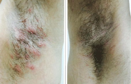
A 29-year-old man presented to our ED with complaints of fever, bilateral localized axillary skin rash, and pain. He stated that his complaints had started two days before admission. At the initial examination, he had tender and polymorphic erythematous papules in the bilateral axillae (Figure 1a and 1b). We did not detect any other signs or symptoms that may cause fever. His vital signs were blood pressure of 120/72 mmHg, pulse rate of 120/minute (regular), temperature of 40oC, SaO2 of 99%, and respiratory rate of 12/ minute. His state of consciousness was assessed as normal (Glasgow coma scale: 15). After the initial examination, blood and urine samples were taken, posterior-anterior and lateral chest x-rays were performed, and the patient was treated with 1 g of IV acetaminophen. The patient’s blood urea, creatinine, sodium, potassium, aspartate aminotransferase, alanine aminotransferase, sedimentation, and urine analysis were all within normal ranges. Chest x-rays revealed no abnormalities. A complete blood count showed leukocytosis (10,970/ mcl; reference value 3,500–10,500/mcl), and the C-reactive protein (CRP) value was increased (6.6 mg/dl; reference value 0–6 mg/dl). We did not detect any other physical examination findings that could be related to infection.

    Figure 1:  (a-left) Skin lesion in the left axilla. (b-right) Skin lesion in the right
axilla.
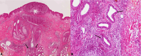
After being prescribed a non-steroidal anti-inflammatory drug (naproxen sodium 550 mg once daily) and systemic antibiotics (amoxicillin/clavulanic acid 1 g twice daily), the patient was referred to the dermatology clinic for further evaluation. During the dermatologic consultation, a 4-mm punch biopsy was performed of the patient’s right axilla. Histopathology revealed neutrophil infiltration, nuclear pyknosis, and cytoplasmic eosinophilia in the eccrine glandular epithelium, which confirmed the diagnosis of NEH. The periadnexal stroma showed a variable infiltrate of neutrophils, lymphocytes, histiocytes, and eosinophils. There was no evidence of vasculitis (Figure 2a and 2b). A mid-potency topical corticosteroid (methylprednisolone aceponate 0.1% twice daily) was initiated for both axillary lesions. One week later, the control examination showed total remission of both axillary lesions and the patient did not mention any fever attacks.

    Figure 2:  (a) Neutrophilic infiltration reaches from the eccrine glands to
the papillary dermis (HE, x20). (b) A dense neutrophil polymorph infiltrate
surrounds the eccrine secretory coils in the deeper reticular dermis (HE,
x100) (black arrows).
Discussion
We present a case of NEH in a young healthy man with complaints of fever and skin rash. The current literature on NEH mainly focuses on the etiology of these cases, and NEH is identified as a ‘distinctive dermatosis occurring in patients with malignancy or undergoing chemotherapy’ [4]. It has been reported that NEH in a healthy adult without any disorders is extremely rare [8]. We suggest that the low rate of NEH in healthy adults may be due to under diagnosis in ED settings.
Dermatologic emergency cases are infrequent compared to other ED admissions. An 18-month retrospective survey reported that 1.8% of total ED visits corresponded to dermatological problems [9], but the rate has been determined to be up to 8%–10% [8]. However, dermatologic emergency applications are relatively fewer, and the diagnoses of most patients are limited in certain cases, including insect bites and urticarial angioedema. We believe that these factors restrict emergency physicians’ experience with primary dermatologic disorders such as NEH.
Skin biopsy and histological examination of the lesion plays a crucial role in the diagnosis of NEH. Before this invasive procedure, detailed history-taking, physical examination, and biochemical tests should be performed. Copaescu et al. emphasized that when considering a diagnosis of NEH, infection must be ruled out [10]. In our case, fever, skin rash, leukocytosis, and elevated CRP were the signs and symptoms related to infection. Although the biopsy results confirmed the definitive diagnosis, we believe that the physical examination and laboratory tests directed the physicians toward the proper treatment with the NEH evaluation. Another important evaluation step is the investigation of skin-tissue cultures from lesions when the infectious origin of NEH is considered [11]. In our case, we did not perform skin-tissue cultures of the lesions and thus could not identify the infectious agent. We acknowledge that this is a limitation of our case presentation.
Our patient reported that the fever and skin rash had occurred simultaneously two days earlier, and fever was his major symptom before ED admission. Wenzel et al. reported that NEH onset is associated with fever [12]. Our patient’s symptoms were in agreement with Wenzel et al.’s report, but it was still unclear whether fever was a result of, or the reason for, the NEH pathogenesis. Another controversial issue regarding NEH is the use of antibiotics on follow-up. In our case, oral antibiotics were used, and we observed a parallel clinical improvement in the patient’s fever and skin rash. Antonovich et al. reported that NEH usually resolves spontaneously, but infectious forms of NEH often require treatment with antibiotics [6]. In addition to Antonovich et al.’s result, we believe that fever is an indicator of the systemic extent of the infection, and may be used as a decision-making criteria for systemic antibiotic use in the treatment of NEH.
Conclusion
NEH is a real dermatologic emergency that emergency physicians must be able to recognize and evaluate in ED settings. Although skin biopsy is the key element of diagnosis, detailed physical examinations, laboratory tests, and skin cultures should be performed. Furthermore, fever should be accepted as a criteria for the administration of systemic antibiotics.
References
- Yeh I, George E, Fleckman P. Eccrine hidradenitis sine neutrophils: a toxic response to chemotherapy. J Cutan Pathol. 2011; 38: 905-910.
- Nijsten TE, Meuleman L, Lambert J. Chronic pruritic neutrophilic eccrine hidradenitis in a patient with Behçet's disease. Br J Dermatol. 2002; 147: 797-800.
- Beutner KR, Packman CH, Markowitch W. Neutrophilic eccrine hidradenitis associated with Hodgkin's disease and chemotherapy. A case report. Arch Dermatol. 1986; 122: 809-811.
- Bhanu P, Santosh KV, Gondi S, Manjunath KG, Rajendaran SC, Raj N. Neutrophilic eccrine hidradenitis: a new culprit-carbamazepine. Indian J Pharmacol. 2013; 45: 91-92.
- EL Sayed F, Ammoury A, Chababi M, Dhaybi R, Bazex J. Neutrophilic eccrine hidradenitis to acetaminophen. J Eur Acad Dermatol Venereol. 2006; 20: 1338-1340.
- Antonovich DD, Berke A, Grant-Kels JM, Fung M. Infectious eccrine hidradenitis caused by Nocardia. J Am Acad Dermatol. 2004; 50: 315-318.
- Vuong CH, Walters R, Stein JA. Infectious eccrine hidradenitis associated with intense sun exposure. Cutis. 2013; 92: 40-45.
- Kambayashi Y, Fujimura T, Yamasaki K, Aiba S. Therapy-resistant, spontaneously remitting generalized neutrophilic eccrine hidradenitis in a healthy patient decreases the expression of dermcidin in affected eccrine glands. Case Rep Dermatol. 2011; 3: 228–234.
- Kaptanoglu AF, Özgöl Y TM. Dermatological Applications to the Emergency Department of an University Hospital in Northern Cyprus. JAEM. 2012; 11: 137–140.
- Copaescu AM, Castilloux JF, Chababi-Atallah M, Sinave C, Bertrand J. A classic clinical case: neutrophilic eccrine hidradenitis. Case Rep Dermatol. 2013; 5: 340-346.
- Bassas-Vila J, Fernández-Figueras MT, Romaní J, Ferrándiz C. Infectious eccrine hidradenitis: a report of 3 cases and a review of the literature. Actas Dermosifiliogr. 2014; 105: e7-12.
- Wenzel FG, Horn TD. Nonneoplastic disorders of the eccrine glands. J Am Acad Dermatol. 1998; 38: 1-17.