
Special Article – Emergency Medicine Case Reports
Austin J Emergency & Crit Care Med. 2017; 4(1): 1052.
Unrecognised Trauma due to Resuscitation in a Pregnant Woman
Dasari P*, Gowri MK and Kumar DM
University Research Co., LLC, Quality & Performance Institute, USA
*Corresponding author: Department of Obstetrics and Gynecology, JIPMER, India
Received: November 28, 2016; Accepted: February 08, 2017; Published: February 10, 2017
Abstract
A 26 year old second para that underwent resuscitation and emergency LSCS at 30 weeks of gestation following convulsions was referred to us after 36 hours of delivery. At admission she was in Glasgow coma scale E1VTM1 and ventilator support was continued and hypernatremia which was detected at admission was corrected. She had cerebral oedema on CT and subcutaneous emphysema of neck and upper chest and massive pneumothorax which was detected after 12-24 hours of transfer to our Institute. She also had right Vocal cord palsy and all these were attributed to the complications of CPR. She was managed with anti-oedema measures and ICD She required prolonged ventilation, underwent tracheostomy and finally decanulated on 23rd day of admission and discharged after 35 days of hospitalization. The diagnosis of post cardiac arrest syndrome was complicated because of her initial history of high grade fever and chills, vomiting and head ache convulsions and also possible diagnosis of eclampsia. It is essential to diagnose and manage the complications arising out of resuscitation so as to increase the chances of survival though the complications cannot be avoided at times. An undetected injury can be life threatening.
Keywords: Pregnancy; CPR; Pneumothorax; Pneumomediastinum; Vocal cord palsy
Introduction
Survival post cardio pulmonary resuscitation is reported to vary and depends on the time efficacy of such procedure and also the disease entity. Complications associated with cardiopulmonary resuscitation are rare but add to morbidity and mortality at times. These include trauma to the heart, chest wall, lungs, liver, spleen and gastrointestinal tract rupture such as stomach, esophagus and colon. It is important to recognize these complications also so that effective management can be undertaken to treat the same. Unrecognized trauma due to resuscitation is described in the case reported here.
Case Presentation
A 26 year old second para that recently underwent emergency LSCS (36 hours) at 30 weeks of gestation for eclampsia was referred to our Institute with ventilator support after resuscitation in another Institute. At arrival she was unconscious and on Ambu bag ventilation, pupils were pin point and sluggishly reacting to light. She was febrile (101.40F) with a pulse rate of 116/min and her BP was 120/70 mmHg. On auscultation there were bilateral crepts with diminished air entry. CVS was normal except for tachycardia. Abdomen showed minimal distension with a recent caesarean section wound and the abdominal drain had 10ml hemorrhagic serous fluid. She was admitted to ICU and her ABG showed hyper-natraemia. Corrective measures were started as per protocol and she was started on broad spectrum antibiotics. Subcutaneous emphysema of neck was noted while insertion of central line and CT head and X ray chest were requested.
Her present Obstetric history was that she had regular antenatal care at nearby primary health centre and was normal till 4 days ago when she developed fever with chills, vomiting and headache for which she was advised hospitalization but she went home and developed a state of confusion, slurred speech and convulsions and hence she was rushed to a medical college nearby and after resuscitative measures she was diagnosed as antepartum eclampsia and treated with injection Magnesium sulphate. After 4 hours of admission she was taken up for emergency Caesarean section and a preterm baby of 940 gm was delivered. Intra-operatively a fall in saturation was noted and she was diagnosed to be having pulmonary oedema and received management for the same and she was kept in ICU on Ventilator for 32 hours after which she was shifted to our Institute because of lack of improvement. During her first pregnancy she had history of hypertension and delivered an IUGR baby weighing 2.2kg which is alive and healthy but during the present pregnancy she had no history recording of hypertension.
At admission, her haemogram showed the following results. Hb-11.7g%, TC-17,000, N-95, L3, Platelets 4.17lakh. Urea 81mg/dl, creat-1.6mg/dl, RBS-202mg/dl, Na-150mEq/L,AST- 56IU/L, ALT- 24IU/L, Bilurubin-0.7mg/dl , PT-INR – 13.8/1.02sec. Urine albumin was negative.
Her CT head which done after 12 hours of admission showed cerebral oedema (Figure 1) and CT Venogram was normal. Fundus examination showed early papilloedema. Neurologist opinion was obtained and she was started on anti-oedema measures as per the advice. X-Ray chest was reported after 24 hours which showed emphysema of neck and massive pneumothorax (Figure 2) same was managed by Inserting Intercostal Drain (ICD). CECT chest and abdomen were obtained which showed Pneumomediastinum, pneumoperitoneum, right upper lobe and left lower lobe collapse (Figure 3). Bronchoscopy was done to rule out bronchial disruption. Over 48 hrs, patient’s GCS had improved significantly. Repeat CT head showed widening of sylvian fissure implying a decrease in the cerebral edema. She required ventilatory support for 10 days as her respiratory efforts were poor following weaning after 10 days. The infectious work –up showed negative blood, unine and cervical swab cultures. Negative for infectious diseases such as Malaria and Dengue fever. Her tracheal aspirate though negative initially grew Entero bacter species which was resistant to all drugs. VAP was treated with injection Colistin. She underwent Tracheostomy on 16th day and decanulated on 23rd day after improvement. There was difficulty in swallowing and hoarseness of voice after recovery and ENT examination showed right Vocal Cord palsy and she needed speech therapy. After multidisciplinary management she was discharged home on 35th day.
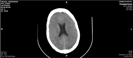
    Figure 1: CT head which done after 12 hours of admission showed cerebral
oedema and CT Venogram was normal.
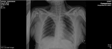
    Figure 2: X-Ray chest was reported after 24 hours which showed emphysema
of neck and massive pneumothorax.
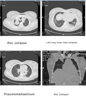
    Figure 3: CECT chest and abdomen were obtained which showed
pneumomediastinum, pneumoperitoneum, right upper lobe and left lower
lobe collapse Bronchoscopy was done to rule out bronchial disruption.
Discussion
The injuries that occur during resuscitation are inevitable in the process and include minor, major as well as life-threatening. The minor injuries are abrasions, ecchymosis, subcutaneous emphysema, subcutaneous haematomas, mucosal lacerations. Major injuries include fracture of ribs, clavicle, thyroid cartilage, dislocation of tempero mandibular joint etc. Rupture of organs such as heart, liver, lungs, spleen gastro-intestinal tract are life threatening and add to mortality. These are related to the duration and quality of resuscitation rather than the age and gender. A retrospecective study of non-traumatic death cases over a 10 year period reported a positive correlation between the injuries and the duration of CPR [1].
Injuries due to CPR can be due to Ventilation (intubation) related or related to Chest compressions [2]. The present lady sustained injuries during resuscitation due to ventilation which are subcutaneous emphysema, pneumothorax and Vocal cord palsy. The duration of chest compressions are reported to be related to thoracic injuries which significantly increase after 60 minutes of chest compression1. This factor could not be ascertained in this case as the details of resuscitation were not available in the referral slip. She was mis-diagnosed as pulmonary oedema on table and managed though she had pneumothorax. With advanced air way technique 40% had injuries and only less than 50% of these were recognized in the retrospective analysis by Umit Kaldrum and collegues [1].
The management of post CPR is complicated by the development of Post-cardiac arrest syndromes and trauma due to CPR. The [3] post cardiac arrest syndromes described include post cardiac arrest brain injury, postcardiac arrest myocardial dysfunction and systemic ischaemia or reperfusion [3]. The occurrence of cerebral oedema in this lady as a post cardiac arrest syndrome is debatable as she had history of convulsions which were attributed to eclampsia which is also a cause for cerebral oedema the vocal cord palsy points out to a difficult intubation and the pneumothorax could have been the complication of intubation as well as chest compression. The reported rate of pneumothorax as a complication of CPR is quite high, the left sided pneumothorax was 19.2% and right sided 5.4% [1]. The recognition of pneumothorax is essential and the management is simple and dramatic improvement results after ICD placement.
The survival following CPR is increasing with the advances in resuscitative techniques but still more than 50% deaths are occurring. Return of Spontaneous Circulation (ROSC) is an important factor that influences survival and also the type of institutional care has an influence in reducing the mortality after CPR. Mortality was observed to be lower in urban, teaching and large hospitals [4] the care delivered after ROSC is important and therapeutic mild hypothermia can play an important role by reducing the ROS (Reactive Oxygen Species), metabolic needs of tissues especially the myocardium [5]. But in practice it is employed only in 5% and presence of protocols and care bundles like Cardio Cerebral Resuscitation (CCR) can improve the survival [6]. It is important to recognise the resuscitation related injuries and distinguish them from natural disease process that occurred prior to resuscitation trauma to deliver optimum care.
Conclusion
Iatrogenic injuries such as pneumothorax, Pneumomediastinum can result during resuscitation. Post cardiac arrest syndromes to be recognized early and management of such conditions reduce the mortality.
References
- Kaldrum U, Toygar M, Karbeyaz K, Arziman I, Tuncer SK, Eyi YE. Complications of Cardiopulmonary resuscitation in traumatic cases and factors affecting complications. Egyptian Journal of Forensic Sciences. 2016; 6: 270-274.
- Hashimoto Y, Moriya F, Furumiya J. Forensic aspects of complications resulting from cardiopulmonary resuscitation. Legal Medicine. 2007; 9: 94-99.
- Nolan JP, Neumar RW, Adrie C, Aibiki M, Berg RA, Böttiger B. Postcardiac arrest syndrome: Epidemiology, pathophysiology, treatment, and prognostication: A Scientific Statement from the International Liaison Committee on Resuscitation; the American Heart Association Emergency Cardiovascular Care Committee; the Council on Cardiovascular Surgery and Anesthesia; the Council on Cardiopulmonary, Perioperative, and Critical Care; the Council on Clinical Cardiology; the Council on Stroke. Resuscitation. 2008; 79: 350-379.
- Carr BG, Goyal M, Band RA, Gaieski DF, Abella BS, Merchant RM. A National analysis of the relationship between hospital factors and post cardiac arrest mortality. Intensive Care Med. 2009; 35: 505–511.
- Yang L, Zhao X, Liu L. Mild hypothermia in improving multiple organ dysfunction after Cardiac arrest. World J Emerg Med. 2010; 1: 196-200.
- Bobrow BJ and Kren KB. Regionalization of post-cardiac arrest care. Curr Opin Crit Care. 2009; 15: 221–227.