
Case Report
Austin J Emergency & Crit Care Med. 2017; 4(1): 1055.
Right Upper Abdominal Pain and Jaundice, Which Brings to Mind Biliary Tract Diseases at First Sight: A Case Report
Vural A¹* and Yesin M²
¹Department of Emergency Medicine, Kars Harakani State Hospital, Kars, Turkey
²Department of Cardiology, Kars Harakani State Hospital, Kars, Turkey
*Corresponding author: Vural A, Emergency Medicine Specialist, Kars Harakani State Hospital, Emergency Departmant, Kars, Turkey
Received: February 06, 2017; Accepted: March 14, 2017; Published: March 20, 2017
Abstract
Ventricular septal defect after myocardial infarction is rare but it is very mortal complication of acute myocardial infarction. Diagnosis can be easily made by using transthoracic Doppler echocardiography. Spontaneous closure of ventricular septal defect is very rare and the treatment is emergency surgery.
In this case we present a 54 years-old male patient who admitted to our emergency service with sub acute myocardial infarction who later diagnosed for rupture of apical ventricular septum and was successfully operated on follow-up.
Keywords: Acute myocardial infarction; Ventricular septal defect; Echocardiograph; Pansystolic murmur; Surgery
Abbreviations
VSD: Ventricular Septal Defect; MI: Myocardial Infarction; ECG: Electrocardiogram; EF: Ejection Fraction; CT: Computed Tomography; IABP: Intra Aortic Ballon Pump; LAD: Left Anterior Descending; CABG: Coronary Artery Bypass Graft
Introduction
Ventricular septal defect (VSD) after myocardial infarction (MI) is rare (1-3%), but very mortal complication [1-3]. Post MI VSD is usually seen in first two weeks after MI [1]. Patients generally admit with complaints of acute heart failure findings, shock and newly developed pansystolic murmur during first week after MI [3,4]. Diagnosis can be easily made by using transthoracic Doppler echocardiography [2-5]. Spontaneous closure of VSD is very rare and these post infarction defects require urgent surgical treatment [1,2].
Case Presantation
A 54-year-old patient with a history of arterial hypertension admitted to our emergency department with complaint of right upper abdominal pain, vomiting and jaundice which started five days before. Physical examination revealed a regular pulse of 90 beats/min. Arterial blood pressure was measured 110/70mmHg. Patient was tachypneic (respiratory rate 22/min) looking toxic. There markable findings on first physical examination were moderete right upper abdominal sensitivity and pain and also mild to moderete jaundice.
On laboratory findings, (pathologically): hemoglobin 17.9g/dL (12-16.8), hematocrit 54.2% (36.2-49.7) and leukocyte 22.6 x 10³ (4- 10.6) were detected in the whole blood count. In biochemistry tests, glucose was found to be 430mg/dL (74-105), urea 191mg/dL (13-43), creatinine 2.2mg/dL (0.7-1.3), AST 155 (0-31), ALT 95 (GGT) 224U/L (11-50), ALP 195U/L (56-119), amylase 306U/L (0-110), total/direct bilirubin 4.46/4.03mg /dl, and sodium 122mmol (135-145). In blood gases, ph: 7.32 (7.37-7.45), PCO2: 28.8 (35-46), HCO3 act: 17 (21-26), BE: -8 (±2mmol/L ).
According to the patient’s physical examination and laboratory findigs, we thought that this condition is due to biliary tract pathologies such as cholecystitis, choledocholithiasis, cholangitis or pancreatitis. But, no pathology was detected in his abdominal computed tomography (CT) and then, detailed physical examination was performed and detailed history of patient was taken.There markable finding on detailed physical examination was 3-4/6 grade parasternal pansystolic murmur best heard at the apex and detailed his story revealed that the patient had chest pain approximately 2 weeks before. The 12-lead electrocardiogram (ECG) showed ST segment elevation in anterior derivations (Figure 1). But, he was not complaining of chest pain at emergency admission. In the differential diagnosis there was no pathology such as cholecystitis, choledo cholithiasis or pancreatitis in his abdominal CT and there was no history of any intoxication, trauma, malignancy or septic infection. That's why we thought that the clinic and laboratory findings of the patient is due to tissue hypoxia after MI. Then, the patient was consulted for transthoracic echo cardiography which revealed apical aneurysm, advanced hypokinetic anterior septum, impaired systolic functions (ejection fraction (EF) : 35%) and also a defect in the apical section (Figure 2). The pansystolic murmur was thought to have a risen due to ventricular septal defect. The patient was transferred to the tertiary center for cardiac surgery. At followup, it was learned that the patient admitted to cardiovascular surgery intensive care unit with cardiogenic shock. Then, positive inotropic agents were started and intra aortic balloon pump (IABP) was inserted to the patient immediately. Preoperative coronary angiography showed that there was single-vesseldisease (Left Anterior Descending (LAD) 100% totally occluded). And then the defect repair successfully and coronary artery bypass graft (CABG) were applied . We learned that the patient was being operated for the second time due to residual defect on follow-up but, unfortunately, the patient died on the 16th day of hospitalization.
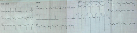
Figure 1: Patients ECG on admission revealed sinus rhythm, Q-waves and ST segment elevation in anterior leads.
Discussion
Post MI VSD is a mechanical complication that is seen due to reperfusion defect in occluded main artery which in turn can lead to hemodynamic instability findings like secondary organ failure, respiratory distress, congestive heart failure and shock [6]. Respiratory distress, hypo perfusion and secondary organ dysfunctions because of decreased ejection fraction were seen in our patient on his admission to emergency department. Described risk factors of Post MI VSD are typically extensive acute MI, hypertension, diabetes and no history of chest pain (2). According to previous studies, post MI VSD is mostly seen in patients who are male, over 65 years old, after anterior MI, who have single vessel disease and experienced MI for the first time [1,3,5]. Same way, our patient had history of hypertension, subacute anterior MI and no complaint of chest pain. Also, our patient had MI first time in his life, and coronary angiography showed no coronary artery occlusion except LAD. Transthoracic echocardiography and Doppler study played key role in diagnosis of VSD in our patient (Figure 2). In study performed by Drobac et al., it is stated that transthoracic echocardiography is effective tool for rapidly diagnosing the presence and localization of a VSD [4]. The treatment of post MI VSD is emergency surgery. First month mortality rate is 80% in patients who did not undergo surgery, but put on medical follow up [3]. Early surgical intervention which is performed before multi organ insufficiency is decreasing postoperative mortality. Therefore, the first choice in treatment of post MI VSD must be surgical approach. IABP is providing time to prepare for the surgery and helps to improving vital signs [5,7]. In our case, IABP was placed before surgery in order to recover impaired cardiac output and stabilize patient hemodynamically.
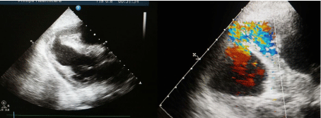
    Figure 2: Transthoracic echocardiographic study image is showing VSD due to rupture of septum in apical segment and doppler echocardiography detected apical
ventricular septum rupture with left to right shunt.
As a conclusion, post MI VSD must be kept on mind as a mechanical complication in patients who admit to emergency department with findings of acute heart failure after uncured extensive acute MI and who have newly developed pansystolic murmur. In case of this complication, patients must be forwarded for emergency surgery. Besides, If the emergency services are very busy, we want to remind physicians that the importance of a detailed physical examination and history of patients to prevent time loss.
References
- Kaya A, Yasemin K, Ordu S, Özköleli M, Özhan H, Dağlar B, et al. Akut Miyokard Enfarktüsü Sonrasi Gelisen Genis Ventriküler Septal Rüptürün Basarili Tedavisi A Successfully Treated Case Of Giant Ventricular Septal Rupture After Acute Myocardial Infarction. Eurasian J Med. 2007; 39: 148– 150.
- Ertas G, Çetinkaya AS, Tatligil S. Apical Ventricular Septal Rupture After Subacute Anterior Myocardial Infarction. Düzce Tip Dergisi. 2013; 15: 54–55.
- Erdoğan MB, Uygur F, Yamak B, Batryaliev T, Kisaciklioğlu B. Surgical Treatment Of Post MI Vsd In Patients Who Were Hemodynamically Stabilized With Preoperative (+) Inotrops And Iabp. Türk Girisimsel Kard Der. 2009; 13: 6–12.
- Drobac M, Gilbert B, Howard R, Baigrie R, Rakowski H. Ventricular Septal Defect After Myocardial Infarction : Diagnosis By Two-Dimensional Contrast Echocardiography. Circulation. 1983; 67: 335–340.
- Orhan G, Yücel O, Biçer Y, Sargin M, Senay S, Ketenci B, et al. Infarktüs Sonrasi Gelisen Ventriküler Septal Defektte Erken Cerrahi Girisim Early Surgical Treatment In Postinfarct Ventricular Septal Defect. Turkish J Thorac Cardiovasc Surg. 2004; 12: 1–5.
- Senocak H, Atesal S, Karakelleoğlu S, Sahin M, Alp N. Akut Miyokard Infarktüsü Sonucu Olusan Ventriküler Septal Defekt. Turkiye Klinikleri J Cardiol. 1992; 5: 190-192.
- Demirtas M, Yapica F, Akar H, Kaplan M, Alhan C, H T, et al. Early Surgical Treatment of Ventricular Septal Rupture in Acute Myocardial infarction. Türk Kardiol Dern Ars. 1996; 24: 234–237.