Clinical Image
Austin J Emergency & Crit Care Med. 2017; 4(2): 1060.
A Patient with Ocular Penetrating Trauma
Heidari SF*
Department of Emergency Medicine, Emam Khomeini Hospital, Medical Faculty, Ilam University of Medical Sciences, Ilam, Iran
*Corresponding author: Seyed Mohammad Reza Hashemian, Chronic Respiratory Disease Research Center (CRDRC), National Research Institute of Tuberculosis and Lung Disease (NRITLD), Shahid Beheshti University of Medical Sciences, Tehran, Iran
Received: June 19, 2017; Accepted: July 19, 2017; Published: July 26, 2017
Clinical Image
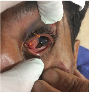
A 43-year-old man presented to the emergency department by EMS because of face trauma. The mechanism of trauma was knocking up a piece of hard wood to the right eye during the break Firewood. Patient had no headache, nausea, vomiting and rhinorrheagia. Past medical history and drug history were negative. Vital sign was stable. On physical examination, he was alert with GCS of 15/15. The patient had no PTA. In physical examination of the right eye, there was severe rupture of cornea and sclera. Uveal tissue is seen outside of eyeball (Uveal prolapse) and, anterior chamber of right eye was flat, that it has been shown in Figure 1. Sight of right eye was in extent of LP. Remnant examinations were unremarkable. After the initial proceedings in emergency department and put the shield on right eye, the patient was transferred to the operation room and repair of laceration of right eye was performed. There is the possibility of extraction or cataract in the damaged eye following trauma.

    Figure 1:  Picture of patient with penetrating trauma to right eye. There is
severe rupture of cornea and sclera. Uveal tissue is seen outside of eyeball
(Uveal prolapse) that were shown with arrows.