Abstract
Ankylosing spondylitis changes the biomechanical properties of the axial spine due to chronic inflammation. The patient might underwent spine fracture and following neurologic sequelae even just low-energy damage. Here we present a young adult with unrevealed ankylosing spondylitis diagnosed with cervical fracture after minimal trauma. Prompt imaging studies establish the diagnosis and subsequently surgical treatment was performed. The postoperative recovery was uneventful.
Keywords: Ankylosing spondylitis; Cervical fracture; Trauma; Computed tomography
Introduction
Ankylosing spondylitis (AS) (also known as Bechterew's disease and Marie Strümpell disease) is a chronic inflammatory disease of unknown cause that primarily affects the axial skeleton. Spinal fractures are up to four times more common in patients with ankylosing spondylitis than the general population, with a lifetime incidence ranging from 5% to 15%. Cervical spine is the most frequent site for acute spinal fractures in AS but was easily delayed or miss diagnosed.
Case Presentation
A 31-year-old young man presented to our clinic with a twoweek history of neck pain after a fall injury. The familial and personal history of the patient wasn’t significant. On examination, he appeared relatively weak. His vital signs were as follows: blood pressure of 124/78mm Hg, heart rate of 78 beats per minute and respiratory rate of 16 breaths per minute. He was a febrile. The neurologic examination revealed marked weaker upper limbs on the right proximally (3/5) and distally (3/5) than the left (3/5 and 3/5). Resting tremor was noticed.Pain and temperature sensation was present and fine touch was intact over shoulders and upper limbs. The neck movement was smooth.
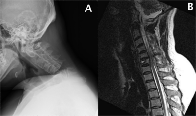
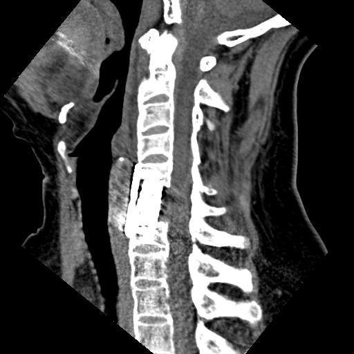
Due to poor response to ketorolac injection, cervical spine X ray was arranged. Plain radiographs incidentally showed ankylosis of the cervical spine and spondylolisthesis of C5 on C6 (Figure 1A). By further magnetic resonance imaging (MRI) of cervical spine, fracture of C6 body and spinal process and mild the cal sac compression of C6 level was disclosed (Figure 1B). Thereafter he was admitted to the trauma service and subsequently underwent C6 corpectomy and titanium mesh cage with anterior cervical plate of C5, C6 and C7 by the neurosurgical service. The human leukocyte antigen B27(HLA-B27) test was positiveeventually. After that the post-operative recovery was uneventful.The Computed Tomography (CT) of cervical spine showed intact alignment with internal instrument fixation over C5-7 with cage fusion at one-month follow-up (Figure 2). The patient was seen in outpatient regular follow-up in half a year and his muscle power gradually recovered after rehabilitation program except mild limitation of neck rotation.

    Figure 1:  Plain radiographs showed ankylosis of the cervical spine and
spondylolisthesis of C5 on C6.
Figure 1B: MRI of cervical spine, fracture of C6 body and spinal process and
mild the cal sac compression of C6 level.

    Figure 2:  Internal instrument fixation over C5-7 with titanium mesh cage
fusion.
Discussion
Ankylosing spondylitis (AS) (also known as Bechterew's disease and Marie Strümpell disease) is a chronic inflammatory disease of unknown cause that primarily affects the axial skeleton. The prevalence of AS was between 0.1% and 1.5% in the United States. The human leukocyte antigen HLA-B27 gene is present in approximately 90% of patients, compared with a prevalence of 1–3/1000 in the general population. It usually found in young adult with the first symptoms in the third decade and male predominant of 3:1 or more was found [1].
Spinal fractures are up to four times more common in patients with ankylosing spondylitis than the general population, with a lifetime incidence ranging from 5% to 15%. AS lead to chronic inflammatory spondyloarthropathy with progressive spinal stiffness that ultimately makes patients susceptible to spinal fractures with traumatic spinal cord injury even from low-energy trauma [2].
Neurologic complication was often presented in AS patient with spinal cord injury at initial presentation [3]. Prompt reduction and stabilization after injury resulted in favorable neurologic outcome [4]. Imaging study of the spine should be considered in patients with AS who complained new or unusual neck and back pain. Immediate identification of stable fractures is substantial in avoiding adverse neurologic outcome result from spine fracture [5]. Computed tomography (CT) may be the first choice with a shorter scanning time, and it can show bonydetails, including fractures and fragments. MR imaging can be much helpful in some patients with diagnostic limitation.6Prompt imaging studies of CT or MRI should be arranged to establish the diagnosis if needed.
AS changes the biomechanical properties of the axial spine due to chronic inflammation. Thereafter the patient with AS might underwent spine fracture and following neurologic sequelae even just minimal trauma [6]. Cervical spine is the most frequent site for acute spinal fractures in AS. Cervical fractures are highly unstable with an increased risk for neurologic deficits and the mortality rate [7]. But it may be neglected by the AS patient, they cannot distinguish the fracture pain from chronic inflammatory pain. Furthermore, the diagnosis may be late diagnosed due to preexisting kyphotic and fusing deformity caused by the disease process [3]. The treatment of spinal fracture in AS should be individualized, accounting for patient preferences and comorbidities [2]. Nonsurgical immobilization is recommended unless spinal dislocation or displacement due to high operative risk and complication rate at specific population [8].
Here we present a young patient with undiagnosed AS underwent cervical fracture after fall injury. Spinal fractures in patients with AS can occur after just minor trauma and may be easily misdiagnosed without detailed history taking and immediate imaging study. In our patient, neck pain with marked weak muscle power and resting tremor may the only clue to early diagnosis with cervical fracture with spondylolisthesis.
References
- Gran JT, Husby G, Hordvik M. Prevalence of ankylosing spondylitis in males and females in a young middle-aged population of tromso, northern norway. Annals of the rheumatic diseases. 1985; 44: 359-367.
- Mathews M, Bolesta MJ. Treatment of spinal fractures in ankylosing spondylitis. Orthopedics. 2013; 36: e1203-1208.
- Chaudhary SB, Hullinger H, Vives MJ. Management of acute spinal fractures in ankylosing spondylitis. ISRN rheumatology. 2011; 2011: 150484.
- Hanson JA, Mirza S. Predisposition for spinal fracture in ankylosing spondylitis. AJR. American journal of roentgenology. 2000; 174: 150.
- Carnell J, Fahimi J, Wills CP. Cervical spine fracture in ankylosing spondylitis. The western journal of emergency medicine. 2009; 10: 267.
- Wang YF, Teng MM, Chang CY, Wu HT, Wang ST. Imaging manifestations of spinal fractures in ankylosing spondylitis. AJNR. American journal of neuroradiology. 2005; 26: 2067-2076.
- Westerveld LA, Verlaan JJ, Oner FC. Spinal fractures in patients with ankylosing spinal disorders: A systematic review of the literature on treatment, neurological status and complications. European spine journal: official publication of the European Spine Society, the European Spinal Deformity Society, and the European Section of the Cervical Spine Research Society. 2009; 18: 145-156.
- Weinstein PR, Karpman RR, Gall EP, Pitt M. Spinal cord injury, spinal fracture, and spinal stenosis in ankylosing spondylitis. Journal of neurosurgery. 1982; 57: 609-616.
