Abstract
A 62-year-old man with history of cigarette smoking presented with fatigue, lightheadedness, exertional dyspnea, lower extremity swelling, ecchymoses, and petechiae. There was no history of trauma, infection, new medications, or abnormal diet. Physical exam revealed red petechial 3-5mm macules and pink-violaceous purpuric indurated patches over the bilateral upper and lower extremities, buttocks, and lower abdomen. Corkscrew hairs were noted on the bilateral lower extremities. Laboratory studies were significant for anemia and elevated acute phase reactants. Thiamine, folate, and vitamin B12 were within normal limits. Workup for thrombocytopenia, platelet dysfunction, coagulopathies, hemolysis, vasculitidies, liver or gastrointestinal disease, rheumatologic disorders, and bone marrow disorders was negative. Skin biopsy and histology revealed dermal extravasated erythrocytes without evidence of vasculitis or thrombi. Ascorbic acid plasma concentration was then tested and below the limit of detection on hospital day six. A diagnosis of scurvy was made, and the patient was discharged on 1000mg Vitamin C daily supplementation. At two-week follow up, constitutional symptoms were resolved, anemia corrected, and cutaneous symptoms improving. The medical, hematology, and dermatology teams did not originally suspect a vitamin C deficiency in this patient. This case emphasizes the importance of the consideration of scurvy on the differential of petechiae, even in patients who do not present with the typical risk factors or features. Medical providers should consider scurvy, particularly in patients at risk for malnutrition due to chronic conditions and/or history of alcohol or tobacco use disorder.
Keywords: Vitamin C; Scurvy; Petechiae
Case Presentation
A 62-year-old man with a history of cigarette smoking presented with seven days of progressive fatigue, lightheadedness, exertional dyspnea, lower extremity swelling, ecchymoses, and petechiae. There was no history of trauma, infection, new medications, or abnormal diet. This was the patient’s third presentation to the hospital with these findings over two years, without diagnosis or improvement. During this period of time he was found to have a macrocytic anemia with hemoglobin between 7.0 - 8.0 requiring six transfusions. Previous work up was extensive, including testing for G6PD deficiency, multiple myeloma, and MGUS. Laboratory testing was previously normal, including results for vitamin B12, folate, thiamine, vitamin D, copper, zinc, closure time, thrombin time, prothrombin time, activated partial thromboplastin time, reticulocytes, direct coombs, bilirubin, haptoglobin, lactate dehydrogenase, cold agglutin, iron studies, antineutrophil cytoplasmic antibodies, myeloperoxidase, uric acid, erythrocyte sedimentation rate, c-reactive protein, and thyroid stimulating hormone. The patient had previously undergone temporal artery biopsy and bone marrow biopsy with normal results. He was followed outpatient by hematology, rheumatology, vascular surgery, and his primary care provider. He was adherent to a vitamin regimen consisting of vitamin B12, folate, and vitamin D supplements.
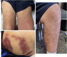
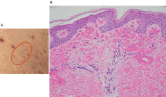
The patient was well-nourished, with a body mass index of 30. Physical exam revealed red petechial 3-5mm macules and pink-violaceous purpuric indurated patches over the bilateral upper and lower extremities, buttocks, and lower abdomen (Figure 1A and 1B). Corkscrew hairs were noted on the bilateral lower extremities (Figure 2A). The lower extremities were swollen bilaterally. There was no hepatosplenomegaly.

    Figure 1: Physical Exam and Perifollicular Hemorrhage. A) Before treatment:
Petechiae on thighs. B) Before treatment: Ecchymoses on buttocks. C) After
two week’s treatment: Lower extremities.

    Figure 2: Corkscrew Hair and Perifollicular Hemorrhage. A) Example of a
corkscrew hair. B) Extravasated erythrocytes and iron laden histiocytes.
Updated laboratory studies were significant for anemia and elevated acute phase reactants. Folate and vitamin B12 were within normal limits. Fecal occult blood test was negative. Repeat workup for platelet dysfunction, coagulopathies, hemolysis, vasculitidies, liver or gastrointestinal disease, rheumatologic disorders, and bone marrow disorders was negative. Histology from a skin biopsy revealed dermal extravasated erythrocytes without evidence of vasculitis or thrombi (Figure 2B). An ascorbic acid level was sent; plasma concentration was below the limit of detection. A diagnosis of scurvy was made and the patient was discharged on 1000mg vitamin C daily. At two-week follow up, constitutional symptoms were resolved, anemia corrected, and cutaneous symptoms improving (Figure 1C). At four-week follow up, cutaneous signs had resolved.
Discussion/Conclusion
Vitamin C deficiency was not originally suspected in the patient presented in this case. The patient kept a relatively normal diet, comprised of eggs, toast, salads, pastas with vegetables, and no alcohol use since quitting 15 years prior. He did not have any disorders of the gastrointestinal tract; his only risk factor for scurvy was his tobacco use. On exam, he lacked the classic gingival findings and follicular hyperkeratosis but had scant perifollicular hemorrhage.
In high-income countries such as the United States, approximately one-third of hospitalized patients are either at risk for malnutrition or are already malnourished at the time of admission [1]. This rate is higher amongst patients with chronic conditions and malabsorptive disorders [2]. Consequently, malnourishment due to micronutrient deficiency is an important consideration in patients with abnormal hematologic findings, such as bruising and petechiae. Importantly, Vitamin C deficiency may occur in patients with chronic alcohol use, psychiatric illness, or gastrointestinal tract disease.
In the case of our patient, it is suspected that his smoking history and remote history of alcohol use disorder may have contributed to malabsorption of ascorbic acid. Smoking is associated with depleted serum levels of ascorbic acid, thought to be related to the oxidative burden it causes [3].
This case emphasizes the importance of the consideration of scurvy in the differential of petechiae, even in patients who do not present with the typical risk factors or features. Medical providers should consider scurvy, particularly in patients at risk for malnutrition due to chronic conditions and those with a history of alcohol or tobacco use disorders. Both inpatient and outpatient providers play an important role in the early recognition of nutritional deficiencies, as cutaneous manifestations often are the first clue to diagnosis. Nutritional deficiencies are common yet underrecognized in hospitalized patients and serve as an independent risk factor for patient morbidity and mortality [4]. Awareness of the cutaneous manifestations of undernutrition, as well as the risk factors for nutritional deficiency, may expedite diagnosis and supplementation. This would improve outcomes for hospitalized patients and by default reduce unnecessary procedures, testing, and health care costs. This case illustrates an atypical presentation of vitamin C deficiency, reviews diagnostic workup, and provides suggestions for supplementation in undernourished patients.
References
- Sorensen J, Kondrup J, Prokopowicz J, Schiesser M, Krähenbühl L, Meier R, et al. EuroOOPS Study Group. EuroOOPS: an international, multicentre study to implement nutritional risk screening and evaluate clinical outcome. Clinical Nutrition. 2008; 27: 340-349.
- Reber E, Gomes F, Bally L, Schuetz P, Stanga Z. Nutritional management of medical inpatients. Journal of Clinical Medicine. 2019; 8: 1130.
- Yanbaeva DG, Dentener MA, Creutzberg EC, Wesseling G, Wouters EF. Systemic effects of smoking. Chest. 2007; 131: 1557-1566.
- Hirschmann JV, Raugi GJ. Adult scurvy. J Am Acad Dermatol. 1999; 41: 895- 910.
