Abstract
Background: Sever’s is the most common cause of heel pain in the pediatric population and increases the risk of calcaneal apophyseal avulsion fractures. This is an uncommon fracture that faces problems with tenuous soft tissue coverage and high shear forces at the fracture site. Multiple fixation techniques have been employed in addressing this injury, with no one method standing out as superior.
Case Presentation: We present a case of a 13-year-old male with a calcaneal apophyseal avulsion fracture in which cannulated headless compression screws were used for fixation. This was found to provide adequate compression across the fracture with minimal hardware prominence. This also allowed for the injection of radiopaque solution through the cannulated screw to rule out subtalar intraarticular penetration. We believe this represents a novel method of addressing these injuries.
Conclusion: Cannulated headless compression screws are a suitable fixation technique for pediatric apophyseal avulsion fractures that provide the added benefit of assessment of intra-articular penetration of the subtalar joint by the injection of radiopaque solution through the cannulated screw.
Keywords: Sever’s; Apophysitis; Apophyseal Avulsion; Headless Compression Screw
Background
This is a case report looking at a 13-year-old male with a right calcaneal avulsion fracture after a long-term history of tight heel cords treated in a novel manner with a technique not previously described in pediatric literature.
Sever’s disease is the most common cause of heel pain in the pediatric population, affecting approximately 3.7 in 1,000 patients.1 This is thought to be due to repetitive stress at the open calcaneal apophysis. This growth plate typically closes around age 14 and, until that time, is vulnerable to repetitive shear forces and subsequent inflammation, primarily due to the pull of the Achilles tendon [1]. Given the path anatomy of the disease process, it is unsurprising that tight heel cords are a predisposing factor to apophysitis. During the period of accelerated growth in early adolescence, the longitudinal growth of the bone often outpaces the elongation ability of the muscle and tendon, inclining the patient toward tight heel cords [2].
Sever’s disease may also result in an increased risk of apophyseal fracture [3]. This is an uncommon injury but one that frequently requires operative intervention. Multiple techniques of fixation have been described in the literature including closed reduction and percutaneous pinning, fragment excision and Achilles tendon advancement, and open reduction with internal fixation [3,4]. To our knowledge, this is the first case described utilizing headless compression screws for open reduction and internal fixation of an acute calcaneal apophyseal avulsion. In addition we also demonstrate a technique that uses radio-opaque dye to confirm that the screws are not intra-articular.
Case Presentation
Our patient is a 13-year-old male who presented to the emergency department with acute right sided heel pain and inability to ambulate after forcefully jumping up and down on a diving board while visiting a family friends’ pool. His mother was at the poolside and assisted him in exiting the pool. The patient sustained no other injuries, and the orthopedics department was consulted after images were obtained.
While the patient was overall healthy, meeting all milestones with no regular medications or prior surgeries, he did have a significant history of “toe-walking” per mom. He was seen by a physiatrist and diagnosed with tight heel cords, right being worse than left. At the time he was prescribed hinged ankle-foot orthoses which have been regularly updated yearly. Although the patient and his mother reported regular use of bracing and stretching, he has continued to toe walk and experience pain in his right heel. They deny any family history or prior fractures. He has not previously been evaluated by an orthopedist.
In the pediatric emergency room, the patient was found to have significant swelling and ecchymosis at the posterior aspect of his lower leg and heel. His skin was intact, and the soft tissue was compressible. He was neurologically intact in regard to sensation in all distributions and he had the ability to fire extensor hallucis longus, flexor hallucis longus and tibialis anterior. He was unable to plantarflex the ankle. He was tender to palpation at the posterior aspect of his lower leg and heel. The calcaneal tendon was palpable and not appreciably disrupted. His contralateral ankle and foot were normal, and he was ankle to dorsiflex several degrees above neutral with the leg extended though he did have a positive silfverskiold. The right heel was placed in a soft, well-padded bulky dressing. There was no immediate/ acute concern for skin threatening, however he was admitted to the orthopedic service with frequent skin checks overnight and scheduled surgery as the first case in the morning.
Initially right calcaneus, ankle and foot radiographs were obtained. The films demonstrated a posterior calcaneal tuberosity avulsion fracture that extended through the physis and through the distal epiphysis (Figure 1). An axial calcaneus radiograph is supplied below (Figure 2). Computed tomography studies were obtained by the emergency department and allowed for better evaluation of the fracture. Several isolated cuts are provided below (Figure 3). The displaced mildly comminuted fracture involved the apophysis but also involved the underlying subcortical bone and exiting through the distal third physis making this a Salter Harris type 4 equivalent, though it functioned clinically as a type 3. The displaced avulsed fragment measured 3.5 cm at its greatest craniocaudal height with the intact calcaneal tendon inserting on this fragment. The fragment was superiorly displaced approximately 1.4 cm.
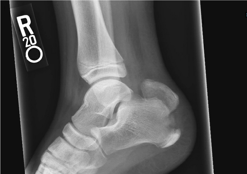
    Figure 1: Lateral radiograph of right ankle/calcaneus demonstrating posterior

calcaneal tuberosity avulsion fracture.
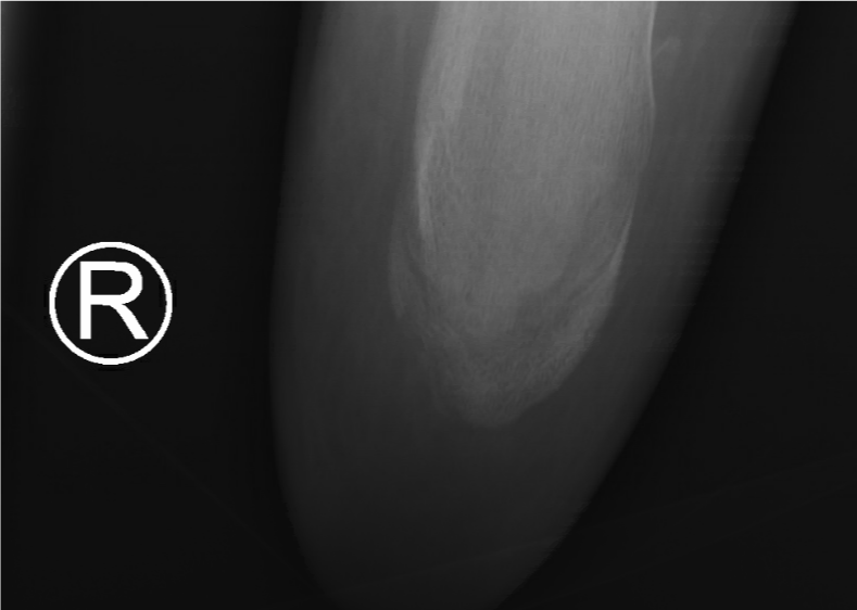
    Figure 2: Axial radiograph of right ankle again demonstrating avulsion

fracture of posterior calcaneal tuberosity.
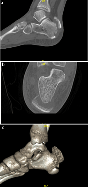
    Figure 3a-c: Selected CT cuts of the posterior calcaneal tuberosity fracture

including 3D reconstruction.
Pre-operative planning included multiple potential interventions. The patient’s history of tight heel cords was taken into account and the patient and family were counselled about the potential need for future intervention for lengthening. While concomitant lengthening was considered, it was the senior author’s opinion that it would be optimal to treat it separately as our post operative splinting, casting, bracing and physical therapy would be contrasting for the two separate issues. We discussed high potential early closure of the physis after this injury. The risk for soft tissue issues post op was also discussed as well as potential left sided injury in the future. When considering treatment nonoperative treatment was decided against given the displacement greater than 1cm, which would leave the patient with a calcaneal defect and weakened ability to plantarflex. Given the fracture pattern, physiologic age of patient and good soft tissue coverage we decided that we would refrain from closed reduction with percutaneous fixation and move forward with open reduction and fixation, especially since outcome correlates directly with quality of reduction. Once the approach was determined, type of fixation was then considered. Options included suture anchors, lag screws and tension band construct. A potential solution was to use headless screw fixation.
While headless screws have been described in the adult population there is a paucity of data regarding its use in the setting of traumatic posterior calcaneal avulsions in the pediatric population. Its benefits include avoiding a potential screw head prominence in the heel which has been described as having a high rate of symptomatic hardware resulting in subsequent surgery for removal. There is a significant cost described in prior literature for the return to the OR [5]. This is important because while the hardware itself may cost more than the additional options, overall it is a cost-effective alternative. In addition, you could spare a child from additional anesthesia exposure and the traumatic experience of surgery. In hand literature a potential concern of use of headless screw fixation is inadequate fixation in the setting of osteoporotic bone.6 In the pediatric population with a healthy child this is not typically an issue, and their bone quality is adequate.
Patient was taken into the operative room, induced, and intubated by anesthesia and placed in the prone position with care taken to ensure all bony prominences were well padded. Antibiotic prophylaxis was administered within one hour of operative start time. A raft of blankets used to position operative extremity. A tourniquet was applied to the right upper thigh. The extremity was prepped and draped in usual, sterile fashion using a Betadine wash. A 4cm longitudinal incision was made over the distal medial calcaneus with #15 blade scalpel. Blunt dissection was carried down to the periosteum. The calcaneal tendon was confirmed to be intact as well as its insertion on the fragment. The fracture site was visualized, and a pointed reduction clamp was used to bring the avulsed fragment distally. A 2.0 K wire was used to hold the reduction (Figure 4). Fluoroscopy was used to confirm the reduction. K wires compatible with the acutrak plus headless screws were placed and the opening drill was used first distally (Figure 5). The appropriate screw was selected and placed. We then repeated the same process at the proximal screw. Imaging was used to confirm reduction and correct placement (Figure 6). To confirm that the screws did not penetrate the subtalar joint we injected omnipaque through the cannulated screw using the included driver and a sterile syringe (Figure 7). We also injected subtalar and did not see any expressed at the screw heads (Figure 8). The wound was thoroughly irrigated and closed with nylon suture. After sterile dressing we placed the patient in a posterior slab splint in 10 degrees of plantarflexion which was the position that the ankle naturally maintained while the ipsilateral knee flexed. The patient was gradually awakened from anesthesia and taken to PACU with no apparent complication and was discharged the same day with a plan to maintain non-weight bearing status and follow up in office.
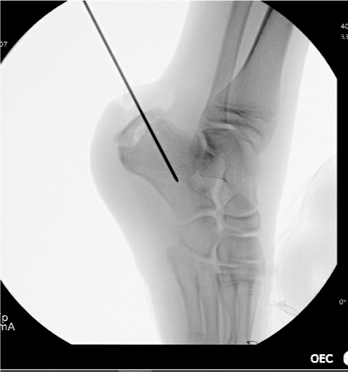
Figure 4: Fracture reduction with 2.0 K wire maintaining the reduction.
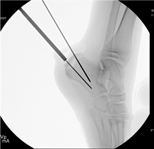
    Figure 5: Placement of acutrak compatible K wire at distal aspect of fragment

across fracture site.
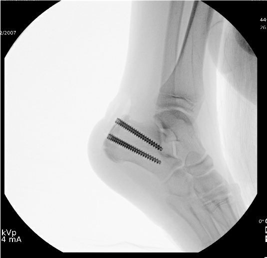
Figure 6: Imaging of both headless screws in place.
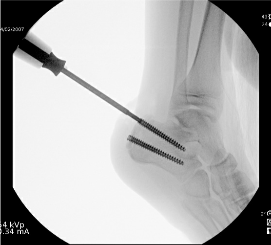
    Figure 7: Injection through cannulated screw using omnipaque with no

subtalar infiltration.
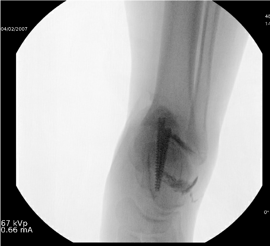
    Figure 8: Imaging of calcaneus after injection of omnipaque into the subtalar

joint with no visualization indicating communication with the screw.
The patient did well on outpatient follow up. His pain was well controlled, and his sutures were removed with no soft tissue complications. He had full sensation and intact plantarflexion. His non-weight bearing status was maintained in a pneumatic walking boot until 6 weeks post op. At that time, he began physical therapy and was eager to return to sports. At his 6-month post op follow up he reported no complaints, and the patient and his mother felt that his toe walking had overall improved since his injury. He had returned to soccer and was at full activity. His dorsiflexion was measured at 15 degrees bilaterally. Radiographs at that time demonstrated hardware intact with healing of the fracture (Figure 9). He is planned to follow up at 9 months post op without any restrictions or bracing.
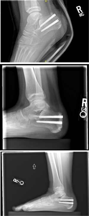
    Figure 9: Two-week, Six-week and Six-month follow up lateral radiographs in

consecutive order from left to right.
Conclusion
This is a case report looking at a novel technique for fixation of an acute, traumatic posterior calcaneal tubercle avulsion in a pediatric patient with a history of minimally treated Severs disease and tight heel cords. Headless screws have been described in the adult literature and in different areas of the body, but this report provides a case with follow up to a period where the child has returned to full activity without any complications.
Headless compression screws have a variety of demonstrated applications. Their use has been validated across many fracture patterns and locations including fractures of the scaphoid, distal femur, capitellum, talus, and others.7 While headless screws are traditionally thought to offer decreased compression and pullout strength, others have shown equivalent biomechanical properties to traditional screws.8 They are commonly employed in areas of cancellous bone, particularly when purchase in the far cortex would be impossible or inappropriate, because the variable pitch of the screw does not rely on bicortical fixation to provide compression.9 An additional benefit of this fixation technique is the lack of hardware prominence, something of particular importance in areas with tenuous soft tissue protection such as the heel [7].
A potential challenge of this case regarding timing of heel cord lengthening ended up being a non-issue. This was surprising given that the child was maintained in a plantarflexed position post op. His recovery in function and improvement in dorsiflexion may have benefitted from his post op physical therapy, though he had had trials in the past.
Another unique aspect of this case is the use of omnipaque dye through the cannulated driver and screw to confirm that the hardware was not intra-articular. This technique has not been described with this system. It allows you to place the longest possible screw safely, confirming the best chance of adequate fixation.
While headless screws have been described in the adult population, children provide an additional subject for their use given the quality of bone and desire to avoid additional surgeries for symptomatic hardware. We provide a case that supports the consideration of headless screw fixation for avulsed calcaneal tuberosity fractures and would recommend the technique described above using radioopaque dye to confirm that there is no intra-articular penetrance.
Consent for Publication
Written informed consent was obtained from the patient for publication of this case report and accompanying images.
Availability of Data and Material
All data underlying the results are available as part of the article and no additional source data are required.
Competing Interests
The authors declare that they have no competing interests.
Funding
No funding was provided for this report.
Author’s Contributions
BL assisted in the surgical intervention and was a major contributor review of the literature and in writing the manuscript.
SB was a major contributor in review of the literature and in writing the manuscript.
DW performed the surgical intervention and was the primary investigator for the report.
All authors read and approved the final manuscript.
References
- Wiegerinck JI, Yntema C, Brouwer HJ, Struijs PAA. Incidence of calcaneal apophysitis in the general population. European Journal of Pediatrics. 2013; 173(5): 677-679. doi:10.1007/s00431-013-2219-9
- Rodríguez-Sanz D, Becerro-de-Bengoa-Vallejo R, López-López D, Calvo- Lobo C, Martínez-Jiménez EM, Perez-Boal E, et al. Slow velocity of the center of pressure and high heel pressures may increase the risk of Sever’s disease: a case-control study. BMC Pediatrics. 2018; 18(1). doi:10.1186/ s12887-018-1318-1
- Lee KT, Young KW, Park YU, Park SY, Kim KC. Neglected Sever’s Disease as a Cause of Calcaneal Apophyseal Avulsion Fracture: Case Report. Foot & Ankle International. 2010; 31(8): 725-728. doi:10.3113/FAI.2010.0725
- Imai Y, Kitano T, Nakagawa K, Takaoka K. Calcaneal apophyseal avulsion fracture. Archives of Orthopaedic and Trauma Surgery. 2007; 127(5): 331- 333. doi:10.1007/s00402-007-0297-8
- Boren M, Steginsky BD, Vora AM, Suhling M. Headless vs. Headed Screws Fixation for Calcaneal Osteotomy and Subtalar Arthrodesis: What’s the Cost?. Foot & Ankle Orthopaedics. 2020; 5(4): 2473011420S2473000142.
- Goodwin JA, Castañeda P, Shelhamer RP, Bosch LC, Edwards SG. A Comparison of Plate Versus Screw Fixation for Segmental Scaphoid Fractures: A Biomechanical Study. HAND. 2019; 14(2): 203-208. doi:10.1177/1558944717732065
- Cheng RZ, Wegner AM, Behn AW, Amanatullah DF. Headless compression screw for horizontal medial malleolus fractures. Clinical Biomechanics. 2018; 55: 1-6. doi:10.1016/j.clinbiomech.2018.03.023
- Capelle JH, Couch CG, Wells KM, Morris RP, Buford WL, Merriman DJ, et al. Fixation Strength of Anteriorly Inserted Headless Screws for Talar Neck Fractures. Foot & Ankle International. 2013; 34(7): 1012-1016. doi:10.1177/1071100713479586
- Reimer H, Kreibich M, Oettinger W. Extended uses for the Herbert/Whipple screw: six case reports out of 35 illustrating technique. Journal of orthopaedic trauma. 1996; 10(1): 7-14. doi:10.1097/00005131-199601000-00002